 ムービー
ムービー コントローラー


+ データを開く

- 基本情報
基本情報
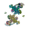
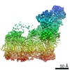
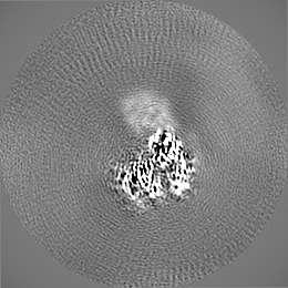
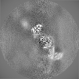
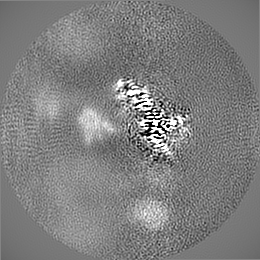
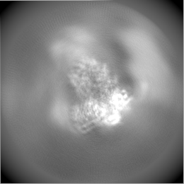
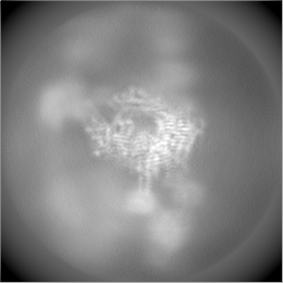
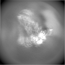
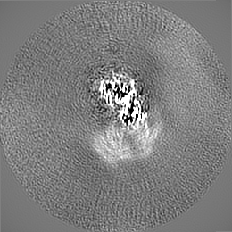
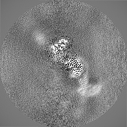
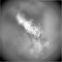
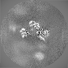
| 登録情報 | データベース: EMDB / ID: EMD-23083 | |||||||||
|---|---|---|---|---|---|---|---|---|---|---|
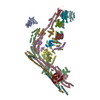
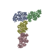
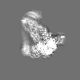
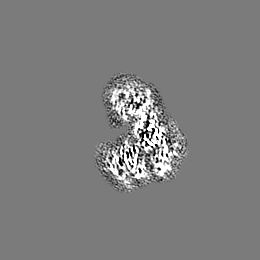
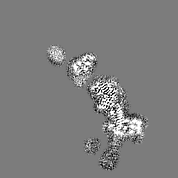
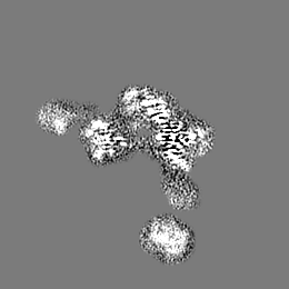
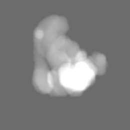
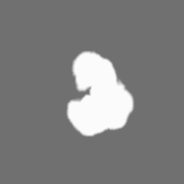
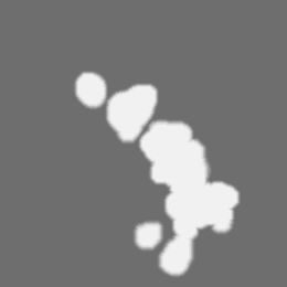
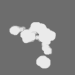
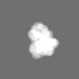
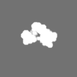
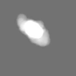
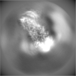
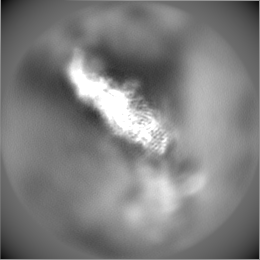
| タイトル | Outer dynein arm core subcomplex from C. reinhardtii | |||||||||
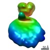
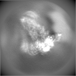
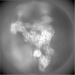
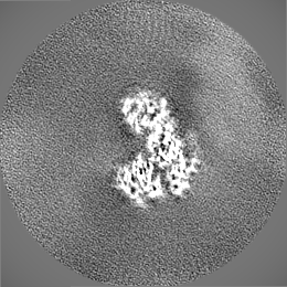
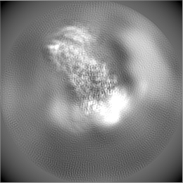
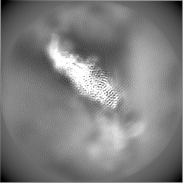
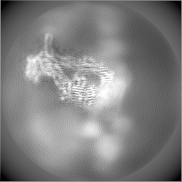
 マップデータ マップデータ | composite ODA core map from focused refinements | |||||||||
 試料 試料 |
| |||||||||
 キーワード キーワード | dynein / microtubule / cilia / MOTOR PROTEIN | |||||||||
| 機能・相同性 |  機能・相同性情報 機能・相同性情報outer dynein arm / outer dynein arm assembly / cilium movement involved in cell motility / 9+2 motile cilium / dynein light chain binding / cilium movement / motile cilium assembly / dynein heavy chain binding / axonemal dynein complex / dynein complex ...outer dynein arm / outer dynein arm assembly / cilium movement involved in cell motility / 9+2 motile cilium / dynein light chain binding / cilium movement / motile cilium assembly / dynein heavy chain binding / axonemal dynein complex / dynein complex / cell projection organization / minus-end-directed microtubule motor activity / dynein light intermediate chain binding / cytoplasmic dynein complex / ciliary plasm / motile cilium / dynein intermediate chain binding / microtubule-based movement / axoneme / microtubule-based process / enzyme regulator activity / microtubule / calcium ion binding / ATP hydrolysis activity / ATP binding / cytoplasm 類似検索 - 分子機能 | |||||||||
| 生物種 |   Chlamydomonas reinhardtii (クラミドモナス) Chlamydomonas reinhardtii (クラミドモナス) | |||||||||
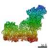
| 手法 | らせん対称体再構成法 / クライオ電子顕微鏡法 / 解像度: 4.0 Å | |||||||||
 データ登録者 データ登録者 | Walton T / Wu H | |||||||||
 引用 引用 | ジャーナル: Nat Commun / 年: 2021 タイトル: Structure of a microtubule-bound axonemal dynein. 著者: Travis Walton / Hao Wu / Alan Brown /  要旨: Axonemal dyneins are tethered to doublet microtubules inside cilia to drive ciliary beating, a process critical for cellular motility and extracellular fluid flow. Axonemal dyneins are evolutionarily ...Axonemal dyneins are tethered to doublet microtubules inside cilia to drive ciliary beating, a process critical for cellular motility and extracellular fluid flow. Axonemal dyneins are evolutionarily and biochemically distinct from cytoplasmic dyneins that transport cargo, and the mechanisms regulating their localization and function are poorly understood. Here, we report a single-particle cryo-EM reconstruction of a three-headed axonemal dynein natively bound to doublet microtubules isolated from cilia. The slanted conformation of the axonemal dynein causes interaction of its motor domains with the neighboring dynein complex. Our structure shows how a heterotrimeric docking complex specifically localizes the linear array of axonemal dyneins to the doublet microtubule by directly interacting with the heavy chains. Our structural analysis establishes the arrangement of conserved heavy, intermediate and light chain subunits, and provides a framework to understand the roles of individual subunits and the interactions between dyneins during ciliary waveform generation. | |||||||||
| 履歴 |
|
- 構造の表示
構造の表示
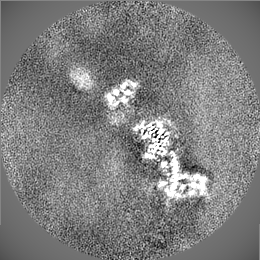
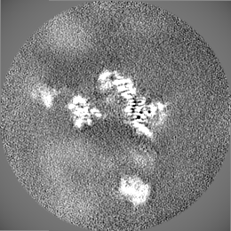
| ムービー |
ムービービューア |
|---|---|
| 構造ビューア | EMマップ: SurfViewMolmilJmol/JSmol |
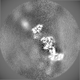
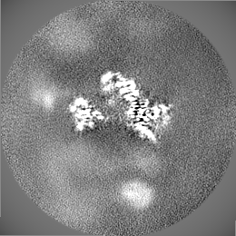
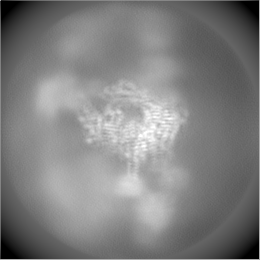
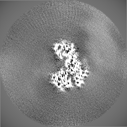
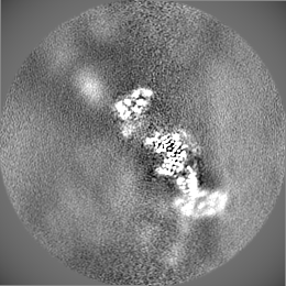
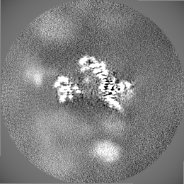
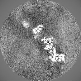
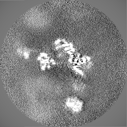
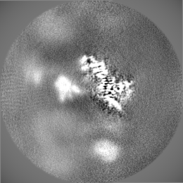
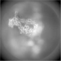
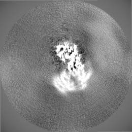
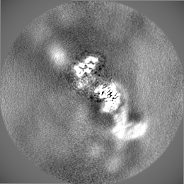
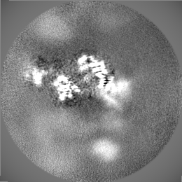
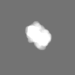
| 添付画像 |

- ダウンロードとリンク
-EMDBアーカイブ
| マップデータ | emd_23083.map.gz | 23.7 MB | EMDBマップデータ形式 | |
|---|---|---|---|---|
| ヘッダ (付随情報) | emd-23083-v30.xmlemd-23083.xml | 76.1 KB 76.1 KB | 表示 表示 | EMDBヘッダ |
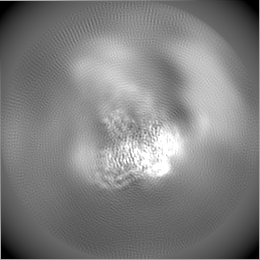
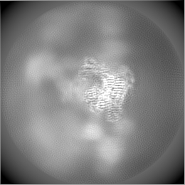
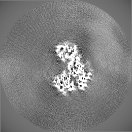
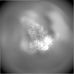
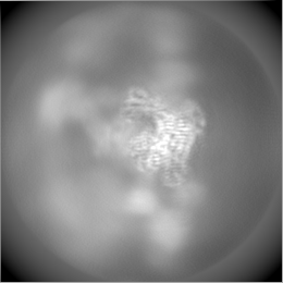
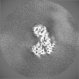
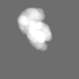
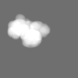
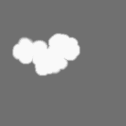
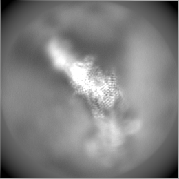
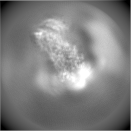
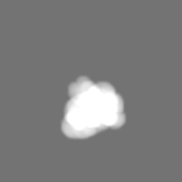
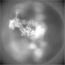
| 画像 | 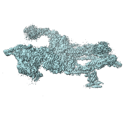 emd_23083.png emd_23083.png | 119.5 KB | ||
| マスクデータ | emd_23083_msk_1.map | 67 MB | マスクマップ | |
| Filedesc metadata | emd-23083.cif.gz | 17.4 KB | ||
| その他 | emd_23083_additional_1.map.gzemd_23083_additional_10.map.gzemd_23083_additional_11.map.gzemd_23083_additional_12.map.gzemd_23083_additional_13.map.gzemd_23083_additional_14.map.gzemd_23083_additional_15.map.gzemd_23083_additional_16.map.gzemd_23083_additional_2.map.gzemd_23083_additional_3.map.gzemd_23083_additional_4.map.gzemd_23083_additional_5.map.gzemd_23083_additional_6.map.gzemd_23083_additional_7.map.gzemd_23083_additional_8.map.gzemd_23083_additional_9.map.gz | 60.3 MB 59.9 MB 60 MB 59.9 MB 6.5 MB 60.1 MB 60.2 MB 354.7 KB 196.1 KB 248.6 KB 59.9 MB 59.8 MB 192.3 KB 59.7 MB 60.3 MB 60.4 MB | ||
| アーカイブディレクトリ |  http://ftp.pdbj.org/pub/emdb/structures/EMD-23083ftp://ftp.pdbj.org/pub/emdb/structures/EMD-23083 http://ftp.pdbj.org/pub/emdb/structures/EMD-23083ftp://ftp.pdbj.org/pub/emdb/structures/EMD-23083 | HTTPS FTP |
-検証レポート
| 文書・要旨 | emd_23083_validation.pdf.gz | 533 KB | 表示 | EMDB検証レポート |
|---|---|---|---|---|
| 文書・詳細版 | emd_23083_full_validation.pdf.gz | 532.6 KB | 表示 | |
| XML形式データ | emd_23083_validation.xml.gz | 5.9 KB | 表示 | |
| CIF形式データ | emd_23083_validation.cif.gz | 6.8 KB | 表示 | |
| アーカイブディレクトリ | https://ftp.pdbj.org/pub/emdb/validation_reports/EMD-23083ftp://ftp.pdbj.org/pub/emdb/validation_reports/EMD-23083 | HTTPS FTP |
-関連構造データ

-リンク
| EMDBのページ | EMDB (EBI/PDBe) / EMDataResource |
|---|---|
| 「今月の分子」の関連する項目 |


-マップ
| ファイル | ダウンロード / ファイル: emd_23083.map.gz / 形式: CCP4 / 大きさ: 67 MB / タイプ: IMAGE STORED AS FLOATING POINT NUMBER (4 BYTES) | ||||||||||||||||||||||||||||||||||||||||||||||||||||||||||||
|---|---|---|---|---|---|---|---|---|---|---|---|---|---|---|---|---|---|---|---|---|---|---|---|---|---|---|---|---|---|---|---|---|---|---|---|---|---|---|---|---|---|---|---|---|---|---|---|---|---|---|---|---|---|---|---|---|---|---|---|---|---|
| 注釈 | composite ODA core map from focused refinements | ||||||||||||||||||||||||||||||||||||||||||||||||||||||||||||
| ボクセルのサイズ | X=Y=Z: 1.36 Å | ||||||||||||||||||||||||||||||||||||||||||||||||||||||||||||
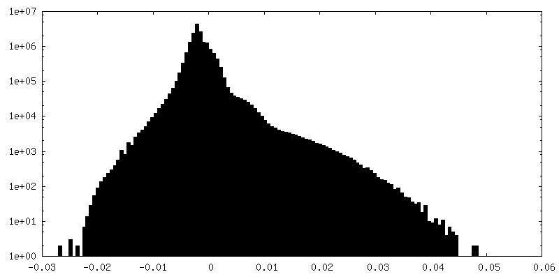
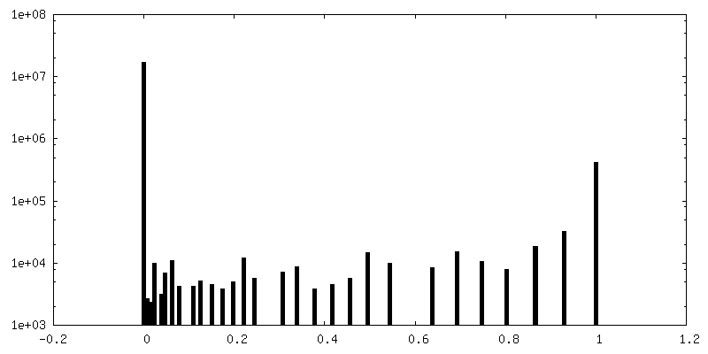
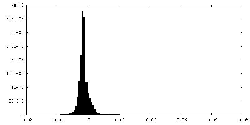
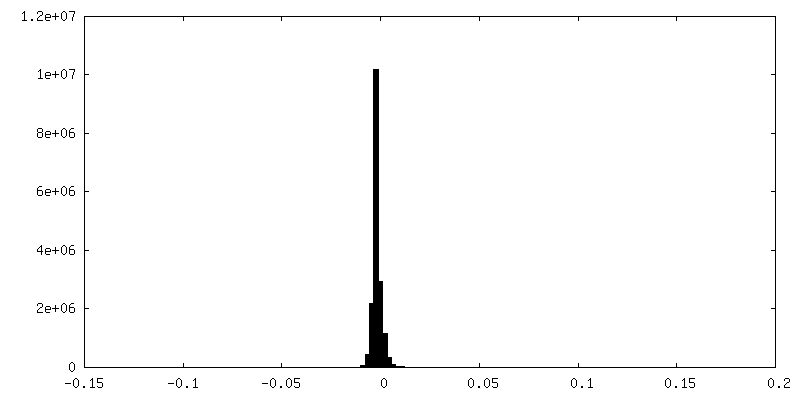
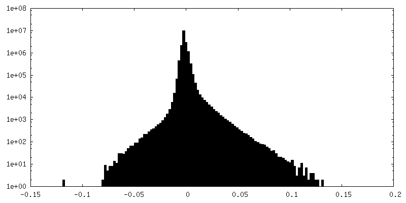
| 密度 |
| ||||||||||||||||||||||||||||||||||||||||||||||||||||||||||||
| 対称性 | 空間群: 1 | ||||||||||||||||||||||||||||||||||||||||||||||||||||||||||||
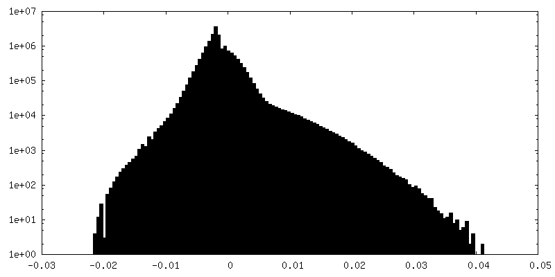
| 詳細 | EMDB XML:
CCP4マップ ヘッダ情報:
| ||||||||||||||||||||||||||||||||||||||||||||||||||||||||||||
-添付データ
+マスク #1
 Z
Z Y
Y X
X


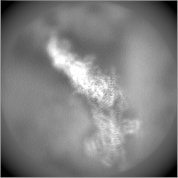
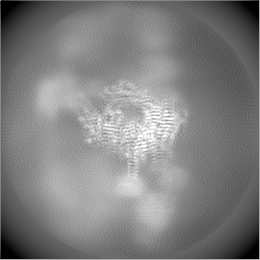
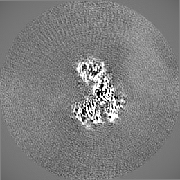
+追加マップ: focused refinement around IC2

+追加マップ: half map 1 for focused refinement around IC1 and LC7a/b


+追加マップ: half map 2 for focused refinement around IC1 and LC7a/b

+追加マップ: half map 2 for focused refinement around IC2


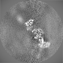
+追加マップ: refinement of the entire ODA core
+追加マップ: half map 2 for refinement of the entire ODA core

+追加マップ: half map 1 for refinement of the entire ODA core
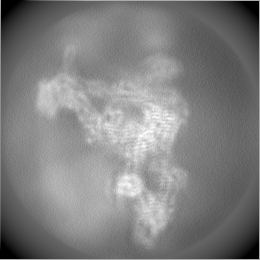

+追加マップ: mask for refinement of the entire ODA core


+追加マップ: mask for focused refinement around IC1 and LC7a/b
+追加マップ: mask for focused refinement around LC8


+追加マップ: half map 1 for focused refinement around IC2


+追加マップ: half map 1 for focused refinement around LC8

+追加マップ: mask for focused refinement around IC2


+追加マップ: half map 2 for focused refinement around LC8

+追加マップ: focused refinement around LC8

+追加マップ: focused refinement around IC1 and LC7a/b


- 試料の構成要素
+全体 : ODA core subcomplex
+超分子 #1: ODA core subcomplex
+分子 #1: Heavy chain alpha
+分子 #2: Flagellar outer dynein arm heavy chain beta
+分子 #3: Dynein gamma chain, flagellar outer arm
+分子 #4: Dynein, 78 kDa intermediate chain, flagellar outer arm
+分子 #5: Dynein, 70 kDa intermediate chain, flagellar outer arm
+分子 #6: Flagellar outer dynein arm light chain 2
+分子 #7: Dynein 18 kDa light chain, flagellar outer arm
+分子 #8: Dynein 11 kDa light chain, flagellar outer arm
+分子 #9: Dynein light chain roadblock LC7a
+分子 #10: Dynein light chain roadblock LC7b
+分子 #11: Dynein 8 kDa light chain, flagellar outer arm
+分子 #12: Dynein light chain 9
+分子 #13: Dynein light chain 10
+分子 #14: DC1
+分子 #15: DC2
+分子 #16: Outer dynein arm-docking complex protein DC3
-実験情報
-構造解析
| 手法 | クライオ電子顕微鏡法 |
|---|---|
 解析 解析 | らせん対称体再構成法 |
| 試料の集合状態 | filament |
-試料調製
| 濃度 | 20 mg/mL | |||||||||||||||||||||
|---|---|---|---|---|---|---|---|---|---|---|---|---|---|---|---|---|---|---|---|---|---|---|
| 緩衝液 | pH: 7.4 構成要素:
詳細: Buffer also contained 1x Protease Arrest (G-Biosciences) | |||||||||||||||||||||
| グリッド | モデル: C-flat-1.2/1.3 / 材質: COPPER / メッシュ: 400 / 前処理 - タイプ: GLOW DISCHARGE / 前処理 - 時間: 30 sec. / 詳細: 15 mA | |||||||||||||||||||||
| 凍結 | 凍結剤: ETHANE / チャンバー内湿度: 100 % / チャンバー内温度: 298 K / 装置: FEI VITROBOT MARK IV 詳細: 2.5 ul of splayed axoneme solution was then dispensed onto glow-discharged C-Flat 1.2/1.3-4Cu grids inside a Vitrobot Mark IV under 100% humidity. After a 10 s delay time, cryo-EM samples ...詳細: 2.5 ul of splayed axoneme solution was then dispensed onto glow-discharged C-Flat 1.2/1.3-4Cu grids inside a Vitrobot Mark IV under 100% humidity. After a 10 s delay time, cryo-EM samples were prepared by first blotting for 10 s with blot force set to 16 and immediately plunged into liquid ethane.. | |||||||||||||||||||||
| 詳細 | Splayed axonemes isolated from Chlamydomonas reinhardtii flagella. |
- 電子顕微鏡法
| 顕微鏡 | TFS KRIOS |
|---|---|
| 特殊光学系 | エネルギーフィルター - 名称: GIF Bioquantum / エネルギーフィルター - スリット幅: 25 eV |
| 撮影 | フィルム・検出器のモデル: GATAN K3 BIOQUANTUM (6k x 4k) 撮影したグリッド数: 2 / 実像数: 20524 / 平均露光時間: 3.7 sec. / 平均電子線量: 61.48 e/Å2 |
| 電子線 | 加速電圧: 300 kV / 電子線源:  FIELD EMISSION GUN FIELD EMISSION GUN |
| 電子光学系 | C2レンズ絞り径: 50.0 µm / 照射モード: FLOOD BEAM / 撮影モード: BRIGHT FIELD / Cs: 2.7 mm |
| 試料ステージ | 試料ホルダーモデル: FEI TITAN KRIOS AUTOGRID HOLDER ホルダー冷却材: NITROGEN |
| 実験機器 |  モデル: Titan Krios / 画像提供: FEI Company |
-画像解析
| 最終 再構成 | 想定した対称性 - らせんパラメータ - Δz: 82.0 Å 想定した対称性 - らせんパラメータ - ΔΦ: 0 ° 想定した対称性 - らせんパラメータ - 軸対称性: C1 (非対称) 解像度のタイプ: BY AUTHOR / 解像度: 4.0 Å / 解像度の算出法: FSC 0.143 CUT-OFF / ソフトウェア - 名称: RELION (ver. 3.1) 詳細: The composite map was generated from three focused refinements of the full ODA core map. The three focused refinements centered on IC1-LC7a/b (3.6 A resolution), IC2 (3.5 A resolution), and ...詳細: The composite map was generated from three focused refinements of the full ODA core map. The three focused refinements centered on IC1-LC7a/b (3.6 A resolution), IC2 (3.5 A resolution), and the LC8s (4.0 A resolution). 使用した粒子像数: 485694 |
|---|---|
| Segment selection | 選択した数: 5584147 / ソフトウェア - 名称: RELION (ver. 3.1) |
| 初期モデル | モデルのタイプ: PDB ENTRY PDBモデル - PDB ID: |
| 最終 角度割当 | タイプ: NOT APPLICABLE / ソフトウェア - 名称: RELION (ver. 3.1) |
-原子モデル構築 1
| 精密化 | 空間: REAL / プロトコル: OTHER / 当てはまり具合の基準: Correlation coefficient |
|---|---|
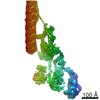
| 得られたモデル |  PDB-7kzn: |
