 Movie
Movie Controller

[English] 日本語
 Yorodumi
- EMDB-2281: Three-dimensional reconstruction of intact human integrin alphaII... -
+ Open data

- Basic information
| Entry | Database: EMDB / ID: EMD-2281 | |||||||||
|---|---|---|---|---|---|---|---|---|---|---|
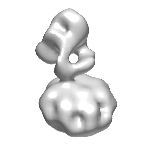
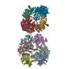
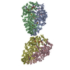
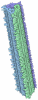
| Title | Three-dimensional reconstruction of intact human integrin alphaIIbbeta3 in a phospholipid bilayer nanodisc | |||||||||
 Map data Map data | Reconstruction of integrin alphaIIbbeta3 in lipid bilayer nanodisc | |||||||||
 Sample Sample |
| |||||||||
 Keywords Keywords | integrin / alphaIIbbeta3 / nanodisc | |||||||||
| Function / homology |  Function and homology information Function and homology informationregulation of serotonin uptake / positive regulation of adenylate cyclase-inhibiting opioid receptor signaling pathway / tube development / alpha9-beta1 integrin-ADAM8 complex / response to platelet-derived growth factor / integrin alphaIIb-beta3 complex / regulation of postsynaptic neurotransmitter receptor diffusion trapping / maintenance of postsynaptic specialization structure / alphav-beta3 integrin-vitronectin complex / platelet alpha granule membrane ...regulation of serotonin uptake / positive regulation of adenylate cyclase-inhibiting opioid receptor signaling pathway / tube development / alpha9-beta1 integrin-ADAM8 complex / response to platelet-derived growth factor / integrin alphaIIb-beta3 complex / regulation of postsynaptic neurotransmitter receptor diffusion trapping / maintenance of postsynaptic specialization structure / alphav-beta3 integrin-vitronectin complex / platelet alpha granule membrane / positive regulation of glomerular mesangial cell proliferation / integrin alphav-beta3 complex / negative regulation of lipoprotein metabolic process / smooth muscle cell migration / alphav-beta3 integrin-PKCalpha complex / fibrinogen binding / positive regulation of leukocyte migration / alphav-beta3 integrin-HMGB1 complex / negative regulation of lipid transport / vascular endothelial growth factor receptor 2 binding / angiogenesis involved in wound healing / positive regulation of vascular endothelial growth factor signaling pathway / regulation of release of sequestered calcium ion into cytosol / Elastic fibre formation / mesodermal cell differentiation / positive regulation of bone resorption / alphav-beta3 integrin-IGF-1-IGF1R complex / platelet-derived growth factor receptor binding / cell-cell adhesion mediated by integrin / filopodium membrane / extracellular matrix binding / glycinergic synapse / positive regulation of fibroblast migration / positive regulation of cell adhesion mediated by integrin / positive regulation of vascular endothelial growth factor receptor signaling pathway / apolipoprotein A-I-mediated signaling pathway / regulation of bone resorption / negative regulation of low-density lipoprotein particle clearance / apoptotic cell clearance / wound healing, spreading of epidermal cells / positive regulation of smooth muscle cell migration / integrin complex / Molecules associated with elastic fibres / heterotypic cell-cell adhesion / cell adhesion mediated by integrin / negative chemotaxis / positive regulation of osteoblast proliferation / Mechanical load activates signaling by PIEZO1 and integrins in osteocytes / cellular response to insulin-like growth factor stimulus / Syndecan interactions / p130Cas linkage to MAPK signaling for integrins / regulation of postsynaptic neurotransmitter receptor internalization / cell-substrate adhesion / protein disulfide isomerase activity / microvillus membrane / PECAM1 interactions / GRB2:SOS provides linkage to MAPK signaling for Integrins / negative regulation of endothelial cell apoptotic process / TGF-beta receptor signaling activates SMADs / lamellipodium membrane / fibronectin binding / negative regulation of macrophage derived foam cell differentiation / negative regulation of lipid storage / Integrin cell surface interactions / ECM proteoglycans / substrate adhesion-dependent cell spreading / positive regulation of T cell migration / cell-matrix adhesion / embryo implantation / coreceptor activity / Integrin signaling / positive regulation of endothelial cell proliferation / positive regulation of smooth muscle cell proliferation / positive regulation of endothelial cell migration / cell adhesion molecule binding / integrin-mediated signaling pathway / Turbulent (oscillatory, disturbed) flow shear stress activates signaling by PIEZO1 and integrins in endothelial cells / response to activity / protein kinase C binding / wound healing / Signal transduction by L1 / cellular response to xenobiotic stimulus / regulation of actin cytoskeleton organization / cell-cell adhesion / cellular response to mechanical stimulus / platelet activation / Signaling by high-kinase activity BRAF mutants / MAP2K and MAPK activation / RUNX1 regulates genes involved in megakaryocyte differentiation and platelet function / platelet aggregation / VEGFA-VEGFR2 Pathway / integrin binding / ruffle membrane / blood coagulation / positive regulation of angiogenesis / Signaling by RAF1 mutants / cell-cell junction / Signaling by moderate kinase activity BRAF mutants / Paradoxical activation of RAF signaling by kinase inactive BRAF / Signaling downstream of RAS mutants Similarity search - Function | |||||||||
| Biological species |  Homo sapiens (human) Homo sapiens (human) | |||||||||
| Method | single particle reconstruction / negative staining / Resolution: 20.5 Å | |||||||||
 Authors Authors | Choi WS / Rice WJ / Stokes DL / Coller BS | |||||||||
 Citation Citation | Journal: Blood / Year: 2013 Title: Three-dimensional reconstruction of intact human integrin αIIbβ3: new implications for activation-dependent ligand binding. Authors: Won-Seok Choi / William J Rice / David L Stokes / Barry S Coller /  Abstract: Integrin αIIbβ3 plays a central role in hemostasis and thrombosis. We provide the first 3-dimensional reconstruction of intact purified αIIbβ3 in a nanodisc lipid bilayer. Unlike previous models, ...Integrin αIIbβ3 plays a central role in hemostasis and thrombosis. We provide the first 3-dimensional reconstruction of intact purified αIIbβ3 in a nanodisc lipid bilayer. Unlike previous models, it shows that the ligand-binding head domain is on top, pointing away from the membrane. Moreover, unlike the crystal structure of the recombinant ectodomain, the lower legs are not parallel, straight, and adjacent. Rather, the αIIb lower leg is bent between the calf-1 and calf-2 domains and the β3 Integrin-Epidermal Growth Factor (I-EGF) 2 to 4 domains are freely coiled rather than in a cleft between the β3 headpiece and the αIIb lower leg. Our data indicate an important role for the region that links the distal calf-2 and β-tail domains to their respective transmembrane (TM) domains in transmitting the conformational changes in the TM domains associated with inside-out activation. | |||||||||
| History |
|
- Structure visualization
| Movie |
Movie viewer |
|---|---|
| Structure viewer | EM map: SurfViewMolmilJmol/JSmol |
| Supplemental images |
- Downloads & links
-EMDB archive
| Map data | emd_2281.map.gz | 8.5 MB | EMDB map data format | |
|---|---|---|---|---|
| Header (meta data) | emd-2281-v30.xmlemd-2281.xml | 14.3 KB 14.3 KB | Display Display | EMDB header |
| Images | 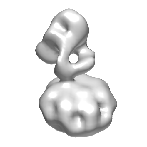 EMD-2281.png EMD-2281.png | 45.9 KB | ||
| Archive directory |  http://ftp.pdbj.org/pub/emdb/structures/EMD-2281ftp://ftp.pdbj.org/pub/emdb/structures/EMD-2281 http://ftp.pdbj.org/pub/emdb/structures/EMD-2281ftp://ftp.pdbj.org/pub/emdb/structures/EMD-2281 | HTTPS FTP |
-Related structure data
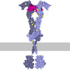
| Related structure data |  4cakMC M: atomic model generated by this map C: citing same article ( |
|---|---|
| Similar structure data |

-Links
| EMDB pages | EMDB (EBI/PDBe) / EMDataResource |
|---|---|
| Related items in Molecule of the Month |







-Map
| File | Download / File: emd_2281.map.gz / Format: CCP4 / Size: 9.4 MB / Type: IMAGE STORED AS FLOATING POINT NUMBER (4 BYTES) | ||||||||||||||||||||||||||||||||||||||||||||||||||||||||||||||||||||
|---|---|---|---|---|---|---|---|---|---|---|---|---|---|---|---|---|---|---|---|---|---|---|---|---|---|---|---|---|---|---|---|---|---|---|---|---|---|---|---|---|---|---|---|---|---|---|---|---|---|---|---|---|---|---|---|---|---|---|---|---|---|---|---|---|---|---|---|---|---|
| Annotation | Reconstruction of integrin alphaIIbbeta3 in lipid bilayer nanodisc | ||||||||||||||||||||||||||||||||||||||||||||||||||||||||||||||||||||
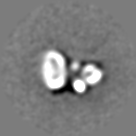
| Projections & slices | Image control
Images are generated by Spider. | ||||||||||||||||||||||||||||||||||||||||||||||||||||||||||||||||||||
| Voxel size | X=Y=Z: 2.96 Å | ||||||||||||||||||||||||||||||||||||||||||||||||||||||||||||||||||||
| Density |
| ||||||||||||||||||||||||||||||||||||||||||||||||||||||||||||||||||||
| Symmetry | Space group: 1 | ||||||||||||||||||||||||||||||||||||||||||||||||||||||||||||||||||||
| Details | EMDB XML:
CCP4 map header:
| ||||||||||||||||||||||||||||||||||||||||||||||||||||||||||||||||||||
 Z (Sec.)
 Y (Row.)
 X (Col.)




-Supplemental data
- Sample components
-Entire : Integrin alphaIIbbeta3 in lipid bilayer nanodisc
| Entire | Name: Integrin alphaIIbbeta3 in lipid bilayer nanodisc |
|---|---|
| Components |
|
-Supramolecule #1000: Integrin alphaIIbbeta3 in lipid bilayer nanodisc
| Supramolecule | Name: Integrin alphaIIbbeta3 in lipid bilayer nanodisc / type: sample / ID: 1000 / Details: The sample was monodisperse. / Oligomeric state: monomer / Number unique components: 2 |
|---|---|
| Molecular weight | Experimental: 230 KDa / Theoretical: 230 KDa / Method: SDS-Page |
-Macromolecule #1: integrin alphaIIb
| Macromolecule | Name: integrin alphaIIb / type: protein_or_peptide / ID: 1 / Name.synonym: GPIIb Details: Native protein purified from platelet, heterodimer with integrin beta3 subunit Number of copies: 1 / Oligomeric state: hetero dimer / Recombinant expression: No |
|---|---|
| Source (natural) | Organism: Homo sapiens (human) / synonym: Human / Tissue: Blood / Cell: Platelet / Location in cell: Plasma membrane |
| Molecular weight | Experimental: 130 KDa / Theoretical: 130 KDa |
| Sequence | UniProtKB: Integrin alpha-IIb |
-Macromolecule #2: Integrin beta3
| Macromolecule | Name: Integrin beta3 / type: protein_or_peptide / ID: 2 / Name.synonym: GPIIIa Details: Native protein purified from platelet, heterodimer with integrin alphaIIb subunit Number of copies: 1 / Oligomeric state: hetero dimer / Recombinant expression: No |
|---|---|
| Source (natural) | Organism: Homo sapiens (human) / synonym: Human / Tissue: Blood / Cell: Platelet / Location in cell: Plasma membrane |
| Molecular weight | Experimental: 100 KDa / Theoretical: 100 KDa |
| Sequence | UniProtKB: Integrin beta-3 |
-Experimental details
-Structure determination
| Method | negative staining |
|---|---|
 Processing Processing | single particle reconstruction |
| Aggregation state | particle |
-Sample preparation
| Concentration | 0.025 mg/mL |
|---|---|
| Buffer | pH: 7.4 Details: 150 mM NaCl, 10 mM HEPES, pH 7.4, 1 mM CaCl2 and 1 mM MgCl2 |
| Staining | Type: NEGATIVE / Details: 2% uranyl acetate |
| Grid | Details: 200 mesh copper grid with thin carbon support, glow discharged in H2/O2 atmosphere in plasma cleaner |
| Vitrification | Cryogen name: NONE / Instrument: OTHER |
- Electron microscopy #1
| Microscopy ID | 1 |
|---|---|
| Microscope | FEI TECNAI F20 |
| Alignment procedure | Legacy - Astigmatism: Objective lens astigmatism was corrected at 250,000 times magnification |
| Details | Low dose package used. CCD magnification is ~1.76 times film magnification |
| Date | Feb 1, 2010 |
| Image recording | Category: CCD / Film or detector model: GENERIC TVIPS (4k x 4k) / Digitization - Sampling interval: 15 µm / Number real images: 1500 / Average electron dose: 13 e/Å2 / Bits/pixel: 16 |
| Tilt angle min | 0 |
| Electron beam | Acceleration voltage: 120 kV / Electron source:  FIELD EMISSION GUN FIELD EMISSION GUN |
| Electron optics | Calibrated magnification: 50592 / Illumination mode: FLOOD BEAM / Imaging mode: BRIGHT FIELD / Cs: 2.0 mm / Nominal defocus max: 1.2 µm / Nominal defocus min: 1.2 µm / Nominal magnification: 29000 |
| Sample stage | Specimen holder model: SIDE ENTRY, EUCENTRIC / Tilt angle max: 50 |
| Experimental equipment | 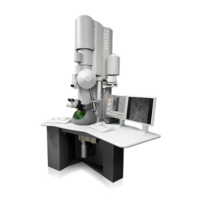 Model: Tecnai F20 / Image courtesy: FEI Company |
-Electron microscopy #2
| Microscopy ID | 2 |
|---|---|
| Microscope | FEI TECNAI F20 |
| Alignment procedure | Legacy - Astigmatism: Objective lens astigmatism was corrected at 250,000 times magnification |
| Details | Low dose package used. CCD magnification is ~1.76 times film magnification |
| Date | Jul 31, 2010 |
| Image recording | Category: CCD / Film or detector model: GENERIC TVIPS (4k x 4k) / Digitization - Sampling interval: 15 µm / Number real images: 1500 / Average electron dose: 13 e/Å2 / Bits/pixel: 16 |
| Tilt angle min | 0 |
| Electron beam | Acceleration voltage: 200 kV / Electron source: FIELD EMISSION GUN |
| Electron optics | Calibrated magnification: 88249 / Illumination mode: FLOOD BEAM / Imaging mode: BRIGHT FIELD / Cs: 2.0 mm / Nominal defocus max: 0.6 µm / Nominal defocus min: 0.6 µm / Nominal magnification: 50000 |
| Sample stage | Specimen holder model: SIDE ENTRY, EUCENTRIC / Tilt angle max: 50 |
| Experimental equipment | Model: Tecnai F20 / Image courtesy: FEI Company |
-Image processing
| Details | 5 random conical tilt reconstructions were made from class averages, then aligned and merged to make an initial model for reference-based alignment. |
|---|---|
| Final reconstruction | Applied symmetry - Point group: C1 (asymmetric) / Algorithm: OTHER / Resolution.type: BY AUTHOR / Resolution: 20.5 Å / Resolution method: FSC 0.5 CUT-OFF / Software - Name: spider Details: Initial model was made from aligning and averaging 5 models made by random conical tilt. Number images used: 25008 |
| Final angle assignment | Details: SPIDER: theta 45 degrees, phi 45 degrees |
| Final two d classification | Number classes: 5 |
-Atomic model buiding 1
| Initial model | PDB ID: Chain - #0 - Chain ID: A / Chain - #1 - Chain ID: B |
|---|---|
| Software | Name: Chimera |
| Details | The individual alphaIIb and beta3 domains were docked into the anchor graph of the EM map so as to maintain the sequence of domains and the distances between domains in the crystal structure. The locations were then optimized by maximizing the cross-correlation and the atomic inclusion of the domain within the EM map. |
| Refinement | Space: REAL / Protocol: RIGID BODY FIT / Target criteria: Cross correlation |
| Output model | PDB-4cak: |

