 Movie
Movie Controller

[English] 日本語
 Yorodumi
- EMDB-22474: Interphotoreceptor retinoid-binding protein (IRBP) in complex wit... -
+ Open data

- Basic information
| Entry | Database: EMDB / ID: EMD-22474 | |||||||||||||||
|---|---|---|---|---|---|---|---|---|---|---|---|---|---|---|---|---|
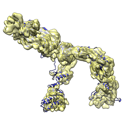
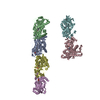
| Title | Interphotoreceptor retinoid-binding protein (IRBP) in complex with a monoclonal antibody (F3F5 mAb5) | |||||||||||||||
 Map data Map data | ||||||||||||||||
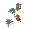
 Sample Sample |
| |||||||||||||||
 Keywords Keywords | IRBP / retinoid / Interphotoreceptor retinoid-binding protein / Antibody / TRANSPORT PROTEIN-IMMUNE SYSTEM complex | |||||||||||||||
| Function / homology |  Function and homology information Function and homology informationcone matrix sheath / interphotoreceptor matrix / all-trans-retinol binding / oleic acid binding / retinal binding / retinol binding / serine-type peptidase activity / proteolysis Similarity search - Function | |||||||||||||||
| Biological species |  | |||||||||||||||
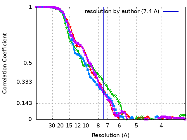
| Method | single particle reconstruction / cryo EM / Resolution: 7.4 Å | |||||||||||||||
 Authors Authors | Sears AE / Albiez S | |||||||||||||||
| Funding support |  United States, United States,  Switzerland, 4 items Switzerland, 4 items
| |||||||||||||||
 Citation Citation | Journal: FASEB J / Year: 2020 Title: Single particle cryo-EM of the complex between interphotoreceptor retinoid-binding protein and a monoclonal antibody. Authors: Avery E Sears / Stefan Albiez / Sahil Gulati / Benlian Wang / Philip Kiser / Lubomir Kovacik / Andreas Engel / Henning Stahlberg / Krzysztof Palczewski / Abstract: Interphotoreceptor retinoid-binding protein (IRBP) is a highly expressed protein secreted by rod and cone photoreceptors that has major roles in photoreceptor homeostasis as well as retinoid and ...Interphotoreceptor retinoid-binding protein (IRBP) is a highly expressed protein secreted by rod and cone photoreceptors that has major roles in photoreceptor homeostasis as well as retinoid and polyunsaturated fatty acid transport between the neural retina and retinal pigment epithelium. Despite two crystal structures reported on fragments of IRBP and decades of research, the overall structure of IRBP and function within the visual cycle remain unsolved. Here, we studied the structure of native bovine IRBP in complex with a monoclonal antibody (mAb5) by cryo-electron microscopy, revealing the tertiary and quaternary structure at sufficient resolution to clearly identify the complex components. Complementary mass spectrometry experiments revealed the structure and locations of N-linked carbohydrate post-translational modifications. This work provides insight into the structure of IRBP, displaying an elongated, flexible three-dimensional architecture not seen among other retinoid-binding proteins. This work is the first step in elucidation of the function of this enigmatic protein. | |||||||||||||||
| History |
|
- Structure visualization
| Movie |
Movie viewer |
|---|---|
| Structure viewer | EM map: SurfViewMolmilJmol/JSmol |
| Supplemental images |
- Downloads & links
-EMDB archive
| Map data | emd_22474.map.gz | 2.9 MB | EMDB map data format | |
|---|---|---|---|---|
| Header (meta data) | emd-22474-v30.xmlemd-22474.xml | 22.3 KB 22.3 KB | Display Display | EMDB header |
| FSC (resolution estimation) | emd_22474_fsc.xmlemd_22474_fsc_2.xmlemd_22474_fsc_3.xmlemd_22474_fsc_4.xml | 8.2 KB 8.3 KB 8.3 KB 8.3 KB | Display Display Display Display | FSC data file |
| Images |  emd_22474.png emd_22474.png | 124.8 KB | ||
| Masks | emd_22474_msk_1.map | 52.7 MB | Mask map | |
| Filedesc metadata | emd-22474.cif.gz | 7.5 KB | ||
| Archive directory |  http://ftp.pdbj.org/pub/emdb/structures/EMD-22474ftp://ftp.pdbj.org/pub/emdb/structures/EMD-22474 http://ftp.pdbj.org/pub/emdb/structures/EMD-22474ftp://ftp.pdbj.org/pub/emdb/structures/EMD-22474 | HTTPS FTP |
-Related structure data
| Related structure data |  7jtiMC M: atomic model generated by this map C: citing same article ( |
|---|---|
| Similar structure data | |
| EM raw data | EMPIAR-10388 (Title: Single particle cryo-EM of the complex between interphotoreceptor retinoid-binding protein and a monoclonal antibody Data size: 729.9 Data #1: Unaligned multi-frame micrographs of IRBP and antibody [micrographs - multiframe]) |

-Links
| EMDB pages | EMDB (EBI/PDBe) / EMDataResource |
|---|
-Map
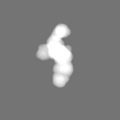
| File | Download / File: emd_22474.map.gz / Format: CCP4 / Size: 52.7 MB / Type: IMAGE STORED AS FLOATING POINT NUMBER (4 BYTES) | ||||||||||||||||||||||||||||||||||||||||||||||||||||||||||||||||||||
|---|---|---|---|---|---|---|---|---|---|---|---|---|---|---|---|---|---|---|---|---|---|---|---|---|---|---|---|---|---|---|---|---|---|---|---|---|---|---|---|---|---|---|---|---|---|---|---|---|---|---|---|---|---|---|---|---|---|---|---|---|---|---|---|---|---|---|---|---|---|
| Projections & slices | Image control
Images are generated by Spider. | ||||||||||||||||||||||||||||||||||||||||||||||||||||||||||||||||||||
| Voxel size | X=Y=Z: 1.662 Å | ||||||||||||||||||||||||||||||||||||||||||||||||||||||||||||||||||||
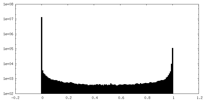
| Density |
| ||||||||||||||||||||||||||||||||||||||||||||||||||||||||||||||||||||
| Symmetry | Space group: 1 | ||||||||||||||||||||||||||||||||||||||||||||||||||||||||||||||||||||
| Details | EMDB XML:
CCP4 map header:
| ||||||||||||||||||||||||||||||||||||||||||||||||||||||||||||||||||||
 Z (Sec.)
 Y (Row.)
 X (Col.)


-Supplemental data
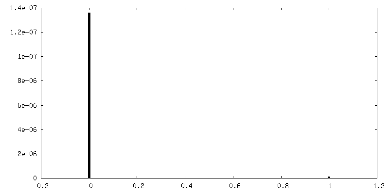
-Mask #1
| File | emd_22474_msk_1.map | ||||||||||||
|---|---|---|---|---|---|---|---|---|---|---|---|---|---|
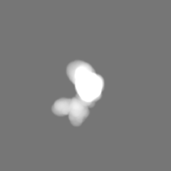
| Projections & Slices |
| ||||||||||||
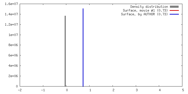
| Density Histograms |


- Sample components
-Entire : Complex between interphotoreceptor retinoid-binding protein and a...
| Entire | Name: Complex between interphotoreceptor retinoid-binding protein and a monoclonal antibody |
|---|---|
| Components |
|
-Supramolecule #1: Complex between interphotoreceptor retinoid-binding protein and a...
| Supramolecule | Name: Complex between interphotoreceptor retinoid-binding protein and a monoclonal antibody type: complex / ID: 1 / Parent: 0 / Macromolecule list: all |
|---|---|
| Molecular weight | Theoretical: 193 KDa |
-Supramolecule #2: monoclonal antibody F3F5 mAb5
| Supramolecule | Name: monoclonal antibody F3F5 mAb5 / type: complex / ID: 2 / Parent: 1 / Macromolecule list: #1-#2 |
|---|---|
| Source (natural) | Organism: |
-Supramolecule #3: Interphotoreceptor retinoid-binding protein (IRBP)
| Supramolecule | Name: Interphotoreceptor retinoid-binding protein (IRBP) / type: complex / ID: 3 / Parent: 1 / Macromolecule list: #3-#6 |
|---|---|
| Source (natural) | Organism: |
-Macromolecule #1: mAb5 Fab light chain
| Macromolecule | Name: mAb5 Fab light chain / type: protein_or_peptide / ID: 1 / Number of copies: 1 / Enantiomer: LEVO |
|---|---|
| Source (natural) | Organism: |
| Molecular weight | Theoretical: 22.596021 KDa |
| Recombinant expression | Organism: |
| Sequence | String: QAVVTQESAL TTSPGETVTL TCRSSTGAVT TSNYANWVQE KPDHLFTGLI GGTNNRAPGV PARFSGSLIG DKAALTITGA QTEDEAIYF CALWYSNHWV FGGGTKLTVL GQPKSSPSVT LFPPSSEELE TNKATLVCTI TDFYPGVVTV DWKVDGTPVT Q GMETTQPS ...String: QAVVTQESAL TTSPGETVTL TCRSSTGAVT TSNYANWVQE KPDHLFTGLI GGTNNRAPGV PARFSGSLIG DKAALTITGA QTEDEAIYF CALWYSNHWV FGGGTKLTVL GQPKSSPSVT LFPPSSEELE TNKATLVCTI TDFYPGVVTV DWKVDGTPVT Q GMETTQPS KQSNNKYMAS SYLTLTARAW ERHSSYSCQV THEGHTVEKS LS |
-Macromolecule #2: mAb5 Fab heavy chain
| Macromolecule | Name: mAb5 Fab heavy chain / type: protein_or_peptide / ID: 2 / Number of copies: 1 / Enantiomer: LEVO |
|---|---|
| Source (natural) | Organism: |
| Molecular weight | Theoretical: 23.777783 KDa |
| Recombinant expression | Organism: |
| Sequence | String: QVQLKESGPG LVAPSQSLSI TCTVSGFLLI SNGVHWVRQP PGKGLEWLGV IWAGGNTNYN SALMSRVSIS KDNSKSQVFL KMKSLQTDD TAMYYCARDF YDYDVFYYAM DYWGQGTSVT VSSAKTTPPS VYPLAPGSAA QTNSMVTLGC LVKGYFPEPV T VTWNSGSL ...String: QVQLKESGPG LVAPSQSLSI TCTVSGFLLI SNGVHWVRQP PGKGLEWLGV IWAGGNTNYN SALMSRVSIS KDNSKSQVFL KMKSLQTDD TAMYYCARDF YDYDVFYYAM DYWGQGTSVT VSSAKTTPPS VYPLAPGSAA QTNSMVTLGC LVKGYFPEPV T VTWNSGSL SSGVHTFPAV LQSDLYTLSS SVTVPSSTWP SETVTCNVAH PASSTKVDKK IVP |
-Macromolecule #3: Retinol-binding protein 3
| Macromolecule | Name: Retinol-binding protein 3 / type: protein_or_peptide / ID: 3 / Number of copies: 1 / Enantiomer: LEVO |
|---|---|
| Source (natural) | Organism: |
| Molecular weight | Theoretical: 33.267301 KDa |
| Sequence | String: VIQRLQEALR EYYTLVDRVP ALLSHLAAMD LSSVVSEDDL VTKLNAGLQA VSEDPRLQVQ VVRPKEASSG PEEEAEEPPE AVPEVPEDE AVRRALVDSV FQVSVLPGNV GYLRFDSFAD ASVLEVLGPY ILHQVWEPLQ DTEHLIMDLR QNPGGPSSAV P LLLSYFQS ...String: VIQRLQEALR EYYTLVDRVP ALLSHLAAMD LSSVVSEDDL VTKLNAGLQA VSEDPRLQVQ VVRPKEASSG PEEEAEEPPE AVPEVPEDE AVRRALVDSV FQVSVLPGNV GYLRFDSFAD ASVLEVLGPY ILHQVWEPLQ DTEHLIMDLR QNPGGPSSAV P LLLSYFQS PDASPVRLFS TYDRRTNITR EHFSQTELLG RPYGTQRGVY LLTSHRTATA AEELAFLMQS LGWATLVGEI TA GSLLHTH TVSLLETPEG GLALTVPVLT FIDNHGECWL GGGVVPDAIV LAEEALDRAQ EVLEFH UniProtKB: Retinol-binding protein 3 |
-Macromolecule #4: Retinol-binding protein 3
| Macromolecule | Name: Retinol-binding protein 3 / type: protein_or_peptide / ID: 4 / Number of copies: 1 / Enantiomer: LEVO |
|---|---|
| Source (natural) | Organism: |
| Molecular weight | Theoretical: 33.386637 KDa |
| Sequence | String: FSPTLIADMA KIFMDNYCSP EKLTGMEEAI DAASSNTEIL SISDPTMLAN VLTDGVKKTI SDSRVKVTYE PDLILAAPPA MPDIPLEHL AAMIKGTVKV EILEGNIGYL KIQHIIGEEM AQKVGPLLLE YIWDKILPTS AMILDFRSTV TGELSGIPYI V SYFTDPEP ...String: FSPTLIADMA KIFMDNYCSP EKLTGMEEAI DAASSNTEIL SISDPTMLAN VLTDGVKKTI SDSRVKVTYE PDLILAAPPA MPDIPLEHL AAMIKGTVKV EILEGNIGYL KIQHIIGEEM AQKVGPLLLE YIWDKILPTS AMILDFRSTV TGELSGIPYI V SYFTDPEP LIHIDSVYDR TADLTIELWS MPTLLGKRYG TSKPLIILTS KDTLGIAEDV AYCLKNLKRA TIVGENTAGG TV KMSKMKV GDTDFYVTVP VAKSINPITG KSWEINGVAP DVDVAAEDAL DAAIAIIKLR AEIPALAQAA AT |
-Macromolecule #5: Retinol-binding protein 3
| Macromolecule | Name: Retinol-binding protein 3 / type: protein_or_peptide / ID: 5 / Number of copies: 1 / Enantiomer: LEVO |
|---|---|
| Source (natural) | Organism: |
| Molecular weight | Theoretical: 32.399768 KDa |
| Sequence | String: SLGELVEGTG RLLEAHYARP EVVGQMGALL RAKLAQGAYR TAVDLESLAS QLTADLQEMS GDHRLLVFHS PGEMVAEEAP PPPPVVPSP EELSYLIEAL FKTEVLPGQL GYLRFDAMAE LETVKAVGPQ LVQLVWQKLV DTAALVVDLR YNPGSYSTAV P LLCSYFFE ...String: SLGELVEGTG RLLEAHYARP EVVGQMGALL RAKLAQGAYR TAVDLESLAS QLTADLQEMS GDHRLLVFHS PGEMVAEEAP PPPPVVPSP EELSYLIEAL FKTEVLPGQL GYLRFDAMAE LETVKAVGPQ LVQLVWQKLV DTAALVVDLR YNPGSYSTAV P LLCSYFFE AEPRRHLYSV FDRATSRVTE VWTLPHVTGQ RYGSHKDLYV LVSHTSGSAA EAFAHTMQDL QRATIIGEPT AG GALSVGI YQVGSSALYA SMPTQMAMSA STGEAWDLAG VEPDITVPMS VALSTARDIV TLR UniProtKB: Retinol-binding protein 3 |
-Macromolecule #6: Retinol-binding protein 3
| Macromolecule | Name: Retinol-binding protein 3 / type: protein_or_peptide / ID: 6 / Number of copies: 1 / Enantiomer: LEVO |
|---|---|
| Source (natural) | Organism: |
| Molecular weight | Theoretical: 32.192508 KDa |
| Sequence | String: PTVLQTAGKL VADNYASPEL GVKMAAELSG LQSRYARVTS EAALAELLQA DLQVLSGDPH LKTAHIPEDA KDRIPGIVPM QIPSPEVFE DLIKFSFHTN VLEGNVGYLR FDMFGDCELL TQVSELLVEH VWKKIVHTDA LIVDMRFNIG GPTSSISALC S YFFDEGPP ...String: PTVLQTAGKL VADNYASPEL GVKMAAELSG LQSRYARVTS EAALAELLQA DLQVLSGDPH LKTAHIPEDA KDRIPGIVPM QIPSPEVFE DLIKFSFHTN VLEGNVGYLR FDMFGDCELL TQVSELLVEH VWKKIVHTDA LIVDMRFNIG GPTSSISALC S YFFDEGPP ILLDKIYNRP NNSVSELWTL SQLEGERYGS KKSMVILTST LTAGAAEEFT YIMKRLGRAL VIGEVTSGGC QP PQTYHVD DTDLYLTIPT ARSVGAADGS SWEGVGVVPD VAVPAEAALT RAQEMLQHT UniProtKB: Retinol-binding protein 3 |
-Experimental details
-Structure determination
| Method | cryo EM |
|---|---|
 Processing Processing | single particle reconstruction |
| Aggregation state | particle |
-Sample preparation
| Concentration | 0.2 mg/mL | |||||||||||||||
|---|---|---|---|---|---|---|---|---|---|---|---|---|---|---|---|---|
| Buffer | pH: 8 Component:
| |||||||||||||||
| Grid | Model: Quantifoil R1.2/1.3 / Material: COPPER / Mesh: 400 / Support film - Material: GRAPHENE OXIDE / Support film - topology: CONTINUOUS / Support film - Film thickness: 1.12 | |||||||||||||||
| Vitrification | Cryogen name: ETHANE / Chamber humidity: 90 % / Chamber temperature: 277 K / Instrument: FEI VITROBOT MARK IV / Details: Blot for 3 seconds before plunging.. | |||||||||||||||
| Details | The sample had minor aggregation but was overall monodisperse. |
- Electron microscopy
| Microscope | FEI TITAN KRIOS |
|---|---|
| Specialist optics | Energy filter - Name: GIF Quantum LS / Energy filter - Slit width: 20 eV |
| Image recording | Film or detector model: GATAN K2 QUANTUM (4k x 4k) / Detector mode: SUPER-RESOLUTION / Digitization - Dimensions - Width: 1000 pixel / Digitization - Dimensions - Height: 1000 pixel / Number grids imaged: 4 / Number real images: 2649 / Average exposure time: 1.0 sec. / Average electron dose: 80.0 e/Å2 Details: Images were collected in movie mode at 35 frames per second. |
| Electron beam | Acceleration voltage: 300 kV / Electron source:  FIELD EMISSION GUN FIELD EMISSION GUN |
| Electron optics | Illumination mode: FLOOD BEAM / Imaging mode: BRIGHT FIELD / Nominal defocus max: 2.2 µm / Nominal defocus min: 1.0 µm / Nominal magnification: 60000 |
| Sample stage | Specimen holder model: GATAN 910 MULTI-SPECIMEN SINGLE TILT CRYO TRANSFER HOLDER Cooling holder cryogen: NITROGEN |
| Experimental equipment |  Model: Titan Krios / Image courtesy: FEI Company |
+Image processing
-Atomic model buiding 1
| Initial model | PDB ID: Chain - Chain ID: A / Chain - Source name: PDB / Chain - Initial model type: experimental model |
|---|---|
| Refinement | Space: REAL / Protocol: RIGID BODY FIT / Overall B value: 156 / Target criteria: Correlation coefficient |
| Output model | PDB-7jti: |

