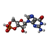
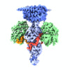
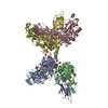
登録情報 データベース : EMDB / ID : EMD-22458タイトル Structure of the Visual Signaling Complex between Transducin and Phosphodiesterase 6 Visual Signaling Complex between Transducin and Phosphodiesterase 6 複合体 : Complex of Transducin and Phosphodiesterase 6複合体 : Rod cGMP-specific 3',5'-cyclic phosphodiesterase subunit alpha (E.C.3.1.4.35), Rod cGMP-specific 3',5'-cyclic phosphodiesterase subunit beta (E.C.3.1.4.35), Retinal rod rhodopsin-sensitive cGMP 3',5'-cyclic phosphodiesterase subunit gamma (E.C.3.1.4.35)タンパク質・ペプチド : Rod cGMP-specific 3',5'-cyclic phosphodiesterase subunit alphaタンパク質・ペプチド : Rod cGMP-specific 3',5'-cyclic phosphodiesterase subunit betaタンパク質・ペプチド : Guanine nucleotide-binding protein G(t) subunit alpha-1, Guanine nucleotide-binding protein G(i) subunit alpha-1 chimera複合体 : Guanine nucleotide-binding protein G(t) subunit alpha-1, Guanine nucleotide-binding protein G(i) subunit alpha-1 chimera,Gt-alpha/Gi1-alpha chimeraタンパク質・ペプチド : Retinal rod rhodopsin-sensitive cGMP 3',5'-cyclic phosphodiesterase subunit gammaリガンド : ZINC IONリガンド : MAGNESIUM IONリガンド : 2-{2-ETHOXY-5-[(4-ETHYLPIPERAZIN-1-YL)SULFONYL]PHENYL}-5-METHYL-7-PROPYLIMIDAZO[5,1-F][1,2,4]TRIAZIN-4(1H)-ONEリガンド : GUANOSINE-3',5'-MONOPHOSPHATEリガンド : GUANOSINE-5'-TRIPHOSPHATE / / / / / / 機能・相同性 分子機能 ドメイン・相同性 構成要素
/ / / / / / / / / / / / / / / / / / / / / / / / / / / / / / / / / / / / / / / / / / / / / / / / / / / / / / / / / / / / / / / / / / / / / / / / / / / / / / 生物種 Bos taurus (ウシ)手法 / / 解像度 : 3.2 Å Gao Y / Eskici G 資金援助 Organization Grant number 国 National Institutes of Health/National Institute of General Medical Sciences (NIH/NIGMS) R35 GM122575 National Institutes of Health/National Cancer Institute (NIH/NCI) R01 CA201402 National Institutes of Health/National Institute of Neurological Disorders and Stroke (NIH/NINDS) R01 NS092695
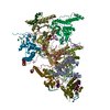
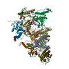
ジャーナル : Mol Cell / 年 : 2020タイトル : Structure of the Visual Signaling Complex between Transducin and Phosphodiesterase 6.著者 : Yang Gao / Gözde Eskici / Sekar Ramachandran / Frédéric Poitevin / Alpay Burak Seven / Ouliana Panova / Georgios Skiniotis / Richard A Cerione / 要旨 : Heterotrimeric G proteins communicate signals from activated G protein-coupled receptors to downstream effector proteins. In the phototransduction pathway responsible for vertebrate vision, the G ... Heterotrimeric G proteins communicate signals from activated G protein-coupled receptors to downstream effector proteins. In the phototransduction pathway responsible for vertebrate vision, the G protein-effector complex is composed of the GTP-bound transducin α subunit (Gα·GTP) and the cyclic GMP (cGMP) phosphodiesterase 6 (PDE6), which stimulates cGMP hydrolysis, leading to hyperpolarization of the photoreceptor cell. Here we report a cryo-electron microscopy (cryoEM) structure of PDE6 complexed to GTP-bound Gα. The structure reveals two Gα·GTP subunits engaging the PDE6 hetero-tetramer at both the PDE6 catalytic core and the PDEγ subunits, driving extensive rearrangements to relieve all inhibitory constraints on enzyme catalysis. Analysis of the conformational ensemble in the cryoEM data highlights the dynamic nature of the contacts between the two Gα·GTP subunits and PDE6 that supports an alternating-site catalytic mechanism. 履歴 登録 2020年8月15日 - ヘッダ(付随情報) 公開 2020年10月21日 - マップ公開 2020年10月21日 - 更新 2024年5月29日 - 現状 2024年5月29日 処理サイト : RCSB / 状態 : 公開
すべて表示 表示を減らす
 データを開く
 基本情報
基本情報 マップデータ
 試料
試料 キーワード
キーワード 機能・相同性情報
 データ登録者
 米国, 3件
  引用
引用 ジャーナル: Mol Cell / 年: 2020
 構造の表示
構造の表示 ムービービューア
 SurfView
 Molmil
 Jmol/JSmol
 ダウンロードとリンク
 emd_22458.map.gz
 EMDBマップデータ形式
 emd-22458-v30.xml
 emd-22458.xml
 EMDBヘッダ
 emd_22458_fsc.xml
 FSCデータファイル
 emd_22458.png
 emd-22458.cif.gz
 http://ftp.pdbj.org/pub/emdb/structures/EMD-22458
 ftp://ftp.pdbj.org/pub/emdb/structures/EMD-22458
 emd_22458_validation.pdf.gz
 EMDB検証レポート
 emd_22458_full_validation.pdf.gz
 emd_22458_validation.xml.gz
 emd_22458_validation.cif.gz
 https://ftp.pdbj.org/pub/emdb/validation_reports/EMD-22458
 ftp://ftp.pdbj.org/pub/emdb/validation_reports/EMD-22458
 リンク
リンク EMDB (EBI/PDBe) /
  EMDataResource
 マップ
マップ ダウンロード / ファイル: emd_22458.map.gz / 形式: CCP4 / 大きさ: 125 MB / タイプ: IMAGE STORED AS FLOATING POINT NUMBER (4 BYTES)
 試料の構成要素
 解析
解析 試料調製
試料調製 電子顕微鏡法
 FIELD EMISSION GUN
 画像解析
画像解析
 ムービー
ムービー コントローラー





 Z (Sec.)
 Y (Row.)
 X (Col.)


