[English] 日本語
Yorodumi
- EMDB-21071: Cryo-EM structure of human pannexin 1 -

+
Open data


ID or keywords:

Loading...

-
Basic information

Entry
Database: EMDB / ID: EMD-21071
TitleCryo-EM structure of human pannexin 1
Map dataStructure of human pannexin 1
Sample
  • Complex: human pannexin 1PANX1
    • Protein or peptide: Pannexin-1
Function / homology
Function and homology information


ATP transmembrane transporter activity / ATP transport / Electric Transmission Across Gap Junctions / leak channel activity / positive regulation of interleukin-1 alpha production / monoatomic anion channel activity / bleb / wide pore channel activity / monoatomic anion transmembrane transport / gap junction channel activity ...ATP transmembrane transporter activity / ATP transport / Electric Transmission Across Gap Junctions / leak channel activity / positive regulation of interleukin-1 alpha production / monoatomic anion channel activity / bleb / wide pore channel activity / monoatomic anion transmembrane transport / gap junction channel activity / gap junction / positive regulation of macrophage cytokine production / oogenesis / response to ATP / monoatomic cation transport / The NLRP3 inflammasome / positive regulation of interleukin-1 beta production / response to ischemia / calcium channel activity / calcium ion transport / actin filament binding / cell-cell signaling / scaffold protein binding / protease binding / transmembrane transporter binding / signaling receptor binding / endoplasmic reticulum membrane / structural molecule activity / endoplasmic reticulum / protein-containing complex / membrane / identical protein binding / plasma membrane
Similarity search - Function
Pannexin / Innexin / Innexin / Pannexin family profile.
Similarity search - Domain/homology
Biological speciesHomo sapiens (human)
Methodsingle particle reconstruction / cryo EM / Resolution: 3.77 Å
AuthorsDeng Z / He Z / Yuan P
CitationJournal: Nat Struct Mol Biol / Year: 2020
Title: Cryo-EM structures of the ATP release channel pannexin 1.
Authors: Zengqin Deng / Zhihui He / Grigory Maksaev / Ryan M Bitter / Michael Rau / James A J Fitzpatrick / Peng Yuan /
Abstract: The plasma membrane adenosine triphosphate (ATP) release channel pannexin 1 (PANX1) has been implicated in many physiological and pathophysiological processes associated with purinergic signaling, ...The plasma membrane adenosine triphosphate (ATP) release channel pannexin 1 (PANX1) has been implicated in many physiological and pathophysiological processes associated with purinergic signaling, including cancer progression, apoptotic cell clearance, inflammation, blood pressure regulation, oocyte development, epilepsy and neuropathic pain. Here we present near-atomic-resolution structures of human and frog PANX1 determined by cryo-electron microscopy that revealed a heptameric channel architecture. Compatible with ATP permeation, the transmembrane pore and cytoplasmic vestibule were exceptionally wide. An extracellular tryptophan ring located at the outer pore created a constriction site, potentially functioning as a molecular sieve that restricts the size of permeable substrates. The amino and carboxyl termini, not resolved in the density map, appeared to be structurally dynamic and might contribute to narrowing of the pore during channel gating. In combination with functional characterization, this work elucidates the previously unknown architecture of pannexin channels and establishes a foundation for understanding their unique channel properties.
History
DepositionDec 5, 2019-
Header (metadata) releaseJan 15, 2020-
Map releaseApr 1, 2020-
UpdateApr 22, 2020-
Current statusApr 22, 2020Processing site: RCSB / Status: Released

-
Structure visualization
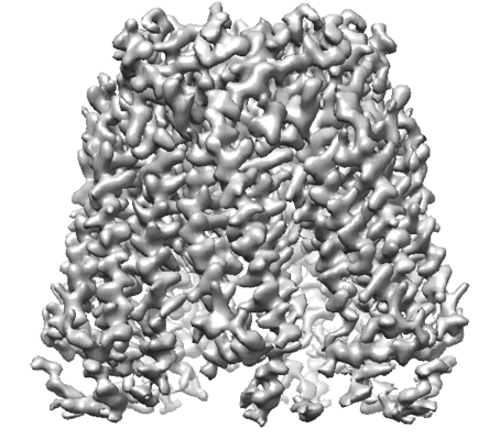
Movie
  • Surface view with section colored by density value
  • Surface level: 0.02
  • Imaged by UCSF Chimera
  • Download
  • Surface view colored by cylindrical radius
  • Surface level: 0.02
  • Imaged by UCSF Chimera
  • Download
  • Surface view with fitted model
  • Atomic models: PDB-6v6d
  • Surface level: 0.02
  • Imaged by UCSF Chimera
  • Download
Movie viewer
Structure viewerEM map:
SurfViewMolmilJmol/JSmol
Supplemental images

Downloads & links

-
Map

FileDownload / File: emd_21071.map.gz / Format: CCP4 / Size: 52.7 MB / Type: IMAGE STORED AS FLOATING POINT NUMBER (4 BYTES)
AnnotationStructure of human pannexin 1
Voxel sizeX=Y=Z: 1.1 Å
Density
Contour LevelBy AUTHOR: 0.01 / Movie #1: 0.02
Minimum - Maximum-0.051843256 - 0.105800465
Average (Standard dev.)0.00014086327 (±0.0038405827)
SymmetrySpace group: 1
Details

EMDB XML:

Map geometry
Axis orderXYZ
Origin000
Dimensions240240240
Spacing240240240
CellA=B=C: 264.0 Å
α=β=γ: 90.0 °

CCP4 map header:

modeImage stored as Reals
Å/pix. X/Y/Z1.11.11.1
M x/y/z240240240
origin x/y/z0.0000.0000.000
length x/y/z264.000264.000264.000
α/β/γ90.00090.00090.000
start NX/NY/NZ000
NX/NY/NZ256256256
MAP C/R/S123
start NC/NR/NS000
NC/NR/NS240240240
D min/max/mean-0.0520.1060.000

-
Supplemental data

-
Sample components

-
Entire : human pannexin 1

EntireName: human pannexin 1PANX1
Components
  • Complex: human pannexin 1PANX1
    • Protein or peptide: Pannexin-1

-
Supramolecule #1: human pannexin 1

SupramoleculeName: human pannexin 1 / type: complex / ID: 1 / Parent: 0 / Macromolecule list: all
Source (natural)Organism: Homo sapiens (human)
Recombinant expressionOrganism: Komagataella pastoris (fungus)

-
Macromolecule #1: Pannexin-1

MacromoleculeName: Pannexin-1 / type: protein_or_peptide / ID: 1 / Number of copies: 7 / Enantiomer: LEVO
Source (natural)Organism: Homo sapiens (human)
Molecular weightTheoretical: 49.11198 KDa
Recombinant expressionOrganism: Komagataella pastoris (fungus)
SequenceString: MAIAQLATEY VFSDFLLKEP TEPKFKGLRL ELAVDKMVTC IAVGLPLLLI SLAFAQEISI GTQISCFSPS SFSWRQAAFV DSYCWAAVQ QKNSLQSESG NLPLWLHKFF PYILLLFAIL LYLPPLFWRF AAAPHICSDL KFIMEELDKV YNRAIKAAKS A RDLDMRDG ...String:
MAIAQLATEY VFSDFLLKEP TEPKFKGLRL ELAVDKMVTC IAVGLPLLLI SLAFAQEISI GTQISCFSPS SFSWRQAAFV DSYCWAAVQ QKNSLQSESG NLPLWLHKFF PYILLLFAIL LYLPPLFWRF AAAPHICSDL KFIMEELDKV YNRAIKAAKS A RDLDMRDG ACSVPGVTEN LGQSLWEVSE SHFKYPIVEQ YLKTKKNSNN LIIKYISCRL LTLIIILLAC IYLGYYFSLS SL SDEFVCS IKSGILRNDS TVPDQFQCKL IAVGIFQLLS VINLVVYVLL APVVVYTLFV PFRQKTDVLK VYEILPTFDV LHF KSEGYN DLSLYNLFLE ENISEVKSYK CLKVLENIKS SGQGIDPMLL LTNLGMIKMD VVDGKTPMSA EMREEQGNQT AELQ GMNID SETKANNGEK NARQRLLDSS CSNSLEVLFQ

-
Experimental details

-
Structure determination

Methodcryo EM
Processingsingle particle reconstruction
Aggregation stateparticle

-
Sample preparation

Concentration6 mg/mL
BufferpH: 8
VitrificationCryogen name: ETHANE

-
Electron microscopy

MicroscopeFEI TITAN KRIOS
Electron beamAcceleration voltage: 300 kV / Electron source: FIELD EMISSION GUN
Electron opticsIllumination mode: OTHER / Imaging mode: OTHER
Image recordingFilm or detector model: GATAN K2 SUMMIT (4k x 4k) / Average electron dose: 62.0 e/Å2
Experimental equipment
Model: Titan Krios / Image courtesy: FEI Company

-
Image processing

Initial angle assignmentType: ANGULAR RECONSTITUTION
Final angle assignmentType: ANGULAR RECONSTITUTION
Final reconstructionResolution.type: BY AUTHOR / Resolution: 3.77 Å / Resolution method: FSC 0.143 CUT-OFF / Number images used: 15796

+
About Yorodumi

-
News

-
Feb 9, 2022. New format data for meta-information of EMDB entries

New format data for meta-information of EMDB entries

  • Version 3 of the EMDB header file is now the official format.
  • The previous official version 1.9 will be removed from the archive.

Related info.:EMDB header

External links:wwPDB to switch to version 3 of the EMDB data model

-
Aug 12, 2020. Covid-19 info

Covid-19 info

URL: https://pdbj.org/emnavi/covid19.php

New page: Covid-19 featured information page in EM Navigator.

Related info.:Covid-19 info / Mar 5, 2020. Novel coronavirus structure data

+
Mar 5, 2020. Novel coronavirus structure data

Novel coronavirus structure data

Related info.:Yorodumi Speices / Aug 12, 2020. Covid-19 info

External links:COVID-19 featured content - PDBj / Molecule of the Month (242):Coronavirus Proteases

+
Jan 31, 2019. EMDB accession codes are about to change! (news from PDBe EMDB page)

EMDB accession codes are about to change! (news from PDBe EMDB page)

  • The allocation of 4 digits for EMDB accession codes will soon come to an end. Whilst these codes will remain in use, new EMDB accession codes will include an additional digit and will expand incrementally as the available range of codes is exhausted. The current 4-digit format prefixed with “EMD-” (i.e. EMD-XXXX) will advance to a 5-digit format (i.e. EMD-XXXXX), and so on. It is currently estimated that the 4-digit codes will be depleted around Spring 2019, at which point the 5-digit format will come into force.
  • The EM Navigator/Yorodumi systems omit the EMD- prefix.

Related info.:Q: What is EMD? / ID/Accession-code notation in Yorodumi/EM Navigator

External links:EMDB Accession Codes are Changing Soon! / Contact to PDBj

+
Jul 12, 2017. Major update of PDB

Major update of PDB

  • wwPDB released updated PDB data conforming to the new PDBx/mmCIF dictionary.
  • This is a major update changing the version number from 4 to 5, and with Remediation, in which all the entries are updated.
  • In this update, many items about electron microscopy experimental information are reorganized (e.g. em_software).
  • Now, EM Navigator and Yorodumi are based on the updated data.

External links:wwPDB Remediation / Enriched Model Files Conforming to OneDep Data Standards Now Available in the PDB FTP Archive

-
Yorodumi

Thousand views of thousand structures

  • Yorodumi is a browser for structure data from EMDB, PDB, SASBDB, etc.
  • This page is also the successor to EM Navigator detail page, and also detail information page/front-end page for Omokage search.
  • The word "yorodu" (or yorozu) is an old Japanese word meaning "ten thousand". "mi" (miru) is to see.

Related info.:EMDB / PDB / SASBDB / Comparison of 3 databanks / Yorodumi Search / Aug 31, 2016. New EM Navigator & Yorodumi / Yorodumi Papers / Jmol/JSmol / Function and homology information / Changes in new EM Navigator and Yorodumi

Read more