National Institutes of Health/National Institute of General Medical Sciences
GM097348
United States
National Institutes of Health/National Institute of General Medical Sciences
GM110533001
United States
Citation
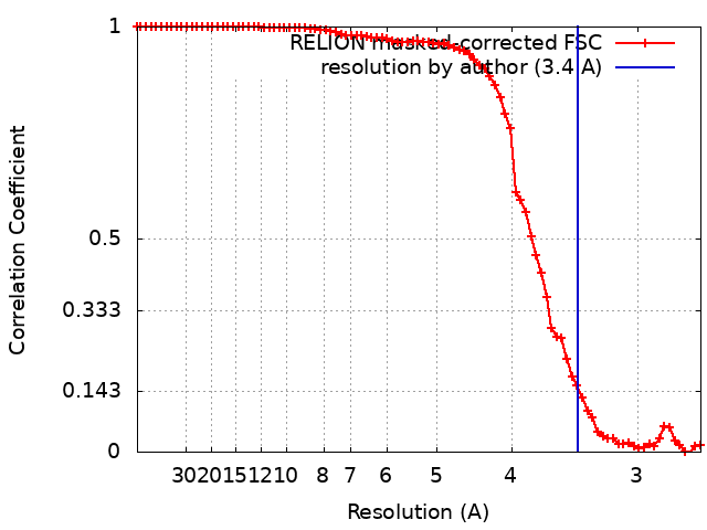
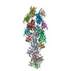
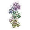
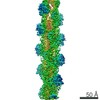
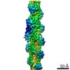
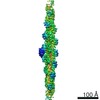
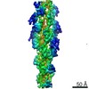
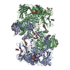
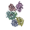
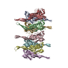
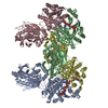
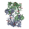
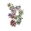
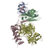
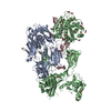
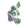
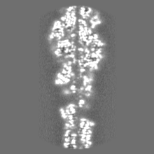
Journal: Proc Natl Acad Sci U S A / Year: 2020 Title: Structures of cofilin-induced structural changes reveal local and asymmetric perturbations of actin filaments. Authors: Andrew R Huehn / Jeffrey P Bibeau / Anthony C Schramm / Wenxiang Cao / Enrique M De La Cruz / Charles V Sindelar / Abstract: Members of the cofilin/ADF family of proteins sever actin filaments, increasing the number of filament ends available for polymerization or depolymerization. Cofilin binds actin filaments with ...Members of the cofilin/ADF family of proteins sever actin filaments, increasing the number of filament ends available for polymerization or depolymerization. Cofilin binds actin filaments with positive cooperativity, forming clusters of contiguously bound cofilin along the filament lattice. Filament severing occurs preferentially at boundaries between bare and cofilin-decorated (cofilactin) segments and is biased at 1 side of a cluster. A molecular understanding of cooperative binding and filament severing has been impeded by a lack of structural data describing boundaries. Here, we apply methods for analyzing filament cryo-electron microscopy (cryo-EM) data at the single subunit level to directly investigate the structure of boundaries within partially decorated cofilactin filaments. Subnanometer resolution maps of isolated, bound cofilin molecules and an actin-cofilactin boundary indicate that cofilin-induced actin conformational changes are local and limited to subunits directly contacting bound cofilin. An isolated, bound cofilin compromises longitudinal filament contacts of 1 protofilament, consistent with a single cofilin having filament-severing activity. An individual, bound phosphomimetic (S3D) cofilin with weak severing activity adopts a unique binding mode that does not perturb actin structure. Cofilin clusters disrupt both protofilaments, consistent with a higher severing activity at boundaries compared to single cofilin. Comparison of these structures indicates that this disruption is substantially greater at pointed end sides of cofilactin clusters than at the barbed end. These structures, with the distribution of bound cofilin clusters, suggest that maximum binding cooperativity is achieved when 2 cofilins occupy adjacent sites. These results reveal the structural origins of cooperative cofilin binding and actin filament severing.
History
Deposition
Sep 12, 2019
-
Header (metadata) release
Jan 15, 2020
-
Map release
Jan 15, 2020
-
Update
Nov 25, 2020
-
Current status
Nov 25, 2020
Processing site: RCSB / Status: Released
-
Structure visualization
Movie
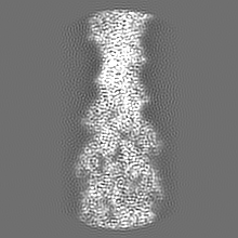
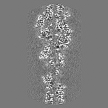
Surface view with section colored by density value
In the structure databanks used in Yorodumi, some data are registered as the other names, "COVID-19 virus" and "2019-nCoV". Here are the details of the virus and the list of structure data.
Jan 31, 2019. EMDB accession codes are about to change! (news from PDBe EMDB page)
EMDB accession codes are about to change! (news from PDBe EMDB page)
The allocation of 4 digits for EMDB accession codes will soon come to an end. Whilst these codes will remain in use, new EMDB accession codes will include an additional digit and will expand incrementally as the available range of codes is exhausted. The current 4-digit format prefixed with “EMD-” (i.e. EMD-XXXX) will advance to a 5-digit format (i.e. EMD-XXXXX), and so on. It is currently estimated that the 4-digit codes will be depleted around Spring 2019, at which point the 5-digit format will come into force.
The EM Navigator/Yorodumi systems omit the EMD- prefix.
Related info.:Q: What is EMD? / ID/Accession-code notation in Yorodumi/EM Navigator
Yorodumi is a browser for structure data from EMDB, PDB, SASBDB, etc.
This page is also the successor to EM Navigator detail page, and also detail information page/front-end page for Omokage search.
The word "yorodu" (or yorozu) is an old Japanese word meaning "ten thousand". "mi" (miru) is to see.
Related info.:EMDB / PDB / SASBDB / Comparison of 3 databanks / Yorodumi Search / Aug 31, 2016. New EM Navigator & Yorodumi / Yorodumi Papers / Jmol/JSmol / Function and homology information / Changes in new EM Navigator and Yorodumi
 Movie
Movie Controller

 Yorodumi
 Open data
 Basic information
 Map data
 Sample
 Function and homology information
 Homo sapiens (human)
 Authors
 United States, 2 items
  Citation
 Structure visualization
 Downloads & links
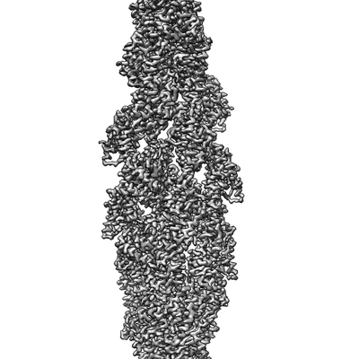 emd_20711.png
 http://ftp.pdbj.org/pub/emdb/structures/EMD-20711


 Z (Sec.)
 Y (Row.)
 X (Col.)


 Sample components
 Processing
 Electron microscopy
 FIELD EMISSION GUN