 Movie
Movie Controller


+ Open data

- Basic information
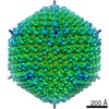
| Entry | Database: EMDB / ID: EMD-20083 | |||||||||
|---|---|---|---|---|---|---|---|---|---|---|
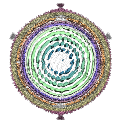
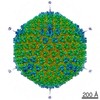
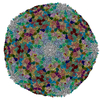
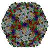
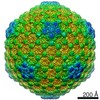
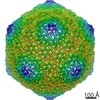
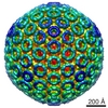
| Title | Cryo-EM reconstruction of Sulfolobus polyhedral virus 1 (SPV1) | |||||||||
 Map data Map data | Cryo-EM reconstruction of Sulfolobus polyhedral virus 1 (SPV1) | |||||||||
 Sample Sample |
| |||||||||
 Keywords Keywords | archaeal pilus / STRUCTURAL PROTEIN / icosahedral symmetry / VIRUS | |||||||||
| Function / homology | : / : / Sulfolobus polyhedral virus 1 capsid protein VP10 / Sulfolobus polyhedral virus 1 major capsid protein VP4 / icosahedral viral capsid / Capsid protein VP10 / Capsid protein VP4 Function and homology information Function and homology information | |||||||||
| Biological species |   Sulfolobus polyhedral virus 1 / Sulfolobus spindle-shaped virus 1 Sulfolobus polyhedral virus 1 / Sulfolobus spindle-shaped virus 1 | |||||||||
| Method | single particle reconstruction / cryo EM / Resolution: 3.7 Å | |||||||||
 Authors Authors | Wang F / Liu Y | |||||||||
| Funding support |  United States, 1 items United States, 1 items
| |||||||||
 Citation Citation | Journal: Proc Natl Acad Sci U S A / Year: 2019 Title: A packing for A-form DNA in an icosahedral virus. Authors: Fengbin Wang / Ying Liu / Zhangli Su / Tomasz Osinski / Guilherme A P de Oliveira / James F Conway / Stefan Schouten / Mart Krupovic / David Prangishvili / Edward H Egelman /   Abstract: Studies on viruses infecting archaea living in the most extreme environments continue to show a remarkable diversity of structures, suggesting that the sampling continues to be very sparse. We have ...Studies on viruses infecting archaea living in the most extreme environments continue to show a remarkable diversity of structures, suggesting that the sampling continues to be very sparse. We have used electron cryo-microscopy to study at 3.7-Å resolution the structure of the polyhedral virus 1 (SPV1), which was originally isolated from a hot, acidic spring in Beppu, Japan. The 2 capsid proteins with variant single jelly-roll folds form pentamers and hexamers which assemble into a = 43 icosahedral shell. In contrast to tailed icosahedral double-stranded DNA (dsDNA) viruses infecting bacteria and archaea, and herpesviruses infecting animals and humans, where naked DNA is packed under very high pressure due to the repulsion between adjacent layers of DNA, the circular dsDNA in SPV1 is fully covered with a viral protein forming a nucleoprotein filament with attractive interactions between layers. Most strikingly, we have been able to show that the DNA is in an A-form, as it is in the filamentous viruses infecting hyperthermophilic acidophiles. Previous studies have suggested that DNA is in the B-form in bacteriophages, and our study is a direct visualization of the structure of DNA in an icosahedral virus. | |||||||||
| History |
|
- Structure visualization
| Movie |
Movie viewer |
|---|---|
| Structure viewer | EM map: SurfViewMolmilJmol/JSmol |
| Supplemental images |
- Downloads & links
-EMDB archive
| Map data | emd_20083.map.gz | 1.7 GB | EMDB map data format | |
|---|---|---|---|---|
| Header (meta data) | emd-20083-v30.xmlemd-20083.xml | 17.3 KB 17.3 KB | Display Display | EMDB header |
| Images |  emd_20083.png emd_20083.png | 248.8 KB | ||
| Filedesc metadata | emd-20083.cif.gz | 6.1 KB | ||
| Archive directory |  http://ftp.pdbj.org/pub/emdb/structures/EMD-20083ftp://ftp.pdbj.org/pub/emdb/structures/EMD-20083 http://ftp.pdbj.org/pub/emdb/structures/EMD-20083ftp://ftp.pdbj.org/pub/emdb/structures/EMD-20083 | HTTPS FTP |
-Related structure data
| Related structure data |  6oj0MC M: atomic model generated by this map C: citing same article ( |
|---|---|
| Similar structure data |

-Links
| EMDB pages | EMDB (EBI/PDBe) / EMDataResource |
|---|
-Map
| File | Download / File: emd_20083.map.gz / Format: CCP4 / Size: 1.9 GB / Type: IMAGE STORED AS FLOATING POINT NUMBER (4 BYTES) | ||||||||||||||||||||||||||||||||||||||||||||||||||||||||||||
|---|---|---|---|---|---|---|---|---|---|---|---|---|---|---|---|---|---|---|---|---|---|---|---|---|---|---|---|---|---|---|---|---|---|---|---|---|---|---|---|---|---|---|---|---|---|---|---|---|---|---|---|---|---|---|---|---|---|---|---|---|---|
| Annotation | Cryo-EM reconstruction of Sulfolobus polyhedral virus 1 (SPV1) | ||||||||||||||||||||||||||||||||||||||||||||||||||||||||||||
| Projections & slices | Image control
Images are generated by Spider. | ||||||||||||||||||||||||||||||||||||||||||||||||||||||||||||
| Voxel size | X=Y=Z: 1.4 Å | ||||||||||||||||||||||||||||||||||||||||||||||||||||||||||||
| Density |
| ||||||||||||||||||||||||||||||||||||||||||||||||||||||||||||
| Symmetry | Space group: 1 | ||||||||||||||||||||||||||||||||||||||||||||||||||||||||||||
| Details | EMDB XML:
CCP4 map header:
| ||||||||||||||||||||||||||||||||||||||||||||||||||||||||||||
 Z (Sec.)
 Y (Row.)
 X (Col.)


-Supplemental data
- Sample components
-Entire : Sulfolobus spindle-shaped virus 1
| Entire | Name: Sulfolobus spindle-shaped virus 1 |
|---|---|
| Components |
|
-Supramolecule #1: Sulfolobus spindle-shaped virus 1
| Supramolecule | Name: Sulfolobus spindle-shaped virus 1 / type: virus / ID: 1 / Parent: 0 / Macromolecule list: all / NCBI-ID: 244589 / Sci species name: Sulfolobus spindle-shaped virus 1 / Virus type: VIRION / Virus isolate: STRAIN / Virus enveloped: Yes / Virus empty: No |
|---|---|
| Host (natural) | Organism: Sulfolobus sp. S38A |
-Macromolecule #1: Structural protein VP4
| Macromolecule | Name: Structural protein VP4 / type: protein_or_peptide / ID: 1 / Number of copies: 42 / Enantiomer: LEVO |
|---|---|
| Source (natural) | Organism: Sulfolobus polyhedral virus 1 |
| Molecular weight | Theoretical: 19.394885 KDa |
| Sequence | String: MSESVTQQVF NFAVTKSQPF GGYVYSTNLT ASTSSAVTST QLTPLNLSIT LGQITLSGNS LVIPATQIWY LTDAYVSVPD YTNITNGAE ADGVILIYKD GVKLMLTTPL ISSMSISNPA RTHLAQAVKY SPQSILTMYF NPTKPATAST SYPNTVYFTV V VVDFSYAQ NPARAVVSAN AVM UniProtKB: Capsid protein VP4 |
-Macromolecule #2: Uncharacterized protein
| Macromolecule | Name: Uncharacterized protein / type: protein_or_peptide / ID: 2 / Number of copies: 1 / Enantiomer: LEVO |
|---|---|
| Source (natural) | Organism: Sulfolobus polyhedral virus 1 |
| Molecular weight | Theoretical: 20.617277 KDa |
| Sequence | String: MLSLDNYSYV HNITTQTNID LSSQQTIHLA SINGKGYIIF LRFFCEGSSA CFTNVKFSVK ANGLVLYSFR YIQLLELGQA IATAIPSSS QGFSTLLSNY NVLISSPIGT LPQLTLYDSY DNRYGAMLQP AFPLPFVNTL SLDVDILPVS QSSYDPIPYS L NDNQISTN APTGKGNISI EYLLYNCLV UniProtKB: Capsid protein VP10 |
-Experimental details
-Structure determination
| Method | cryo EM |
|---|---|
 Processing Processing | single particle reconstruction |
| Aggregation state | particle |
-Sample preparation
| Buffer | pH: 4 |
|---|---|
| Grid | Details: unspecified |
| Vitrification | Cryogen name: ETHANE |
- Electron microscopy
| Microscope | FEI TITAN KRIOS |
|---|---|
| Image recording | Film or detector model: FEI FALCON III (4k x 4k) / Average electron dose: 20.0 e/Å2 |
| Electron beam | Acceleration voltage: 300 kV / Electron source:  FIELD EMISSION GUN FIELD EMISSION GUN |
| Electron optics | Illumination mode: FLOOD BEAM / Imaging mode: BRIGHT FIELD |
| Experimental equipment |  Model: Titan Krios / Image courtesy: FEI Company |
