ムービー
ムービー コントローラー


+ データを開く

- 基本情報
基本情報
| 登録情報 | データベース: EMDB / ID: EMD-1989 | |||||||||
|---|---|---|---|---|---|---|---|---|---|---|
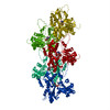
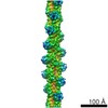
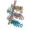
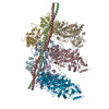
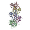
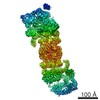
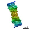
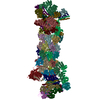
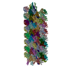
| タイトル | Structure of the Actin-Tropomyosin-Myosin Complex (rigor ATM 1) | |||||||||
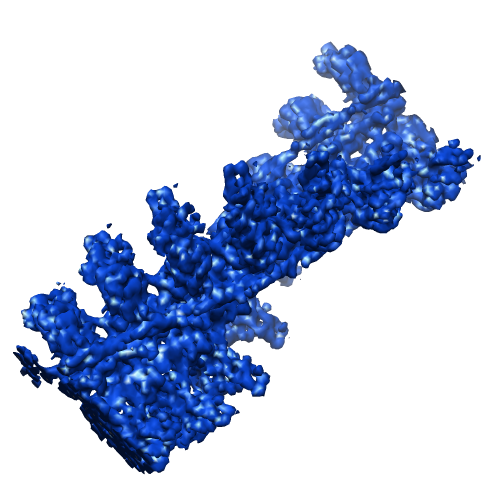
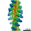
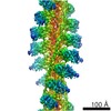
 マップデータ マップデータ | Conformation 1 of the F-actin-myo1E-tropomyosin complex | |||||||||
 試料 試料 |
| |||||||||
 キーワード キーワード | structural protein / cytoskeleton / contractile filament / motor activity / myosin binding / actin binding / ATP catabolic process / rigor state | |||||||||
| 機能・相同性 |  機能・相同性情報 機能・相同性情報: / macropinocytic cup membrane / pseudopodium membrane / actin wave / macropinocytic cup cytoskeleton / myosin I complex / chemotaxis to cAMP / macropinocytic cup / muscle thin filament tropomyosin / leading edge membrane ...: / macropinocytic cup membrane / pseudopodium membrane / actin wave / macropinocytic cup cytoskeleton / myosin I complex / chemotaxis to cAMP / macropinocytic cup / muscle thin filament tropomyosin / leading edge membrane / actin filament-based movement / early phagosome / myosin complex / phagocytosis, engulfment / cytoskeletal motor activator activity / microfilament motor activity / myosin heavy chain binding / tropomyosin binding / actin filament bundle / troponin I binding / filamentous actin / mesenchyme migration / phosphatidylinositol-3,4,5-trisphosphate binding / microvillus / cell leading edge / pseudopodium / skeletal muscle myofibril / actin filament bundle assembly / striated muscle thin filament / skeletal muscle thin filament assembly / actin monomer binding / phagocytosis / phagocytic cup / skeletal muscle fiber development / stress fiber / titin binding / actin filament polymerization / actin filament organization / filopodium / actin filament / 加水分解酵素; 酸無水物に作用; 酸無水物に作用・細胞または細胞小器官の運動に関与 / endocytosis / calcium-dependent protein binding / actin filament binding / lamellipodium / actin cytoskeleton / actin binding / cell body / calmodulin binding / protein heterodimerization activity / protein domain specific binding / hydrolase activity / calcium ion binding / positive regulation of gene expression / magnesium ion binding / protein homodimerization activity / ATP binding / identical protein binding / plasma membrane / cytoplasm / cytosol 類似検索 - 分子機能 | |||||||||
| 生物種 |    | |||||||||
| 手法 | らせん対称体再構成法 / クライオ電子顕微鏡法 / 解像度: 8.1 Å | |||||||||
 データ登録者 データ登録者 | Behrmann E / Mueller M / Penczek PA / Mannherz HG / Manstein DJ / Raunser S | |||||||||
 引用 引用 | ジャーナル: Cell / 年: 2012 タイトル: Structure of the rigor actin-tropomyosin-myosin complex. 著者: Elmar Behrmann / Mirco Müller / Pawel A Penczek / Hans Georg Mannherz / Dietmar J Manstein / Stefan Raunser /  要旨: Regulation of myosin and filamentous actin interaction by tropomyosin is a central feature of contractile events in muscle and nonmuscle cells. However, little is known about molecular interactions ...Regulation of myosin and filamentous actin interaction by tropomyosin is a central feature of contractile events in muscle and nonmuscle cells. However, little is known about molecular interactions within the complex and the trajectory of tropomyosin movement between its "open" and "closed" positions on the actin filament. Here, we report the 8 Å resolution structure of the rigor (nucleotide-free) actin-tropomyosin-myosin complex determined by cryo-electron microscopy. The pseudoatomic model of the complex, obtained from fitting crystal structures into the map, defines the large interface involving two adjacent actin monomers and one tropomyosin pseudorepeat per myosin contact. Severe forms of hereditary myopathies are linked to mutations that critically perturb this interface. Myosin binding results in a 23 Å shift of tropomyosin along actin. Complex domain motions occur in myosin, but not in actin. Based on our results, we propose a structural model for the tropomyosin-dependent modulation of myosin binding to actin. | |||||||||
| 履歴 |
|
- 構造の表示
構造の表示
| ムービー |
ムービービューア |
|---|---|
| 構造ビューア | EMマップ: SurfViewMolmilJmol/JSmol |
| 添付画像 |
- ダウンロードとリンク
-EMDBアーカイブ
| マップデータ | emd_1989.map.gz | 5.2 MB | EMDBマップデータ形式 | |
|---|---|---|---|---|
| ヘッダ (付随情報) | emd-1989-v30.xmlemd-1989.xml | 14.7 KB 14.7 KB | 表示 表示 | EMDBヘッダ |
| 画像 |  emd_1989.png emd_1989.png | 157.9 KB | ||
| アーカイブディレクトリ |  http://ftp.pdbj.org/pub/emdb/structures/EMD-1989ftp://ftp.pdbj.org/pub/emdb/structures/EMD-1989 http://ftp.pdbj.org/pub/emdb/structures/EMD-1989ftp://ftp.pdbj.org/pub/emdb/structures/EMD-1989 | HTTPS FTP |
-関連構造データ
-リンク
| EMDBのページ | EMDB (EBI/PDBe) / EMDataResource |
|---|---|
| 「今月の分子」の関連する項目 |


-マップ
| ファイル | ダウンロード / ファイル: emd_1989.map.gz / 形式: CCP4 / 大きさ: 21 MB / タイプ: IMAGE STORED AS FLOATING POINT NUMBER (4 BYTES) | ||||||||||||||||||||||||||||||||||||||||||||||||||||||||||||||||||||
|---|---|---|---|---|---|---|---|---|---|---|---|---|---|---|---|---|---|---|---|---|---|---|---|---|---|---|---|---|---|---|---|---|---|---|---|---|---|---|---|---|---|---|---|---|---|---|---|---|---|---|---|---|---|---|---|---|---|---|---|---|---|---|---|---|---|---|---|---|---|
| 注釈 | Conformation 1 of the F-actin-myo1E-tropomyosin complex | ||||||||||||||||||||||||||||||||||||||||||||||||||||||||||||||||||||
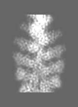
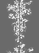
| 投影像・断面図 | 画像のコントロール
画像は Spider により作成 これらの図は立方格子座標系で作成されたものです | ||||||||||||||||||||||||||||||||||||||||||||||||||||||||||||||||||||
| ボクセルのサイズ | X=Y=Z: 1.84 Å | ||||||||||||||||||||||||||||||||||||||||||||||||||||||||||||||||||||
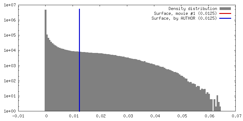
| 密度 |
| ||||||||||||||||||||||||||||||||||||||||||||||||||||||||||||||||||||
| 対称性 | 空間群: 1 | ||||||||||||||||||||||||||||||||||||||||||||||||||||||||||||||||||||
| 詳細 | EMDB XML:
CCP4マップ ヘッダ情報:
| ||||||||||||||||||||||||||||||||||||||||||||||||||||||||||||||||||||
 Z (Sec.)
 Y (Row.)
 X (Col.)


-添付データ
- 試料の構成要素
-全体 : F-actin-myo1E-tropomyosin complex (conformation 1)
| 全体 | 名称: F-actin-myo1E-tropomyosin complex (conformation 1) |
|---|---|
| 要素 |
|
-超分子 #1000: F-actin-myo1E-tropomyosin complex (conformation 1)
| 超分子 | 名称: F-actin-myo1E-tropomyosin complex (conformation 1) / タイプ: sample / ID: 1000 / 詳細: 7 to 7 to 1 (actin to myosin to tropomyosin) / 集合状態: Pentameric / Number unique components: 3 |
|---|
-分子 #1: F-actin
| 分子 | 名称: F-actin / タイプ: protein_or_peptide / ID: 1 / Name.synonym: actin filament / コピー数: 14 / 集合状態: filament / 組換発現: No / データベース: NCBI |
|---|---|
| 由来(天然) | 生物種: |
| 配列 | GO: GO: 0042643 / InterPro: Actin family |
-分子 #2: tropomyosin 1 (alpha)
| 分子 | 名称: tropomyosin 1 (alpha) / タイプ: protein_or_peptide / ID: 2 / Name.synonym: tropomyosin 1 (alpha) / コピー数: 2 / 集合状態: filament of dimers / 組換発現: Yes |
|---|---|
| 由来(天然) | 生物種: |
| 組換発現 | 生物種: Escherichia coli (strain BL21 DE3) / 組換プラスミド: pJC20 |
| 配列 | GO: muscle thin filament tropomyosin / InterPro: Tropomyosin |
-分子 #3: myoE
| 分子 | 名称: myoE / タイプ: protein_or_peptide / ID: 3 / Name.synonym: myosin 1E / コピー数: 14 / 集合状態: filament / 組換発現: Yes |
|---|---|
| 由来(天然) | 生物種: 株: AX2 / 別称: Slime mold |
| 組換発現 | 生物種: 組換プラスミド: pDXA-3H |
| 配列 | GO: myosin I complex / InterPro: Myosin head, motor domain |
-実験情報
-構造解析
| 手法 | クライオ電子顕微鏡法 |
|---|---|
 解析 解析 | らせん対称体再構成法 |
| 試料の集合状態 | filament |
-試料調製
| 濃度 | 0.01 mg/mL |
|---|---|
| 緩衝液 | pH: 7.2 詳細: 5mM Tris, 100mM KCl, 2mM MgCl2, 50mM glutamine, 50mM arginine |
| グリッド | 詳細: C-Flat CF-2/1-4C copper 400 mesh |
| 凍結 | 凍結剤: ETHANE / チャンバー内湿度: 90 % / チャンバー内温度: 101 K / 装置: GATAN CRYOPLUNGE 3 / 詳細: Vitrification instrument: Gatan Cryoplunge 3 / 手法: Manual blotting for approximately 15 seconds |
- 電子顕微鏡法
| 顕微鏡 | JEOL 3200FSC |
|---|---|
| 温度 | 平均: 77 K |
| アライメント法 | Legacy - 非点収差: objective lens astigmatism was corrected at 150,000 times magnification |
| 特殊光学系 | エネルギーフィルター - 名称: in-column Omega filter エネルギーフィルター - エネルギー下限: 0.0 eV エネルギーフィルター - エネルギー上限: 12.0 eV |
| 撮影 | カテゴリ: CCD フィルム・検出器のモデル: TVIPS TEMCAM-F816 (8k x 8k) デジタル化 - サンプリング間隔: 15.6 µm / 実像数: 836 / 平均電子線量: 17 e/Å2 詳細: Over 3000 images were taken of which only the best 836 were used for processing ビット/ピクセル: 14 |
| 電子線 | 加速電圧: 200 kV / 電子線源:  FIELD EMISSION GUN FIELD EMISSION GUN |
| 電子光学系 | 倍率(補正後): 169644 / 照射モード: FLOOD BEAM / 撮影モード: BRIGHT FIELD / Cs: 4.1 mm / 最大 デフォーカス(公称値): 1.5 µm / 最小 デフォーカス(公称値): 0.75 µm / 倍率(公称値): 80000 |
| 試料ステージ | 試料ホルダー: cryogenic stage with side entry access / 試料ホルダーモデル: JEOL 3200FSC CRYOHOLDER |
-画像解析
| 詳細 | Particles were selected by hand using e2helixboxer |
|---|---|
| 最終 再構成 | 想定した対称性 - らせんパラメータ - Δz: 27.6 Å 想定した対称性 - らせんパラメータ - ΔΦ: 166.5 ° アルゴリズム: OTHER / 解像度のタイプ: BY AUTHOR / 解像度: 8.1 Å / 解像度の算出法: FSC 0.5 CUT-OFF / ソフトウェア - 名称: SPARX / 詳細: Particles were classified using CODIM |
| CTF補正 | 詳細: each particle |
-原子モデル構築 1
| 初期モデル | PDB ID: Chain - Chain ID: C |
|---|---|
| ソフトウェア | 名称: DireX |
| 詳細 | PDBEntryID_givenInChain. Protocol: geometry-based conformational sampling using Deformable Elastic Network (DEN) approach. Initial placement was performed using rigid-body fitting in Chimera |
| 精密化 | 空間: REAL / プロトコル: FLEXIBLE FIT |
| 得られたモデル | 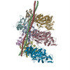 PDB-4a7l: |
-原子モデル構築 2
| 初期モデル | PDB ID: |
|---|---|
| ソフトウェア | 名称: DireX |
| 詳細 | Protocol: geometry-based conformational sampling using Deformable Elastic Network (DEN) approach. Initial placement was performed using rigid-body fitting in Chimera |
| 精密化 | 空間: REAL / プロトコル: FLEXIBLE FIT |
| 得られたモデル | PDB-4a7l: |