Movie
Movie Controller


+ Open data

- Basic information
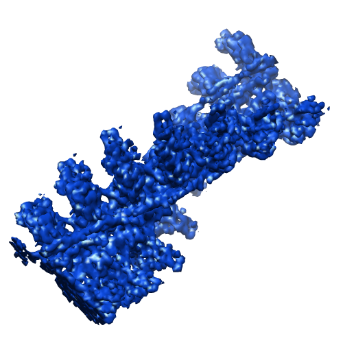
| Entry | Database: EMDB / ID: EMD-1989 | |||||||||
|---|---|---|---|---|---|---|---|---|---|---|
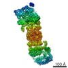
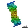
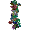
| Title | Structure of the Actin-Tropomyosin-Myosin Complex (rigor ATM 1) | |||||||||
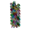
 Map data Map data | Conformation 1 of the F-actin-myo1E-tropomyosin complex | |||||||||
 Sample Sample |
| |||||||||
 Keywords Keywords | structural protein / cytoskeleton / contractile filament / motor activity / myosin binding / actin binding / ATP catabolic process / rigor state | |||||||||
| Function / homology |  Function and homology information Function and homology information: / macropinocytic cup membrane / pseudopodium membrane / actin wave / macropinocytic cup cytoskeleton / myosin I complex / chemotaxis to cAMP / muscle thin filament tropomyosin / macropinocytic cup / actin filament-based movement ...: / macropinocytic cup membrane / pseudopodium membrane / actin wave / macropinocytic cup cytoskeleton / myosin I complex / chemotaxis to cAMP / muscle thin filament tropomyosin / macropinocytic cup / actin filament-based movement / leading edge membrane / early phagosome / myosin complex / cytoskeletal motor activator activity / microfilament motor activity / phagocytosis, engulfment / tropomyosin binding / myosin heavy chain binding / mesenchyme migration / troponin I binding / cell leading edge / pseudopodium / filamentous actin / actin filament bundle / phosphatidylinositol-3,4,5-trisphosphate binding / skeletal muscle thin filament assembly / actin filament bundle assembly / striated muscle thin filament / phagocytic cup / skeletal muscle myofibril / actin monomer binding / phagocytosis / skeletal muscle fiber development / stress fiber / muscle contraction / titin binding / actin filament polymerization / actin filament organization / filopodium / actin filament / Hydrolases; Acting on acid anhydrides; Acting on acid anhydrides to facilitate cellular and subcellular movement / endocytosis / calcium-dependent protein binding / actin filament binding / actin cytoskeleton / lamellipodium / actin binding / cell body / calmodulin binding / hydrolase activity / protein heterodimerization activity / protein domain specific binding / calcium ion binding / positive regulation of gene expression / magnesium ion binding / protein homodimerization activity / ATP binding / identical protein binding / plasma membrane / cytosol / cytoplasm Similarity search - Function | |||||||||
| Biological species |    | |||||||||
| Method | helical reconstruction / cryo EM / Resolution: 8.1 Å | |||||||||
 Authors Authors | Behrmann E / Mueller M / Penczek PA / Mannherz HG / Manstein DJ / Raunser S | |||||||||
 Citation Citation | Journal: Cell / Year: 2012 Title: Structure of the rigor actin-tropomyosin-myosin complex. Authors: Elmar Behrmann / Mirco Müller / Pawel A Penczek / Hans Georg Mannherz / Dietmar J Manstein / Stefan Raunser /  Abstract: Regulation of myosin and filamentous actin interaction by tropomyosin is a central feature of contractile events in muscle and nonmuscle cells. However, little is known about molecular interactions ...Regulation of myosin and filamentous actin interaction by tropomyosin is a central feature of contractile events in muscle and nonmuscle cells. However, little is known about molecular interactions within the complex and the trajectory of tropomyosin movement between its "open" and "closed" positions on the actin filament. Here, we report the 8 Å resolution structure of the rigor (nucleotide-free) actin-tropomyosin-myosin complex determined by cryo-electron microscopy. The pseudoatomic model of the complex, obtained from fitting crystal structures into the map, defines the large interface involving two adjacent actin monomers and one tropomyosin pseudorepeat per myosin contact. Severe forms of hereditary myopathies are linked to mutations that critically perturb this interface. Myosin binding results in a 23 Å shift of tropomyosin along actin. Complex domain motions occur in myosin, but not in actin. Based on our results, we propose a structural model for the tropomyosin-dependent modulation of myosin binding to actin. | |||||||||
| History |
|
- Structure visualization
| Movie |
Movie viewer |
|---|---|
| Structure viewer | EM map: SurfViewMolmilJmol/JSmol |
| Supplemental images |
- Downloads & links
-EMDB archive
| Map data | emd_1989.map.gz | 5.2 MB | EMDB map data format | |
|---|---|---|---|---|
| Header (meta data) | emd-1989-v30.xmlemd-1989.xml | 14.7 KB 14.7 KB | Display Display | EMDB header |
| Images |  emd_1989.png emd_1989.png | 157.9 KB | ||
| Archive directory |  http://ftp.pdbj.org/pub/emdb/structures/EMD-1989ftp://ftp.pdbj.org/pub/emdb/structures/EMD-1989 http://ftp.pdbj.org/pub/emdb/structures/EMD-1989ftp://ftp.pdbj.org/pub/emdb/structures/EMD-1989 | HTTPS FTP |
-Validation report
| Summary document | emd_1989_validation.pdf.gz | 216.2 KB | Display | EMDB validaton report |
|---|---|---|---|---|
| Full document | emd_1989_full_validation.pdf.gz | 215.3 KB | Display | |
| Data in XML | emd_1989_validation.xml.gz | 4.3 KB | Display | |
| Arichive directory | https://ftp.pdbj.org/pub/emdb/validation_reports/EMD-1989ftp://ftp.pdbj.org/pub/emdb/validation_reports/EMD-1989 | HTTPS FTP |
-Related structure data
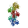
| Related structure data | 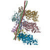 4a7lMC 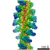 1987C 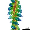 1988C 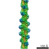 1990C 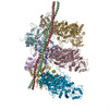 4a7fC 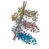 4a7hC 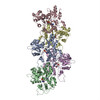 4a7nC M: atomic model generated by this map C: citing same article ( |
|---|---|
| Similar structure data |
-Links
| EMDB pages | EMDB (EBI/PDBe) / EMDataResource |
|---|---|
| Related items in Molecule of the Month |


-Map
| File | Download / File: emd_1989.map.gz / Format: CCP4 / Size: 21 MB / Type: IMAGE STORED AS FLOATING POINT NUMBER (4 BYTES) | ||||||||||||||||||||||||||||||||||||||||||||||||||||||||||||||||||||
|---|---|---|---|---|---|---|---|---|---|---|---|---|---|---|---|---|---|---|---|---|---|---|---|---|---|---|---|---|---|---|---|---|---|---|---|---|---|---|---|---|---|---|---|---|---|---|---|---|---|---|---|---|---|---|---|---|---|---|---|---|---|---|---|---|---|---|---|---|---|
| Annotation | Conformation 1 of the F-actin-myo1E-tropomyosin complex | ||||||||||||||||||||||||||||||||||||||||||||||||||||||||||||||||||||
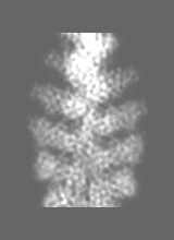
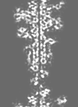
| Projections & slices | Image control
Images are generated by Spider. generated in cubic-lattice coordinate | ||||||||||||||||||||||||||||||||||||||||||||||||||||||||||||||||||||
| Voxel size | X=Y=Z: 1.84 Å | ||||||||||||||||||||||||||||||||||||||||||||||||||||||||||||||||||||
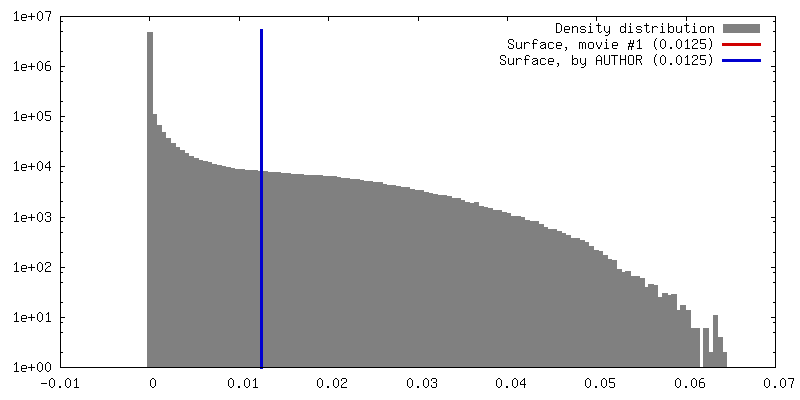
| Density |
| ||||||||||||||||||||||||||||||||||||||||||||||||||||||||||||||||||||
| Symmetry | Space group: 1 | ||||||||||||||||||||||||||||||||||||||||||||||||||||||||||||||||||||
| Details | EMDB XML:
CCP4 map header:
| ||||||||||||||||||||||||||||||||||||||||||||||||||||||||||||||||||||
 Z (Sec.)
 Y (Row.)
 X (Col.)


-Supplemental data
- Sample components
-Entire : F-actin-myo1E-tropomyosin complex (conformation 1)
| Entire | Name: F-actin-myo1E-tropomyosin complex (conformation 1) |
|---|---|
| Components |
|
-Supramolecule #1000: F-actin-myo1E-tropomyosin complex (conformation 1)
| Supramolecule | Name: F-actin-myo1E-tropomyosin complex (conformation 1) / type: sample / ID: 1000 / Details: 7 to 7 to 1 (actin to myosin to tropomyosin) / Oligomeric state: Pentameric / Number unique components: 3 |
|---|
-Macromolecule #1: F-actin
| Macromolecule | Name: F-actin / type: protein_or_peptide / ID: 1 / Name.synonym: actin filament / Number of copies: 14 / Oligomeric state: filament / Recombinant expression: No / Database: NCBI |
|---|---|
| Source (natural) | Organism: |
| Sequence | GO: GO: 0042643 / InterPro: Actin family |
-Macromolecule #2: tropomyosin 1 (alpha)
| Macromolecule | Name: tropomyosin 1 (alpha) / type: protein_or_peptide / ID: 2 / Name.synonym: tropomyosin 1 (alpha) / Number of copies: 2 / Oligomeric state: filament of dimers / Recombinant expression: Yes |
|---|---|
| Source (natural) | Organism: |
| Recombinant expression | Organism: Escherichia coli (strain BL21 DE3) / Recombinant plasmid: pJC20 |
| Sequence | GO: muscle thin filament tropomyosin / InterPro: Tropomyosin |
-Macromolecule #3: myoE
| Macromolecule | Name: myoE / type: protein_or_peptide / ID: 3 / Name.synonym: myosin 1E / Number of copies: 14 / Oligomeric state: filament / Recombinant expression: Yes |
|---|---|
| Source (natural) | Organism: |
| Recombinant expression | Organism: |
| Sequence | GO: myosin I complex / InterPro: Myosin head, motor domain |
-Experimental details
-Structure determination
| Method | cryo EM |
|---|---|
 Processing Processing | helical reconstruction |
| Aggregation state | filament |
-Sample preparation
| Concentration | 0.01 mg/mL |
|---|---|
| Buffer | pH: 7.2 Details: 5mM Tris, 100mM KCl, 2mM MgCl2, 50mM glutamine, 50mM arginine |
| Grid | Details: C-Flat CF-2/1-4C copper 400 mesh |
| Vitrification | Cryogen name: ETHANE / Chamber humidity: 90 % / Chamber temperature: 101 K / Instrument: GATAN CRYOPLUNGE 3 / Details: Vitrification instrument: Gatan Cryoplunge 3 / Method: Manual blotting for approximately 15 seconds |
- Electron microscopy
| Microscope | JEOL 3200FSC |
|---|---|
| Temperature | Average: 77 K |
| Alignment procedure | Legacy - Astigmatism: objective lens astigmatism was corrected at 150,000 times magnification |
| Specialist optics | Energy filter - Name: in-column Omega filter / Energy filter - Lower energy threshold: 0.0 eV / Energy filter - Upper energy threshold: 12.0 eV |
| Image recording | Category: CCD / Film or detector model: TVIPS TEMCAM-F816 (8k x 8k) / Digitization - Sampling interval: 15.6 µm / Number real images: 836 / Average electron dose: 17 e/Å2 Details: Over 3000 images were taken of which only the best 836 were used for processing Bits/pixel: 14 |
| Electron beam | Acceleration voltage: 200 kV / Electron source:  FIELD EMISSION GUN FIELD EMISSION GUN |
| Electron optics | Calibrated magnification: 169644 / Illumination mode: FLOOD BEAM / Imaging mode: BRIGHT FIELD / Cs: 4.1 mm / Nominal defocus max: 1.5 µm / Nominal defocus min: 0.75 µm / Nominal magnification: 80000 |
| Sample stage | Specimen holder: cryogenic stage with side entry access / Specimen holder model: JEOL 3200FSC CRYOHOLDER |
-Image processing
| Details | Particles were selected by hand using e2helixboxer |
|---|---|
| Final reconstruction | Applied symmetry - Helical parameters - Δz: 27.6 Å Applied symmetry - Helical parameters - Δ&Phi: 166.5 ° Algorithm: OTHER / Resolution.type: BY AUTHOR / Resolution: 8.1 Å / Resolution method: FSC 0.5 CUT-OFF / Software - Name: SPARX / Details: Particles were classified using CODIM |
| CTF correction | Details: each particle |
-Atomic model buiding 1
| Initial model | PDB ID: Chain - Chain ID: C |
|---|---|
| Software | Name: DireX |
| Details | PDBEntryID_givenInChain. Protocol: geometry-based conformational sampling using Deformable Elastic Network (DEN) approach. Initial placement was performed using rigid-body fitting in Chimera |
| Refinement | Space: REAL / Protocol: FLEXIBLE FIT |
| Output model | PDB-4a7l: |
-Atomic model buiding 2
| Initial model | PDB ID: |
|---|---|
| Software | Name: DireX |
| Details | Protocol: geometry-based conformational sampling using Deformable Elastic Network (DEN) approach. Initial placement was performed using rigid-body fitting in Chimera |
| Refinement | Space: REAL / Protocol: FLEXIBLE FIT |
| Output model | PDB-4a7l: |