[English] 日本語
 Yorodumi
- EMDB-19584: CryoEM structure of M. smegmatis GMP reductase in complex with GM... -
+ Open data
- Basic information
| Entry |  | |||||||||
|---|---|---|---|---|---|---|---|---|---|---|
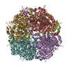
| Title | CryoEM structure of M. smegmatis GMP reductase in complex with GMP at pH 6.6, compressed conformation. | |||||||||
 Map data Map data | LAFTER de-noised | |||||||||
 Sample Sample |
| |||||||||
 Keywords Keywords | GMP reductase / GuaB1 / CBS domain / Mycobacterium smegmatis / OXIDOREDUCTASE | |||||||||
| Function / homology |  Function and homology information Function and homology informationGMP reductase / GMP reductase activity / IMP salvage / IMP dehydrogenase activity / purine ribonucleoside salvage / cytosol Similarity search - Function | |||||||||
| Biological species |  Mycolicibacterium smegmatis (bacteria) Mycolicibacterium smegmatis (bacteria) | |||||||||
| Method | single particle reconstruction / cryo EM / Resolution: 3.85 Å | |||||||||
 Authors Authors | Dolezal M / Kouba T / Pichova I | |||||||||
| Funding support | European Union, 1 items
| |||||||||
 Citation Citation |  Journal: To Be Published Journal: To Be PublishedTitle: Structural basis for allosteric regulation of mycobacterial guanosine 5'-monophosphate reductase with ATP and GTP. Authors: Dolezal M / Knejzlik Z / Kouba T / Filimonenko A / Svachova H / Klima M / Pichova I | |||||||||
| History |
|
- Structure visualization
| Supplemental images |
|---|
- Downloads & links
-EMDB archive
| Map data |  emd_19584.map.gz emd_19584.map.gz | 80.2 MB |  EMDB map data format EMDB map data format | |
|---|---|---|---|---|
| Header (meta data) |  emd-19584-v30.xml emd-19584-v30.xml emd-19584.xml emd-19584.xml | 20.5 KB 20.5 KB | Display Display |  EMDB header EMDB header |
| FSC (resolution estimation) |  emd_19584_fsc.xml emd_19584_fsc.xml | 10 KB | Display |  FSC data file FSC data file |
| Images |  emd_19584.png emd_19584.png | 102.5 KB | ||
| Masks |  emd_19584_msk_1.map emd_19584_msk_1.map | 83.7 MB |  Mask map Mask map | |
| Filedesc metadata |  emd-19584.cif.gz emd-19584.cif.gz | 6.4 KB | ||
| Others |  emd_19584_additional_1.map.gz emd_19584_additional_1.map.gz emd_19584_half_map_1.map.gz emd_19584_half_map_1.map.gz emd_19584_half_map_2.map.gz emd_19584_half_map_2.map.gz | 65 MB 65.2 MB 65.2 MB | ||
| Archive directory |  http://ftp.pdbj.org/pub/emdb/structures/EMD-19584 http://ftp.pdbj.org/pub/emdb/structures/EMD-19584 ftp://ftp.pdbj.org/pub/emdb/structures/EMD-19584 ftp://ftp.pdbj.org/pub/emdb/structures/EMD-19584 | HTTPS FTP |
-Related structure data
| Related structure data |  8ry1MC  8ry0C  8ry3C  8ry4C  8ry5C  8ry6C  8ry7C  8ry8C  8ry9C  8ryaC  8rybC  9hfzC  9hg0C  9hg1C  9hg2C  9hg3C M: atomic model generated by this map C: citing same article ( |
|---|---|
| Similar structure data | Similarity search - Function & homology  F&H Search F&H Search |
- Links
Links
| EMDB pages |  EMDB (EBI/PDBe) / EMDB (EBI/PDBe) /  EMDataResource EMDataResource |
|---|---|
| Related items in Molecule of the Month |
- Map
Map
| File |  Download / File: emd_19584.map.gz / Format: CCP4 / Size: 83.7 MB / Type: IMAGE STORED AS FLOATING POINT NUMBER (4 BYTES) Download / File: emd_19584.map.gz / Format: CCP4 / Size: 83.7 MB / Type: IMAGE STORED AS FLOATING POINT NUMBER (4 BYTES) | ||||||||||||||||||||||||||||||||||||
|---|---|---|---|---|---|---|---|---|---|---|---|---|---|---|---|---|---|---|---|---|---|---|---|---|---|---|---|---|---|---|---|---|---|---|---|---|---|
| Annotation | LAFTER de-noised | ||||||||||||||||||||||||||||||||||||
| Projections & slices | Image control
Images are generated by Spider. | ||||||||||||||||||||||||||||||||||||
| Voxel size | X=Y=Z: 0.7717 Å | ||||||||||||||||||||||||||||||||||||
| Density |
| ||||||||||||||||||||||||||||||||||||
| Symmetry | Space group: 1 | ||||||||||||||||||||||||||||||||||||
| Details | EMDB XML:
|
-Supplemental data
-Mask #1
| File |  emd_19584_msk_1.map emd_19584_msk_1.map | ||||||||||||
|---|---|---|---|---|---|---|---|---|---|---|---|---|---|
| Projections & Slices |
| ||||||||||||
| Density Histograms |
-Additional map: RELION refined
| File | emd_19584_additional_1.map | ||||||||||||
|---|---|---|---|---|---|---|---|---|---|---|---|---|---|
| Annotation | RELION refined | ||||||||||||
| Projections & Slices |
| ||||||||||||
| Density Histograms |
-Half map: #2
| File | emd_19584_half_map_1.map | ||||||||||||
|---|---|---|---|---|---|---|---|---|---|---|---|---|---|
| Projections & Slices |
| ||||||||||||
| Density Histograms |
-Half map: #1
| File | emd_19584_half_map_2.map | ||||||||||||
|---|---|---|---|---|---|---|---|---|---|---|---|---|---|
| Projections & Slices |
| ||||||||||||
| Density Histograms |
- Sample components
-Entire : Msm GMPR with GMP at pH 6.6
| Entire | Name: Msm GMPR with GMP at pH 6.6 |
|---|---|
| Components |
|
-Supramolecule #1: Msm GMPR with GMP at pH 6.6
| Supramolecule | Name: Msm GMPR with GMP at pH 6.6 / type: complex / ID: 1 / Parent: 0 / Macromolecule list: #1 |
|---|---|
| Source (natural) | Organism:  Mycolicibacterium smegmatis (bacteria) / Strain: ATCC 700084 / mc(2)155 Mycolicibacterium smegmatis (bacteria) / Strain: ATCC 700084 / mc(2)155 |
-Macromolecule #1: GMP reductase
| Macromolecule | Name: GMP reductase / type: protein_or_peptide / ID: 1 / Details: Guanosine 5'-monophosphate reductase / Number of copies: 8 / Enantiomer: LEVO / EC number: GMP reductase |
|---|---|
| Source (natural) | Organism:  Mycolicibacterium smegmatis (bacteria) / Strain: ATCC 700084 / mc(2)155 Mycolicibacterium smegmatis (bacteria) / Strain: ATCC 700084 / mc(2)155 |
| Molecular weight | Theoretical: 51.782434 KDa |
| Recombinant expression | Organism:  |
| Sequence | String: MVRFLDGHTP AYDLTYNDVF VVPGRSDVAS RFDVDLSTVD GSGTTIPVVV ANMTAVAGRR MAETVARRGG IVVLPQDLPI TAVSETVDF VKSRDLVVDT PVTLSPEDSV SDANALLHKR AHGAAVVVFE GRPIGLVTEA NCAGVDRFAR VRDIALSDFV T APVGTDPR ...String: MVRFLDGHTP AYDLTYNDVF VVPGRSDVAS RFDVDLSTVD GSGTTIPVVV ANMTAVAGRR MAETVARRGG IVVLPQDLPI TAVSETVDF VKSRDLVVDT PVTLSPEDSV SDANALLHKR AHGAAVVVFE GRPIGLVTEA NCAGVDRFAR VRDIALSDFV T APVGTDPR EVFDLLEHAP IDVAVMTAPD GTLAGVLTRT GAIRAGIYTP AVDAKGRLRI AAAVGINGDV GAKAQALAEA GA DLLVIDT AHGHQAKMLD AIKAVASLDL GLPLVAGNVV SAEGTRDLIE AGASIVKVGV GPGAMCTTRM MTGVGRPQFS AVV ECAAAA RQLGGHVWAD GGVRHPRDVA LALAAGASNV MIGSWFAGTY ESPGDLLFDR DDRPYKESYG MASKRAVAAR TAGD SSFDR ARKGLFEEGI STSRMSLDPA RGGVEDLLDH ITSGVRSTCT YVGAANLPEL HEKVVLGVQS AAGFAEGHPL PAGWT AAAK EDLEHHHHHH HH UniProtKB: GMP reductase |
-Macromolecule #2: GUANOSINE-5'-MONOPHOSPHATE
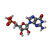
| Macromolecule | Name: GUANOSINE-5'-MONOPHOSPHATE / type: ligand / ID: 2 / Number of copies: 16 / Formula: 5GP |
|---|---|
| Molecular weight | Theoretical: 363.221 Da |
| Chemical component information |  ChemComp-5GP: |
-Experimental details
-Structure determination
| Method | cryo EM |
|---|---|
 Processing Processing | single particle reconstruction |
| Aggregation state | particle |
- Sample preparation
| Buffer | pH: 6.6 Component:
| ||||||||||||
|---|---|---|---|---|---|---|---|---|---|---|---|---|---|
| Vitrification | Cryogen name: ETHANE | ||||||||||||
| Details | 20 mg/ml Msm GMPR with 2 mM GMP in the storage buffer (50 mM Tris, pH 8.0, 2.5 mM TCEP) was diluted with the cryoEM buffer (50 mM HEPES, pH 6.6, 100 mM KCl, 2 mM MgCl2). |
- Electron microscopy
| Microscope | FEI TALOS ARCTICA |
|---|---|
| Image recording | Film or detector model: GATAN K2 SUMMIT (4k x 4k) / Average electron dose: 40.0 e/Å2 |
| Electron beam | Acceleration voltage: 200 kV / Electron source:  FIELD EMISSION GUN FIELD EMISSION GUN |
| Electron optics | Illumination mode: FLOOD BEAM / Imaging mode: BRIGHT FIELD / Nominal defocus max: 3.0 µm / Nominal defocus min: 0.7000000000000001 µm |
| Sample stage | Cooling holder cryogen: NITROGEN |
| Experimental equipment |  Model: Talos Arctica / Image courtesy: FEI Company |
+ Image processing
-Atomic model buiding 1
| Details | The initial structure was obtained by rigid-body fitting an appropriate model into the cryo-EM map using ChimeraX. Phenix.real_space_refine with tight reference model restraints to the starting model was then used to remove the worst clashes. |
|---|---|
| Refinement | Space: REAL |
| Output model |  PDB-8ry1: |
 Movie
Movie Controller


 Z (Sec.)
 Y (Row.)
 X (Col.)


