 Movie
Movie Controller

[English] 日本語
 Yorodumi
- PDB-7r50: Crystal structure of GMP reductase from mycobacterium smegmatis i... -
+ Open data

- Basic information
| Entry | Database: PDB / ID: 7r50 | ||||||
|---|---|---|---|---|---|---|---|
| Title | Crystal structure of GMP reductase from mycobacterium smegmatis in complex with GMP. | ||||||
 Components Components | GMP reductase | ||||||
 Keywords Keywords | OXIDOREDUCTASE / GMP reductase GMPR oxidoreductase GuaB1 octamer CBS domain Bateman domain Mycobacterium smegmatis | ||||||
| Function / homology |  Function and homology information Function and homology informationGMP reductase / GMP reductase activity / IMP salvage / IMP dehydrogenase activity / purine ribonucleoside salvage / cytosol Similarity search - Function | ||||||
| Biological species |  Mycolicibacterium smegmatis (bacteria) Mycolicibacterium smegmatis (bacteria) | ||||||
| Method |  X-RAY DIFFRACTION / SYNCHROTRON / MOLECULAR REPLACEMENT / Resolution: 2.5 Å X-RAY DIFFRACTION / SYNCHROTRON / MOLECULAR REPLACEMENT / Resolution: 2.5 Å | ||||||
 Authors Authors | Dolezal, M. / Klima, M. / Pichova, I. | ||||||
| Funding support |  Czech Republic, 1items Czech Republic, 1items
| ||||||
 Citation Citation | Journal: Febs J. / Year: 2022 Title: The mycobacterial guaB1 gene encodes a guanosine 5'-monophosphate reductase with a cystathionine-beta-synthase domain. Authors: Knejzlik, Z. / Dolezal, M. / Herkommerova, K. / Clarova, K. / Klima, M. / Dedola, M. / Zbornikova, E. / Rejman, D. / Pichova, I. | ||||||
| History |
|
- Structure visualization
| Structure viewer | Molecule: MolmilJmol/JSmol |
|---|
- Downloads & links
-Download
| PDBx/mmCIF format | 7r50.cif.gz | 1.3 MB | Display | PDBx/mmCIF format |
|---|---|---|---|---|
| PDB format | pdb7r50.ent.gz | 1 MB | Display | PDB format |
| PDBx/mmJSON format | 7r50.json.gz | Tree view | PDBx/mmJSON format | |
| Others |  Other downloads Other downloads |
-Validation report
| Arichive directory | https://data.pdbj.org/pub/pdb/validation_reports/r5/7r50ftp://data.pdbj.org/pub/pdb/validation_reports/r5/7r50 | HTTPS FTP |
|---|
-Related structure data
| Related structure data | 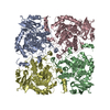 7oy9C 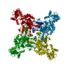 1zfjS C: citing same article ( S: Starting model for refinement |
|---|---|
| Similar structure data |
-Links
 PDBj
PDBj


- Assembly
| Deposited unit | 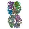
| ||||||||||||
|---|---|---|---|---|---|---|---|---|---|---|---|---|---|
| 1 | 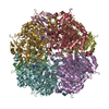
| ||||||||||||
| 2 | 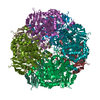
| ||||||||||||
| Unit cell |
|
-Components
| #1: Protein | Mass: 51782.434 Da / Num. of mol.: 16 Source method: isolated from a genetically manipulated source Source: (gene. exp.) Mycolicibacterium smegmatis (bacteria) / Strain: ATCC 700084 / mc(2)155 / Gene: guaB1, MSMEG_3634, MSMEI_3548 / Production host: #2: Chemical | ChemComp-5GP /   Mass: 363.221 Da / Num. of mol.: 16 / Source method: obtained synthetically / Formula: C10H14N5O8P / Feature type: SUBJECT OF INVESTIGATION Mass: 363.221 Da / Num. of mol.: 16 / Source method: obtained synthetically / Formula: C10H14N5O8P / Feature type: SUBJECT OF INVESTIGATIONHas ligand of interest | Y | Has protein modification | N | |
|---|
-Experimental details
-Experiment
| Experiment | Method: X-RAY DIFFRACTION / Number of used crystals: 1 |
|---|
- Sample preparation
| Crystal | Density Matthews: 2.06 Å3/Da / Density % sol: 40.18 % |
|---|---|
| Crystal grow | Temperature: 291 K / Method: vapor diffusion, sitting drop / pH: 7.5 Details: 0.2 M Sodium chloride 20% (v/v) PEG 3000 0.1 M HEPES, pH 7.5 |
-Data collection
| Diffraction | Mean temperature: 100 K / Serial crystal experiment: N |
|---|---|
| Diffraction source | Source: SYNCHROTRON / Site: BESSY  / Beamline: 14.1 / Wavelength: 0.9184 Å / Beamline: 14.1 / Wavelength: 0.9184 Å |
| Detector | Type: DECTRIS PILATUS3 S 6M / Detector: PIXEL / Date: Dec 1, 2018 |
| Radiation | Protocol: SINGLE WAVELENGTH / Monochromatic (M) / Laue (L): M / Scattering type: x-ray |
| Radiation wavelength | Wavelength: 0.9184 Å / Relative weight: 1 |
| Reflection | Resolution: 2.5→47.69 Å / Num. obs: 208443 / % possible obs: 90.06 % / Redundancy: 2.1 % / Biso Wilson estimate: 48.86 Å2 / CC1/2: 0.992 / CC star: 0.998 / Rmerge(I) obs: 0.1364 / Rpim(I) all: 0.1241 / Rrim(I) all: 0.185 / Net I/σ(I): 5.27 |
| Reflection shell | Resolution: 2.5→2.589 Å / Redundancy: 2.1 % / Rmerge(I) obs: 1.632 / Mean I/σ(I) obs: 0.58 / Num. unique obs: 21247 / CC1/2: 0.293 / CC star: 0.673 / Rpim(I) all: 1.495 / Rrim(I) all: 2.22 / % possible all: 86.46 |
- Processing
| Software |
| |||||||||||||||||||||||||||||||||||||||||||||||||||||||||||||||||||||||||||||||||||||||||||||||||||||||||||||||||||||||||||||||||||||||||||||||||||||||||||||||||||||||||||||||||||||||||||||||||||||||||||||||||||||||||
|---|---|---|---|---|---|---|---|---|---|---|---|---|---|---|---|---|---|---|---|---|---|---|---|---|---|---|---|---|---|---|---|---|---|---|---|---|---|---|---|---|---|---|---|---|---|---|---|---|---|---|---|---|---|---|---|---|---|---|---|---|---|---|---|---|---|---|---|---|---|---|---|---|---|---|---|---|---|---|---|---|---|---|---|---|---|---|---|---|---|---|---|---|---|---|---|---|---|---|---|---|---|---|---|---|---|---|---|---|---|---|---|---|---|---|---|---|---|---|---|---|---|---|---|---|---|---|---|---|---|---|---|---|---|---|---|---|---|---|---|---|---|---|---|---|---|---|---|---|---|---|---|---|---|---|---|---|---|---|---|---|---|---|---|---|---|---|---|---|---|---|---|---|---|---|---|---|---|---|---|---|---|---|---|---|---|---|---|---|---|---|---|---|---|---|---|---|---|---|---|---|---|---|---|---|---|---|---|---|---|---|---|---|---|---|---|---|---|---|
| Refinement | Method to determine structure: MOLECULAR REPLACEMENT Starting model: 1zfj Resolution: 2.5→47.6888947297 Å / SU ML: 0.552059369981 / Cross valid method: FREE R-VALUE / σ(F): 1.95835944193 / Phase error: 38.9829274508 Stereochemistry target values: GeoStd + Monomer Library + CDL v1.2
| |||||||||||||||||||||||||||||||||||||||||||||||||||||||||||||||||||||||||||||||||||||||||||||||||||||||||||||||||||||||||||||||||||||||||||||||||||||||||||||||||||||||||||||||||||||||||||||||||||||||||||||||||||||||||
| Solvent computation | Shrinkage radii: 0.9 Å / VDW probe radii: 1.11 Å / Solvent model: FLAT BULK SOLVENT MODEL | |||||||||||||||||||||||||||||||||||||||||||||||||||||||||||||||||||||||||||||||||||||||||||||||||||||||||||||||||||||||||||||||||||||||||||||||||||||||||||||||||||||||||||||||||||||||||||||||||||||||||||||||||||||||||
| Displacement parameters | Biso mean: 57.9802898605 Å2 | |||||||||||||||||||||||||||||||||||||||||||||||||||||||||||||||||||||||||||||||||||||||||||||||||||||||||||||||||||||||||||||||||||||||||||||||||||||||||||||||||||||||||||||||||||||||||||||||||||||||||||||||||||||||||
| Refinement step | Cycle: LAST / Resolution: 2.5→47.6888947297 Å
| |||||||||||||||||||||||||||||||||||||||||||||||||||||||||||||||||||||||||||||||||||||||||||||||||||||||||||||||||||||||||||||||||||||||||||||||||||||||||||||||||||||||||||||||||||||||||||||||||||||||||||||||||||||||||
| Refine LS restraints |
| |||||||||||||||||||||||||||||||||||||||||||||||||||||||||||||||||||||||||||||||||||||||||||||||||||||||||||||||||||||||||||||||||||||||||||||||||||||||||||||||||||||||||||||||||||||||||||||||||||||||||||||||||||||||||
| LS refinement shell |
|
