ムービー
ムービー コントローラー


+ データを開く

- 基本情報
基本情報
| 登録情報 | 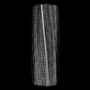 | |||||||||
|---|---|---|---|---|---|---|---|---|---|---|
| タイトル | CryoEM structure of recombinant DeltaN7 alpha-synuclein in PBS | |||||||||
 マップデータ マップデータ | Postprocessed sharpened cryoEM map for the aSyn-DN7 fibril structure | |||||||||
 試料 試料 |
| |||||||||
 キーワード キーワード | synuclein / Parkinson's disease / neurodegeneration / amyloid / helical / fibril / PROTEIN FIBRIL | |||||||||
| 機能・相同性 |  機能・相同性情報 機能・相同性情報negative regulation of mitochondrial electron transport, NADH to ubiquinone / : / response to desipramine / neutral lipid metabolic process / regulation of acyl-CoA biosynthetic process / negative regulation of dopamine uptake involved in synaptic transmission / negative regulation of norepinephrine uptake / positive regulation of SNARE complex assembly / positive regulation of hydrogen peroxide catabolic process / supramolecular fiber ...negative regulation of mitochondrial electron transport, NADH to ubiquinone / : / response to desipramine / neutral lipid metabolic process / regulation of acyl-CoA biosynthetic process / negative regulation of dopamine uptake involved in synaptic transmission / negative regulation of norepinephrine uptake / positive regulation of SNARE complex assembly / positive regulation of hydrogen peroxide catabolic process / supramolecular fiber / mitochondrial membrane organization / regulation of synaptic vesicle recycling / negative regulation of chaperone-mediated autophagy / regulation of reactive oxygen species biosynthetic process / negative regulation of platelet-derived growth factor receptor signaling pathway / positive regulation of protein localization to cell periphery / negative regulation of exocytosis / regulation of glutamate secretion / SNARE complex assembly / positive regulation of neurotransmitter secretion / dopamine biosynthetic process / response to iron(II) ion / regulation of norepinephrine uptake / negative regulation of dopamine metabolic process / regulation of locomotion / mitochondrial ATP synthesis coupled electron transport / regulation of macrophage activation / positive regulation of inositol phosphate biosynthetic process / synaptic vesicle priming / transporter regulator activity / negative regulation of microtubule polymerization / synaptic vesicle transport / positive regulation of receptor recycling / dopamine uptake involved in synaptic transmission / protein kinase inhibitor activity / dynein complex binding / regulation of dopamine secretion / negative regulation of thrombin-activated receptor signaling pathway / nuclear outer membrane / cuprous ion binding / positive regulation of exocytosis / synaptic vesicle exocytosis / response to magnesium ion / positive regulation of endocytosis / kinesin binding / synaptic vesicle endocytosis / enzyme inhibitor activity / cysteine-type endopeptidase inhibitor activity / negative regulation of serotonin uptake / regulation of presynapse assembly / response to type II interferon / alpha-tubulin binding / beta-tubulin binding / phospholipase binding / behavioral response to cocaine / supramolecular fiber organization / cellular response to copper ion / phospholipid metabolic process / cellular response to fibroblast growth factor stimulus / inclusion body / axon terminus / cellular response to epinephrine stimulus / Hsp70 protein binding / response to interleukin-1 / regulation of microtubule cytoskeleton organization / SNARE binding / positive regulation of release of sequestered calcium ion into cytosol / adult locomotory behavior / excitatory postsynaptic potential / phosphoprotein binding / protein tetramerization / fatty acid metabolic process / microglial cell activation / synapse organization / regulation of long-term neuronal synaptic plasticity / ferrous iron binding / protein destabilization / PKR-mediated signaling / phospholipid binding / receptor internalization / tau protein binding / long-term synaptic potentiation / terminal bouton / positive regulation of inflammatory response / synaptic vesicle membrane / actin cytoskeleton / actin binding / growth cone / cellular response to oxidative stress / cell cortex / neuron apoptotic process / microtubule binding / response to lipopolysaccharide / chemical synaptic transmission / molecular adaptor activity / amyloid fibril formation / histone binding / negative regulation of neuron apoptotic process / mitochondrial outer membrane / lysosome 類似検索 - 分子機能 | |||||||||
| 生物種 |  Homo sapiens (ヒト) Homo sapiens (ヒト) | |||||||||
| 手法 | らせん対称体再構成法 / クライオ電子顕微鏡法 / 解像度: 2.5 Å | |||||||||
 データ登録者 データ登録者 | Thacker D / Wilkinson M / Dewison KM / Ranson NA / Brockwell DJ / Radford SE | |||||||||
| 資金援助 |  英国, 2件 英国, 2件
| |||||||||
 引用 引用 | ジャーナル: Proc Natl Acad Sci U S A / 年: 2024 タイトル: Residues 2 to 7 of α-synuclein regulate amyloid formation via lipid-dependent and lipid-independent pathways. 著者: Katherine M Dewison / Benjamin Rowlinson / Jonathan M Machin / Joel A Crossley / Dev Thacker / Martin Wilkinson / Sabine M Ulamec / G Nasir Khan / Neil A Ranson / Patricija van Oosten-Hawle / ...著者: Katherine M Dewison / Benjamin Rowlinson / Jonathan M Machin / Joel A Crossley / Dev Thacker / Martin Wilkinson / Sabine M Ulamec / G Nasir Khan / Neil A Ranson / Patricija van Oosten-Hawle / David J Brockwell / Sheena E Radford /  要旨: Amyloid formation by α-synuclein (αSyn) occurs in Parkinson's disease, multiple system atrophy, and dementia with Lewy bodies. Deciphering the residues that regulate αSyn amyloid fibril formation ...Amyloid formation by α-synuclein (αSyn) occurs in Parkinson's disease, multiple system atrophy, and dementia with Lewy bodies. Deciphering the residues that regulate αSyn amyloid fibril formation will not only provide mechanistic insight but may also reveal targets to prevent and treat disease. Previous investigations have identified several regions of αSyn to be important in the regulation of amyloid formation, including the non-amyloid-β component (NAC), P1 region (residues 36 to 42), and residues in the C-terminal domain. Recent studies have also indicated the importance of the N-terminal region of αSyn for both its physiological and pathological roles. Here, the role of residues 2 to 7 in the N-terminal region of αSyn is investigated in terms of their ability to regulate amyloid fibril formation in vitro and in vivo. Deletion of these residues (αSynΔN7) slows the rate of fibril formation in vitro and reduces the capacity of the protein to be recruited by wild-type (αSynWT) fibril seeds, despite cryo-EM showing a fibril structure consistent with those of full-length αSyn. Strikingly, fibril formation of αSynΔN7 is not induced by liposomes, despite the protein binding to liposomes with similar affinity to αSynWT. A model also showed that αSynΔN7::YFP forms few puncta and lacks motility and lifespan defects typified by expression of αSynWT::YFP. Together, the results demonstrate the involvement of residues 2 to 7 of αSyn in amyloid formation, revealing a target for the design of amyloid inhibitors that may leave the functional role of the protein in membrane binding unperturbed. | |||||||||
| 履歴 |
|
- 構造の表示
構造の表示
| 添付画像 |
|---|

- ダウンロードとリンク
-EMDBアーカイブ
| マップデータ | emd_18570.map.gz | 22.6 MB | EMDBマップデータ形式 | |
|---|---|---|---|---|
| ヘッダ (付随情報) | emd-18570-v30.xmlemd-18570.xml | 18.5 KB 18.5 KB | 表示 表示 | EMDBヘッダ |
| FSC (解像度算出) | emd_18570_fsc.xml | 10.6 KB | 表示 | FSCデータファイル |
| 画像 |  emd_18570.png emd_18570.png | 100.1 KB | ||
| Filedesc metadata | emd-18570.cif.gz | 6.3 KB | ||
| その他 | emd_18570_half_map_1.map.gzemd_18570_half_map_2.map.gz | 89.5 MB 80.7 MB | ||
| アーカイブディレクトリ |  http://ftp.pdbj.org/pub/emdb/structures/EMD-18570ftp://ftp.pdbj.org/pub/emdb/structures/EMD-18570 http://ftp.pdbj.org/pub/emdb/structures/EMD-18570ftp://ftp.pdbj.org/pub/emdb/structures/EMD-18570 | HTTPS FTP |
-検証レポート
| 文書・要旨 | emd_18570_validation.pdf.gz | 906.5 KB | 表示 | EMDB検証レポート |
|---|---|---|---|---|
| 文書・詳細版 | emd_18570_full_validation.pdf.gz | 906.1 KB | 表示 | |
| XML形式データ | emd_18570_validation.xml.gz | 17.6 KB | 表示 | |
| CIF形式データ | emd_18570_validation.cif.gz | 23.2 KB | 表示 | |
| アーカイブディレクトリ | https://ftp.pdbj.org/pub/emdb/validation_reports/EMD-18570ftp://ftp.pdbj.org/pub/emdb/validation_reports/EMD-18570 | HTTPS FTP |
-関連構造データ
-リンク
| EMDBのページ | EMDB (EBI/PDBe) / EMDataResource |
|---|
-マップ
| ファイル | ダウンロード / ファイル: emd_18570.map.gz / 形式: CCP4 / 大きさ: 103 MB / タイプ: IMAGE STORED AS FLOATING POINT NUMBER (4 BYTES) | ||||||||||||||||||||||||||||||||||||
|---|---|---|---|---|---|---|---|---|---|---|---|---|---|---|---|---|---|---|---|---|---|---|---|---|---|---|---|---|---|---|---|---|---|---|---|---|---|
| 注釈 | Postprocessed sharpened cryoEM map for the aSyn-DN7 fibril structure | ||||||||||||||||||||||||||||||||||||
| 投影像・断面図 | 画像のコントロール
画像は Spider により作成 | ||||||||||||||||||||||||||||||||||||
| ボクセルのサイズ | X=Y=Z: 0.95 Å | ||||||||||||||||||||||||||||||||||||
| 密度 |
| ||||||||||||||||||||||||||||||||||||
| 対称性 | 空間群: 1 | ||||||||||||||||||||||||||||||||||||
| 詳細 | EMDB XML:
|
 Z (Sec.)
 Y (Row.)
 X (Col.)


-添付データ
-ハーフマップ: halfmap2 for the aSyn-DN7 fibril structure
| ファイル | emd_18570_half_map_1.map | ||||||||||||
|---|---|---|---|---|---|---|---|---|---|---|---|---|---|
| 注釈 | halfmap2 for the aSyn-DN7 fibril structure | ||||||||||||
| 投影像・断面図 |
| ||||||||||||
| 密度ヒストグラム |


-ハーフマップ: halfmap1 for the aSyn-DN7 fibril structure
| ファイル | emd_18570_half_map_2.map | ||||||||||||
|---|---|---|---|---|---|---|---|---|---|---|---|---|---|
| 注釈 | halfmap1 for the aSyn-DN7 fibril structure | ||||||||||||
| 投影像・断面図 |
| ||||||||||||
| 密度ヒストグラム |


- 試料の構成要素
-全体 : alpha-synuclein DeltaN7 amyloid fibrils
| 全体 | 名称: alpha-synuclein DeltaN7 amyloid fibrils |
|---|---|
| 要素 |
|
-超分子 #1: alpha-synuclein DeltaN7 amyloid fibrils
| 超分子 | 名称: alpha-synuclein DeltaN7 amyloid fibrils / タイプ: complex / ID: 1 / 親要素: 0 / 含まれる分子: all |
|---|---|
| 由来(天然) | 生物種: Homo sapiens (ヒト) |
-分子 #1: Alpha-synuclein
| 分子 | 名称: Alpha-synuclein / タイプ: protein_or_peptide / ID: 1 詳細: DeltaN7, technically residues 2-7 are deleted as the N-terminal Methionine was required for bacterial expression. コピー数: 12 / 光学異性体: LEVO |
|---|---|
| 由来(天然) | 生物種: Homo sapiens (ヒト) |
| 分子量 | 理論値: 13.797286 KDa |
| 組換発現 | 生物種:  |
| 配列 | 文字列: MLSKAKEGVV AAAEKTKQGV AEAAGKTKEG VLYVGSKTKE GVVHGVATVA EKTKEQVTNV GGAVVTGVTA VAQKTVEGAG SIAAATGFV KKDQLGKNEE GAPQEGILED MPVDPDNEAY EMPSEEGYQD YEPEA UniProtKB: Alpha-synuclein |
-実験情報
-構造解析
| 手法 | クライオ電子顕微鏡法 |
|---|---|
 解析 解析 | らせん対称体再構成法 |
| 試料の集合状態 | filament |
-試料調製
| 緩衝液 | pH: 7.4 詳細: 137 mM NaCl, 2.7 mM KCl, 8.1 mM Na2HPO4 and 1.5 mM KH2PO4; pH 7.4 |
|---|---|
| グリッド | 材質: COPPER / メッシュ: 300 / 支持フィルム - 材質: CARBON / 支持フィルム - トポロジー: LACEY / 前処理 - タイプ: PLASMA CLEANING / 前処理 - 時間: 60 sec. |
| 凍結 | 凍結剤: ETHANE / チャンバー内湿度: 90 % / チャンバー内温度: 277 K / 装置: FEI VITROBOT MARK IV |
- 電子顕微鏡法
| 顕微鏡 | FEI TITAN KRIOS |
|---|---|
| 特殊光学系 | エネルギーフィルター - 名称: TFS Selectris / エネルギーフィルター - スリット幅: 10 eV |
| 撮影 | フィルム・検出器のモデル: FEI FALCON IV (4k x 4k) デジタル化 - サイズ - 横: 4096 pixel / デジタル化 - サイズ - 縦: 4096 pixel / 撮影したグリッド数: 1 / 実像数: 5464 / 平均電子線量: 45.0 e/Å2 |
| 電子線 | 加速電圧: 300 kV / 電子線源:  FIELD EMISSION GUN FIELD EMISSION GUN |
| 電子光学系 | C2レンズ絞り径: 50.0 µm / 照射モード: FLOOD BEAM / 撮影モード: BRIGHT FIELD / Cs: 2.7 mm / 最大 デフォーカス(公称値): 2.6 µm 最小 デフォーカス(公称値): 1.4000000000000001 µm 倍率(公称値): 130000 |
| 試料ステージ | 試料ホルダーモデル: FEI TITAN KRIOS AUTOGRID HOLDER |
| 実験機器 |  モデル: Titan Krios / 画像提供: FEI Company |
+画像解析

-原子モデル構築 1
| 初期モデル | PDB ID: Chain - Chain ID: A / Chain - Residue range: 42-92 / Chain - Source name: PDB / Chain - Initial model type: experimental model |
|---|---|
| 詳細 | see methods, rigid body docked chain of PDB: 6osl, manual fitting in coot, real-space refine in phenix |
| 精密化 | 空間: REAL / プロトコル: RIGID BODY FIT / 温度因子: 106 当てはまり具合の基準: cross-correlation coefficient |
| 得られたモデル | 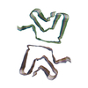 PDB-8qpz: |