+ データを開く
- 基本情報
基本情報
| 登録情報 |  | |||||||||
|---|---|---|---|---|---|---|---|---|---|---|
| タイトル | Cryo-EM Structure of Pre-B Complex (core part) | |||||||||
 マップデータ マップデータ | ||||||||||
 試料 試料 |
| |||||||||
 キーワード キーワード | spliceosome / SPLICING | |||||||||
| 機能・相同性 |  機能・相同性情報 機能・相同性情報ribonucleoprotein complex localization / U4atac snRNP / positive regulation of cytotoxic T cell differentiation / maturation of 5S rRNA / RNA localization / U4atac snRNA binding / R-loop processing / cis assembly of pre-catalytic spliceosome / RNA splicing, via transesterification reactions / U2-type catalytic step 1 spliceosome ...ribonucleoprotein complex localization / U4atac snRNP / positive regulation of cytotoxic T cell differentiation / maturation of 5S rRNA / RNA localization / U4atac snRNA binding / R-loop processing / cis assembly of pre-catalytic spliceosome / RNA splicing, via transesterification reactions / U2-type catalytic step 1 spliceosome / U4 snRNA binding / snRNP binding / spliceosomal tri-snRNP complex / U2-type precatalytic spliceosome / U2-type spliceosomal complex / mRNA cis splicing, via spliceosome / U2-type prespliceosome assembly / U2-type catalytic step 2 spliceosome / U4 snRNP / U2 snRNP / U2-type prespliceosome / K63-linked polyubiquitin modification-dependent protein binding / precatalytic spliceosome / spliceosomal complex assembly / mRNA Splicing - Minor Pathway / mRNA 3'-splice site recognition / negative regulation of mRNA splicing, via spliceosome / MLL1 complex / spliceosomal tri-snRNP complex assembly / protein deubiquitination / U5 snRNA binding / U5 snRNP / U2 snRNA binding / U6 snRNA binding / pre-mRNA intronic binding / spliceosomal snRNP assembly / ribonucleoprotein complex binding / Cajal body / U1 snRNA binding / U4/U6 x U5 tri-snRNP complex / catalytic step 2 spliceosome / mRNA Splicing - Major Pathway / RNA splicing / response to cocaine / spliceosomal complex / mRNA splicing, via spliceosome / mRNA processing / cellular response to xenobiotic stimulus / cellular response to tumor necrosis factor / cellular response to lipopolysaccharide / protein-macromolecule adaptor activity / ubiquitinyl hydrolase 1 / nucleic acid binding / hydrolase activity / RNA helicase activity / nuclear speck / ciliary basal body / RNA helicase / cell division / intracellular membrane-bounded organelle / GTPase activity / mRNA binding / centrosome / chromatin / GTP binding / nucleolus / Golgi apparatus / positive regulation of transcription by RNA polymerase II / ATP hydrolysis activity / RNA binding / extracellular exosome / zinc ion binding / nucleoplasm / ATP binding / identical protein binding / nucleus / membrane / cytosol / cytoplasm 類似検索 - 分子機能 | |||||||||
| 生物種 |  Homo sapiens (ヒト) Homo sapiens (ヒト) | |||||||||
| 手法 | 単粒子再構成法 / クライオ電子顕微鏡法 / 解像度: 3.5 Å | |||||||||
 データ登録者 データ登録者 | Zhang Z / Kumar V / Dybkov O / Will CL / Zhong J / Ludwig S / Urlaub H / Kastner B / Stark H / Luehrmann R | |||||||||
| 資金援助 |  ドイツ, 1件 ドイツ, 1件
| |||||||||
 引用 引用 |  ジャーナル: Nature / 年: 2024 ジャーナル: Nature / 年: 2024タイトル: Structural insights into the cross-exon to cross-intron spliceosome switch. 著者: Zhenwei Zhang / Vinay Kumar / Olexandr Dybkov / Cindy L Will / Jiayun Zhong / Sebastian E J Ludwig / Henning Urlaub / Berthold Kastner / Holger Stark / Reinhard Lührmann /   要旨: Early spliceosome assembly can occur through an intron-defined pathway, whereby U1 and U2 small nuclear ribonucleoprotein particles (snRNPs) assemble across the intron. Alternatively, it can occur ...Early spliceosome assembly can occur through an intron-defined pathway, whereby U1 and U2 small nuclear ribonucleoprotein particles (snRNPs) assemble across the intron. Alternatively, it can occur through an exon-defined pathway, whereby U2 binds the branch site located upstream of the defined exon and U1 snRNP interacts with the 5' splice site located directly downstream of it. The U4/U6.U5 tri-snRNP subsequently binds to produce a cross-intron (CI) or cross-exon (CE) pre-B complex, which is then converted to the spliceosomal B complex. Exon definition promotes the splicing of upstream introns and plays a key part in alternative splicing regulation. However, the three-dimensional structure of exon-defined spliceosomal complexes and the molecular mechanism of the conversion from a CE-organized to a CI-organized spliceosome, a pre-requisite for splicing catalysis, remain poorly understood. Here cryo-electron microscopy analyses of human CE pre-B complex and B-like complexes reveal extensive structural similarities with their CI counterparts. The results indicate that the CE and CI spliceosome assembly pathways converge already at the pre-B stage. Add-back experiments using purified CE pre-B complexes, coupled with cryo-electron microscopy, elucidate the order of the extensive remodelling events that accompany the formation of B complexes and B-like complexes. The molecular triggers and roles of B-specific proteins in these rearrangements are also identified. We show that CE pre-B complexes can productively bind in trans to a U1 snRNP-bound 5' splice site. Together, our studies provide new mechanistic insights into the CE to CI switch during spliceosome assembly and its effect on pre-mRNA splice site pairing at this stage. | |||||||||
| 履歴 |
|
- 構造の表示
構造の表示
| 添付画像 |
|---|
- ダウンロードとリンク
-EMDBアーカイブ
| マップデータ |  emd_18544.map.gz emd_18544.map.gz | 380.8 MB |  EMDBマップデータ形式 EMDBマップデータ形式 | |
|---|---|---|---|---|
| ヘッダ (付随情報) |  emd-18544-v30.xml emd-18544-v30.xml emd-18544.xml emd-18544.xml | 34.5 KB 34.5 KB | 表示 表示 |  EMDBヘッダ EMDBヘッダ |
| FSC (解像度算出) |  emd_18544_fsc.xml emd_18544_fsc.xml | 16.9 KB | 表示 |  FSCデータファイル FSCデータファイル |
| 画像 |  emd_18544.png emd_18544.png | 53.6 KB | ||
| Filedesc metadata |  emd-18544.cif.gz emd-18544.cif.gz | 12.2 KB | ||
| その他 |  emd_18544_half_map_1.map.gz emd_18544_half_map_1.map.gz emd_18544_half_map_2.map.gz emd_18544_half_map_2.map.gz | 332.6 MB 331.9 MB | ||
| アーカイブディレクトリ |  http://ftp.pdbj.org/pub/emdb/structures/EMD-18544 http://ftp.pdbj.org/pub/emdb/structures/EMD-18544 ftp://ftp.pdbj.org/pub/emdb/structures/EMD-18544 ftp://ftp.pdbj.org/pub/emdb/structures/EMD-18544 | HTTPS FTP |
-検証レポート
| 文書・要旨 |  emd_18544_validation.pdf.gz emd_18544_validation.pdf.gz | 814.4 KB | 表示 |  EMDB検証レポート EMDB検証レポート |
|---|---|---|---|---|
| 文書・詳細版 |  emd_18544_full_validation.pdf.gz emd_18544_full_validation.pdf.gz | 813.9 KB | 表示 | |
| XML形式データ |  emd_18544_validation.xml.gz emd_18544_validation.xml.gz | 24.3 KB | 表示 | |
| CIF形式データ |  emd_18544_validation.cif.gz emd_18544_validation.cif.gz | 32.5 KB | 表示 | |
| アーカイブディレクトリ |  https://ftp.pdbj.org/pub/emdb/validation_reports/EMD-18544 https://ftp.pdbj.org/pub/emdb/validation_reports/EMD-18544 ftp://ftp.pdbj.org/pub/emdb/validation_reports/EMD-18544 ftp://ftp.pdbj.org/pub/emdb/validation_reports/EMD-18544 | HTTPS FTP |
-関連構造データ
| 関連構造データ | 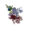 8qp8MC 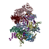 8qozC  8qp9C 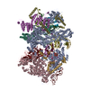 8qpaC  8qpbC 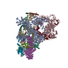 8qpeC  8qpkC 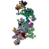 8qxdC 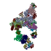 8qzsC 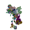 8r08C 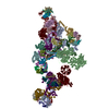 8r09C 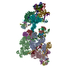 8r0aC 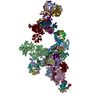 8r0bC 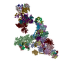 8rm5C M: このマップから作成された原子モデル C: 同じ文献を引用 ( |
|---|---|
| 類似構造データ | 類似検索 - 機能・相同性  F&H 検索 F&H 検索 |
- リンク
リンク
| EMDBのページ |  EMDB (EBI/PDBe) / EMDB (EBI/PDBe) /  EMDataResource EMDataResource |
|---|---|
| 「今月の分子」の関連する項目 |
- マップ
マップ
| ファイル |  ダウンロード / ファイル: emd_18544.map.gz / 形式: CCP4 / 大きさ: 421.9 MB / タイプ: IMAGE STORED AS FLOATING POINT NUMBER (4 BYTES) ダウンロード / ファイル: emd_18544.map.gz / 形式: CCP4 / 大きさ: 421.9 MB / タイプ: IMAGE STORED AS FLOATING POINT NUMBER (4 BYTES) | ||||||||||||||||||||||||||||||||||||
|---|---|---|---|---|---|---|---|---|---|---|---|---|---|---|---|---|---|---|---|---|---|---|---|---|---|---|---|---|---|---|---|---|---|---|---|---|---|
| 投影像・断面図 | 画像のコントロール
画像は Spider により作成 | ||||||||||||||||||||||||||||||||||||
| ボクセルのサイズ | X=Y=Z: 1.35 Å | ||||||||||||||||||||||||||||||||||||
| 密度 |
| ||||||||||||||||||||||||||||||||||||
| 対称性 | 空間群: 1 | ||||||||||||||||||||||||||||||||||||
| 詳細 | EMDB XML:
|
-添付データ
-ハーフマップ: #2
| ファイル | emd_18544_half_map_1.map | ||||||||||||
|---|---|---|---|---|---|---|---|---|---|---|---|---|---|
| 投影像・断面図 |
| ||||||||||||
| 密度ヒストグラム |
-ハーフマップ: #1
| ファイル | emd_18544_half_map_2.map | ||||||||||||
|---|---|---|---|---|---|---|---|---|---|---|---|---|---|
| 投影像・断面図 |
| ||||||||||||
| 密度ヒストグラム |
- 試料の構成要素
+全体 : human spliceosomal cross-exon pre-B complex
+超分子 #1: human spliceosomal cross-exon pre-B complex
+分子 #1: Splicing factor 3A subunit 1
+分子 #2: Probable ATP-dependent RNA helicase DDX23
+分子 #6: U4/U6.U5 tri-snRNP-associated protein 1
+分子 #7: 116 kDa U5 small nuclear ribonucleoprotein component
+分子 #8: Ubiquitin carboxyl-terminal hydrolase 39
+分子 #9: Pre-mRNA-processing-splicing factor 8
+分子 #10: RNA-binding protein 42
+分子 #11: Pre-mRNA-processing factor 6
+分子 #12: U4/U6 small nuclear ribonucleoprotein Prp31
+分子 #13: U4/U6 small nuclear ribonucleoprotein Prp3
+分子 #14: Thioredoxin-like protein 4A
+分子 #15: U4/U6.U5 small nuclear ribonucleoprotein 27 kDa protein
+分子 #3: U6 snRNA
+分子 #4: U5 snRNA
+分子 #5: U4 snRNA
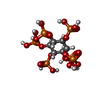
+分子 #16: INOSITOL HEXAKISPHOSPHATE
-実験情報
-構造解析
| 手法 | クライオ電子顕微鏡法 |
|---|---|
 解析 解析 | 単粒子再構成法 |
| 試料の集合状態 | particle |
- 試料調製
試料調製
| 緩衝液 | pH: 7.9 |
|---|---|
| 凍結 | 凍結剤: ETHANE |
- 電子顕微鏡法
| 顕微鏡 | FEI TITAN KRIOS |
|---|---|
| 撮影 | フィルム・検出器のモデル: FEI FALCON III (4k x 4k) 平均電子線量: 45.0 e/Å2 |
| 電子線 | 加速電圧: 300 kV / 電子線源:  FIELD EMISSION GUN FIELD EMISSION GUN |
| 電子光学系 | 照射モード: FLOOD BEAM / 撮影モード: BRIGHT FIELD / 最大 デフォーカス(公称値): 5.0 µm / 最小 デフォーカス(公称値): 1.5 µm |
| 実験機器 |  モデル: Titan Krios / 画像提供: FEI Company |
 ムービー
ムービー コントローラー


 Z (Sec.)
 Y (Row.)
 X (Col.)


