Journal: Nat Struct Mol Biol / Year: 2023 Title: A four-point molecular handover during Okazaki maturation. Authors: Margherita M Botto / Alessandro Borsellini / Meindert H Lamers / Abstract: DNA replication introduces thousands of RNA primers into the lagging strand that need to be removed for replication to be completed. In Escherichia coli when the replicative DNA polymerase Pol IIIα ...DNA replication introduces thousands of RNA primers into the lagging strand that need to be removed for replication to be completed. In Escherichia coli when the replicative DNA polymerase Pol IIIα terminates at a previously synthesized RNA primer, DNA Pol I takes over and continues DNA synthesis while displacing the downstream RNA primer. The displaced primer is subsequently excised by an endonuclease, followed by the sealing of the nick by a DNA ligase. Yet how the sequential actions of Pol IIIα, Pol I polymerase, Pol I endonuclease and DNA ligase are coordinated is poorly defined. Here we show that each enzymatic activity prepares the DNA substrate for the next activity, creating an efficient four-point molecular handover. The cryogenic-electron microscopy structure of Pol I bound to a DNA substrate with both an upstream and downstream primer reveals how it displaces the primer in a manner analogous to the monomeric helicases. Moreover, we find that in addition to its flap-directed nuclease activity, the endonuclease domain of Pol I also specifically cuts at the RNA-DNA junction, thus marking the end of the RNA primer and creating a 5' end that is a suitable substrate for the ligase activity of LigA once all RNA has been removed.
In the structure databanks used in Yorodumi, some data are registered as the other names, "COVID-19 virus" and "2019-nCoV". Here are the details of the virus and the list of structure data.
Jan 31, 2019. EMDB accession codes are about to change! (news from PDBe EMDB page)
EMDB accession codes are about to change! (news from PDBe EMDB page)
The allocation of 4 digits for EMDB accession codes will soon come to an end. Whilst these codes will remain in use, new EMDB accession codes will include an additional digit and will expand incrementally as the available range of codes is exhausted. The current 4-digit format prefixed with “EMD-” (i.e. EMD-XXXX) will advance to a 5-digit format (i.e. EMD-XXXXX), and so on. It is currently estimated that the 4-digit codes will be depleted around Spring 2019, at which point the 5-digit format will come into force.
The EM Navigator/Yorodumi systems omit the EMD- prefix.
Related info.:Q: What is EMD? / ID/Accession-code notation in Yorodumi/EM Navigator
Yorodumi is a browser for structure data from EMDB, PDB, SASBDB, etc.
This page is also the successor to EM Navigator detail page, and also detail information page/front-end page for Omokage search.
The word "yorodu" (or yorozu) is an old Japanese word meaning "ten thousand". "mi" (miru) is to see.
Related info.:EMDB / PDB / SASBDB / Comparison of 3 databanks / Yorodumi Search / Aug 31, 2016. New EM Navigator & Yorodumi / Yorodumi Papers / Jmol/JSmol / Function and homology information / Changes in new EM Navigator and Yorodumi
 Movie
Movie Controller

 Yorodumi
 Open data
 Basic information
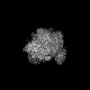
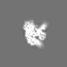
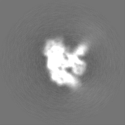
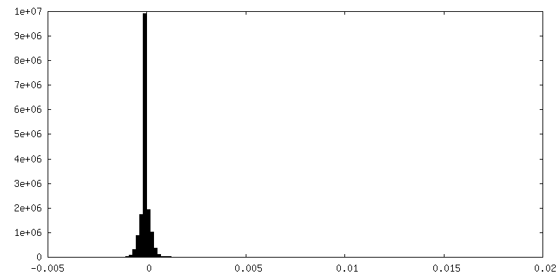
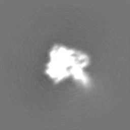
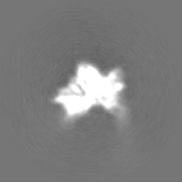
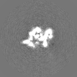
 Map data
 Sample
 Keywords
 Function and homology information
 Authors
 Citation


 Structure visualization
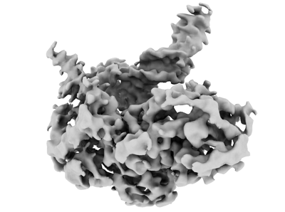
 Downloads & links
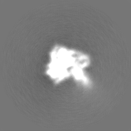
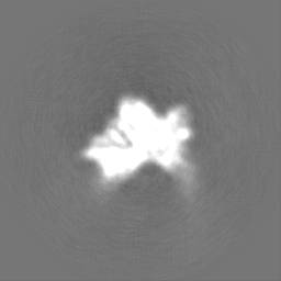
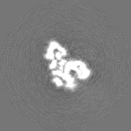
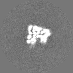
 emd_17033.png
 http://ftp.pdbj.org/pub/emdb/structures/EMD-17033


 Z (Sec.)
 Y (Row.)
 X (Col.)


 Sample components
 Processing
 Electron microscopy
 FIELD EMISSION GUN

