 Movie
Movie Controller

[English] 日本語
 Yorodumi
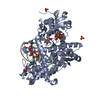
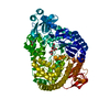
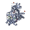
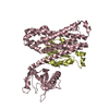
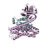
- PDB-1kln: DNA POLYMERASE I KLENOW FRAGMENT (E.C.2.7.7.7) MUTANT/DNA COMPLEX -
+ Open data

- Basic information
| Entry | Database: PDB / ID: 1kln | ||||||
|---|---|---|---|---|---|---|---|
| Title | DNA POLYMERASE I KLENOW FRAGMENT (E.C.2.7.7.7) MUTANT/DNA COMPLEX | ||||||
 Components Components |
| ||||||
 Keywords Keywords | TRANSFERASE/DNA / PROTEIN-DNA COMPLEX / DOUBLE HELIX / TRANSFERASE-DNA COMPLEX | ||||||
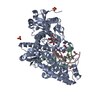
| Function / homology |  Function and homology information Function and homology information5'-3' exonuclease activity / 3'-5' exonuclease activity / base-excision repair / DNA-templated DNA replication / double-strand break repair / DNA-directed DNA polymerase / DNA-directed DNA polymerase activity / DNA replication / DNA repair / DNA binding ...5'-3' exonuclease activity / 3'-5' exonuclease activity / base-excision repair / DNA-templated DNA replication / double-strand break repair / DNA-directed DNA polymerase / DNA-directed DNA polymerase activity / DNA replication / DNA repair / DNA binding / cytoplasm / cytosol Similarity search - Function | ||||||
| Biological species |  | ||||||
| Method |  X-RAY DIFFRACTION / Resolution: 3.2 Å X-RAY DIFFRACTION / Resolution: 3.2 Å | ||||||
 Authors Authors | Beese, L.S. / Derbyshire, V. / Steitz, T.A. | ||||||
 Citation Citation | Journal: Science / Year: 1993 Title: Structure of DNA polymerase I Klenow fragment bound to duplex DNA. Authors: Beese, L.S. / Derbyshire, V. / Steitz, T.A. #1: Journal: Embo J. / Year: 1991Title: Structural Basis for the 3'-5' Exonuclease Activity of Escherichia Coli DNA Polymerase I: A Two Metal Ion Mechanism Authors: Beese, L.S. / Steitz, T.A. #2: Journal: Proc.Natl.Acad.Sci.USA / Year: 1988Title: Co-Crystal Structure of an Editing Complex of Klenow Fragment with DNA Authors: Freemont, P.S. / Friedman, J.M. / Beese, L.S. / Steitz, T.A. #3: Journal: Nature / Year: 1985Title: Structure of Large Fragment of Escherichia Coli DNA Polymerase I Complexed with dTMP Authors: Ollis, D.L. / Brick, P. / Hamlin, R. / Xuong, N.G. / Steitz, T.A. | ||||||
| History |
|
- Structure visualization
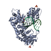
| Structure viewer | Molecule: MolmilJmol/JSmol |
|---|
- Downloads & links
-Download
| PDBx/mmCIF format | 1kln.cif.gz | 130.9 KB | Display | PDBx/mmCIF format |
|---|---|---|---|---|
| PDB format | pdb1kln.ent.gz | 94.8 KB | Display | PDB format |
| PDBx/mmJSON format | 1kln.json.gz | Tree view | PDBx/mmJSON format | |
| Others |  Other downloads Other downloads |
-Validation report
| Arichive directory | https://data.pdbj.org/pub/pdb/validation_reports/kl/1klnftp://data.pdbj.org/pub/pdb/validation_reports/kl/1kln | HTTPS FTP |
|---|
-Related structure data
| Similar structure data |
|---|


-Links
 PDBj
PDBj


- Assembly
| Deposited unit | 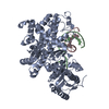
| ||||||||
|---|---|---|---|---|---|---|---|---|---|
| 1 |
| ||||||||
| Unit cell |
|
-Components
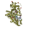
| #1: DNA chain | Mass: 3969.561 Da / Num. of mol.: 1 / Source method: obtained synthetically |
|---|---|
| #2: DNA chain | Mass: 3071.006 Da / Num. of mol.: 1 / Source method: obtained synthetically |
| #3: Protein | Mass: 68117.680 Da / Num. of mol.: 1 / Mutation: D355A / Source method: isolated from a natural source / Source: (natural) Keywords: MUTANT D355A / References: UniProt: P00582 |
| #4: Chemical | ChemComp-ZN /   Mass: 65.409 Da / Num. of mol.: 1 / Source method: obtained synthetically / Formula: Zn Mass: 65.409 Da / Num. of mol.: 1 / Source method: obtained synthetically / Formula: Zn |
-Experimental details
-Experiment
| Experiment | Method: X-RAY DIFFRACTION |
|---|
- Sample preparation
| Crystal | Density Matthews: 3.1 Å3/Da / Density % sol: 60.27 % | ||||||||||||||||||||||||||||||||||||||||||
|---|---|---|---|---|---|---|---|---|---|---|---|---|---|---|---|---|---|---|---|---|---|---|---|---|---|---|---|---|---|---|---|---|---|---|---|---|---|---|---|---|---|---|---|
| Crystal grow | Temperature: 290 K / Method: vapor diffusion / pH: 6.8 / Details: pH 6.80, VAPOR DIFFUSION, temperature 290.00K | ||||||||||||||||||||||||||||||||||||||||||
| Components of the solutions |
| ||||||||||||||||||||||||||||||||||||||||||
| Crystal grow | *PLUS Temperature: 17 ℃ / Method: vapor diffusion / pH: 6.8 | ||||||||||||||||||||||||||||||||||||||||||
| Components of the solutions | *PLUS
|
-Data collection
| Detector | Type: UCSD MARK II / Detector: AREA DETECTOR |
|---|---|
| Radiation | Protocol: SINGLE WAVELENGTH / Monochromatic (M) / Laue (L): M / Scattering type: x-ray |
| Radiation wavelength | Relative weight: 1 |
- Processing
| Software | Name: X-PLOR / Classification: refinement | ||||||||||||
|---|---|---|---|---|---|---|---|---|---|---|---|---|---|
| Refinement | Resolution: 3.2→10 Å / Rfactor Rwork: 0.23 | ||||||||||||
| Refinement step | Cycle: LAST / Resolution: 3.2→10 Å
| ||||||||||||
| Software | *PLUS Name: X-PLOR / Classification: refinement | ||||||||||||
| Refinement | *PLUS Highest resolution: 3.2 Å / Lowest resolution: 10 Å / Rfactor Rwork: 0.23 | ||||||||||||
| Solvent computation | *PLUS | ||||||||||||
| Displacement parameters | *PLUS | ||||||||||||
| Refine LS restraints | *PLUS
|
