Movie
Movie Controller

[English] 日本語
 Yorodumi
- EMDB-17010: CryoEM Structure INO80core Hexasome complex Rvb core refinement state1 -
+ Open data

- Basic information
| Entry |  | |||||||||||||||
|---|---|---|---|---|---|---|---|---|---|---|---|---|---|---|---|---|
| Title | CryoEM Structure INO80core Hexasome complex Rvb core refinement state1 | |||||||||||||||
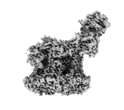
 Map data Map data | ctINO80-hexasome focused refinement Rvb1/2-Ino80 insert-Ies2-Arp5-Ies6 core sharpened map | |||||||||||||||
 Sample Sample |
| |||||||||||||||
 Keywords Keywords | ATP-dependent chromatin remodeler / DNA BINDING PROTEIN | |||||||||||||||
| Function / homology |  Function and homology information Function and homology informationDASH complex / protein transport along microtubule to mitotic spindle pole body / mitotic sister chromatid biorientation / attachment of spindle microtubules to kinetochore / Swr1 complex / attachment of mitotic spindle microtubules to kinetochore / Ino80 complex / DNA helicase activity / kinetochore / mitotic spindle ...DASH complex / protein transport along microtubule to mitotic spindle pole body / mitotic sister chromatid biorientation / attachment of spindle microtubules to kinetochore / Swr1 complex / attachment of mitotic spindle microtubules to kinetochore / Ino80 complex / DNA helicase activity / kinetochore / mitotic spindle / chromatin organization / DNA helicase / chromatin remodeling / DNA repair / ATP hydrolysis activity / ATP binding Similarity search - Function | |||||||||||||||
| Biological species |  Thermochaetoides thermophila (fungus) Thermochaetoides thermophila (fungus) | |||||||||||||||
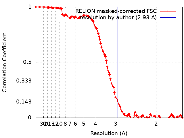
| Method | single particle reconstruction / cryo EM / Resolution: 2.93 Å | |||||||||||||||
 Authors Authors | Zhang M / Jungblut A / Hoffmann T / Eustermann S | |||||||||||||||
| Funding support |  Germany, European Union, 4 items Germany, European Union, 4 items
| |||||||||||||||
 Citation Citation | Journal: Acta Crystallogr D Biol Crystallogr / Year: 2010 Title: Features and development of Coot. Authors: P Emsley / B Lohkamp / W G Scott / K Cowtan /  Abstract: Coot is a molecular-graphics application for model building and validation of biological macromolecules. The program displays electron-density maps and atomic models and allows model manipulations ...Coot is a molecular-graphics application for model building and validation of biological macromolecules. The program displays electron-density maps and atomic models and allows model manipulations such as idealization, real-space refinement, manual rotation/translation, rigid-body fitting, ligand search, solvation, mutations, rotamers and Ramachandran idealization. Furthermore, tools are provided for model validation as well as interfaces to external programs for refinement, validation and graphics. The software is designed to be easy to learn for novice users, which is achieved by ensuring that tools for common tasks are 'discoverable' through familiar user-interface elements (menus and toolbars) or by intuitive behaviour (mouse controls). Recent developments have focused on providing tools for expert users, with customisable key bindings, extensions and an extensive scripting interface. The software is under rapid development, but has already achieved very widespread use within the crystallographic community. The current state of the software is presented, with a description of the facilities available and of some of the underlying methods employed. | |||||||||||||||
| History |
|
- Structure visualization
| Supplemental images |
|---|
- Downloads & links
-EMDB archive
| Map data | emd_17010.map.gz | 5.4 MB | EMDB map data format | |
|---|---|---|---|---|
| Header (meta data) | emd-17010-v30.xmlemd-17010.xml | 35.1 KB 35.1 KB | Display Display | EMDB header |
| FSC (resolution estimation) | emd_17010_fsc.xml | 12.8 KB | Display | FSC data file |
| Images |  emd_17010.png emd_17010.png | 71.9 KB | ||
| Filedesc metadata | emd-17010.cif.gz | 10.1 KB | ||
| Others | emd_17010_additional_1.map.gzemd_17010_half_map_1.map.gzemd_17010_half_map_2.map.gz | 140.5 MB 140.8 MB 140.8 MB | ||
| Archive directory |  http://ftp.pdbj.org/pub/emdb/structures/EMD-17010ftp://ftp.pdbj.org/pub/emdb/structures/EMD-17010 http://ftp.pdbj.org/pub/emdb/structures/EMD-17010ftp://ftp.pdbj.org/pub/emdb/structures/EMD-17010 | HTTPS FTP |
-Related structure data
| Related structure data |  8oocMC  8oo7C 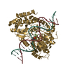 8oo9C 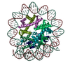 8ooaC 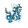 8oofC 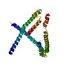 8ookC  8oopC 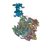 8oorC 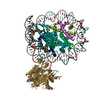 8oosC 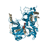 8ootC C: citing same article ( M: atomic model generated by this map |
|---|---|
| Similar structure data |


-Links
| EMDB pages | EMDB (EBI/PDBe) / EMDataResource |
|---|---|
| Related items in Molecule of the Month |


-Map
| File | Download / File: emd_17010.map.gz / Format: CCP4 / Size: 178 MB / Type: IMAGE STORED AS FLOATING POINT NUMBER (4 BYTES) | ||||||||||||||||||||||||||||||||||||
|---|---|---|---|---|---|---|---|---|---|---|---|---|---|---|---|---|---|---|---|---|---|---|---|---|---|---|---|---|---|---|---|---|---|---|---|---|---|
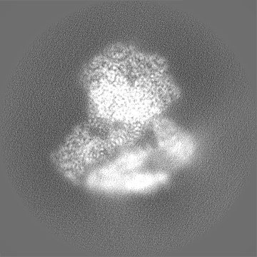
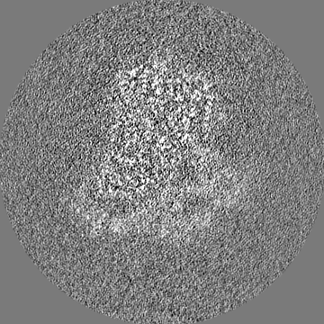
| Annotation | ctINO80-hexasome focused refinement Rvb1/2-Ino80 insert-Ies2-Arp5-Ies6 core sharpened map | ||||||||||||||||||||||||||||||||||||
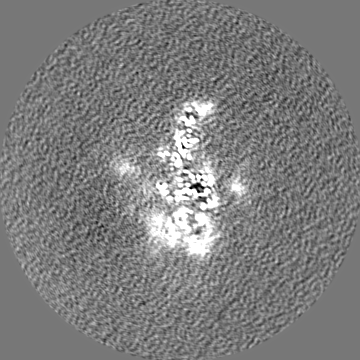
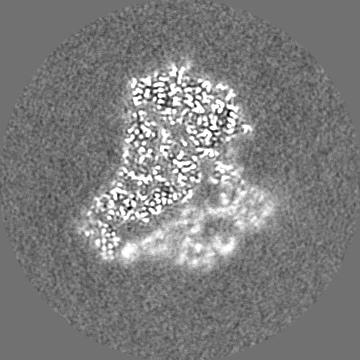
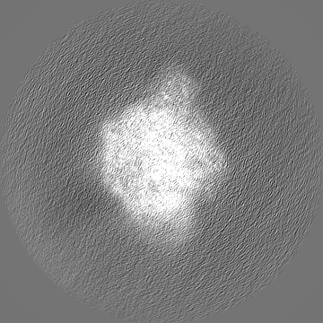
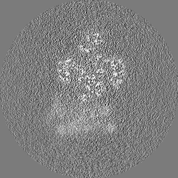
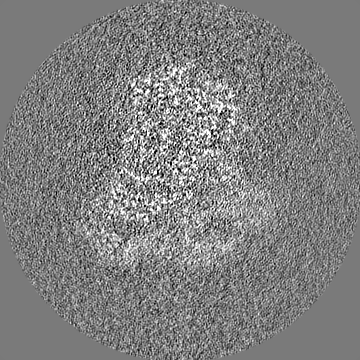
| Projections & slices | Image control
Images are generated by Spider. | ||||||||||||||||||||||||||||||||||||
| Voxel size | X=Y=Z: 0.822 Å | ||||||||||||||||||||||||||||||||||||
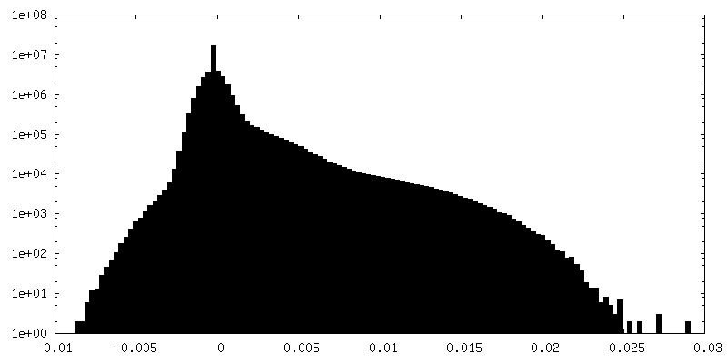
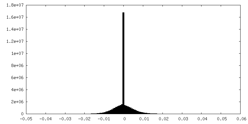
| Density |
| ||||||||||||||||||||||||||||||||||||
| Symmetry | Space group: 1 | ||||||||||||||||||||||||||||||||||||
| Details | EMDB XML:
|
 Z (Sec.)
 Y (Row.)
 X (Col.)


-Supplemental data
-Additional map: ctINO80-hexasome focused refinement Rvb1/2-Ino80 insert-Ies2-Arp5-Ies6 core unsharpened map...
| File | emd_17010_additional_1.map | ||||||||||||
|---|---|---|---|---|---|---|---|---|---|---|---|---|---|
| Annotation | ctINO80-hexasome focused refinement Rvb1/2-Ino80 insert-Ies2-Arp5-Ies6 core unsharpened map | ||||||||||||
| Projections & Slices |
| ||||||||||||
| Density Histograms |


-Half map: ctINO80-hexasome focused refinement Rvb1/2-Ino80 insert-Ies2-Arp5-Ies6 core half map...
| File | emd_17010_half_map_1.map | ||||||||||||
|---|---|---|---|---|---|---|---|---|---|---|---|---|---|
| Annotation | ctINO80-hexasome focused refinement Rvb1/2-Ino80 insert-Ies2-Arp5-Ies6 core half map | ||||||||||||
| Projections & Slices |
| ||||||||||||
| Density Histograms |


-Half map: ctINO80-hexasome focused refinement Rvb1/2-Ino80 insert-Ies2-Arp5-Ies6 core half map...
| File | emd_17010_half_map_2.map | ||||||||||||
|---|---|---|---|---|---|---|---|---|---|---|---|---|---|
| Annotation | ctINO80-hexasome focused refinement Rvb1/2-Ino80 insert-Ies2-Arp5-Ies6 core half map | ||||||||||||
| Projections & Slices |
| ||||||||||||
| Density Histograms |


- Sample components
+Entire : INO80 core module in complex with hexasome
+Supramolecule #1: INO80 core module in complex with hexasome
+Macromolecule #1: RuvB-like helicase
 Trichoplusia ni (cabbage looper)
+Macromolecule #2: RuvB-like helicase
+Macromolecule #3: Chromatin-remodeling ATPase Ino80
+Macromolecule #4: INO80 complex subunit B-like conserved region domain-containing p...
+Macromolecule #5: Vps72/YL1 C-terminal domain-containing protein
+Macromolecule #6: DASH complex subunit DAD4
+Macromolecule #7: ADENOSINE-5'-DIPHOSPHATE

+Macromolecule #8: MAGNESIUM ION
+Macromolecule #9: ADENOSINE-5'-TRIPHOSPHATE

-Experimental details
-Structure determination
| Method | cryo EM |
|---|---|
 Processing Processing | single particle reconstruction |
| Aggregation state | particle |
-Sample preparation
| Concentration | 0.88 mg/mL |
|---|---|
| Buffer | pH: 7.5 Details: 30mM HEPES, pH7.5 50mM NaCl 0.25mM CaCl2 0.25mM DTT 2mM ADP 3.3mM MgCl2 10mM NaF 2mM AlCl3 0.05% octyl-beta-glucoside |
| Grid | Model: Quantifoil R2/1 / Material: COPPER / Mesh: 200 / Support film - Material: CARBON / Support film - topology: HOLEY / Pretreatment - Type: PLASMA CLEANING / Pretreatment - Time: 15 sec. / Details: 10% Oxygene 90% Argon |
| Vitrification | Cryogen name: ETHANE / Chamber humidity: 100 % / Chamber temperature: 281 K / Instrument: FEI VITROBOT MARK IV Details: wait time of 5s, blot force at 3, and a blot time of 2s with Whatman blotting paper (Cytiva, CAT No. 10311807). |
| Details | 11-subunit ctINO80 reconstituted with hexasome |
- Electron microscopy
| Microscope | FEI TITAN KRIOS |
|---|---|
| Image recording | Film or detector model: GATAN K3 (6k x 4k) / Number real images: 15384 / Average electron dose: 50.36 e/Å2 |
| Electron beam | Acceleration voltage: 300 kV / Electron source:  FIELD EMISSION GUN FIELD EMISSION GUN |
| Electron optics | Illumination mode: FLOOD BEAM / Imaging mode: BRIGHT FIELD / Nominal defocus max: 2.0 µm / Nominal defocus min: 0.8 µm |
| Sample stage | Specimen holder model: FEI TITAN KRIOS AUTOGRID HOLDER / Cooling holder cryogen: NITROGEN |
| Experimental equipment |  Model: Titan Krios / Image courtesy: FEI Company |