 Movie
Movie Controller

[English] 日本語
 Yorodumi
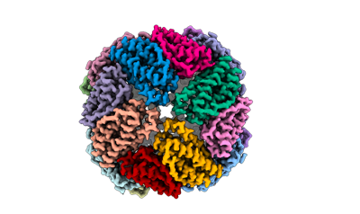
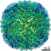
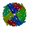
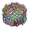
- EMDB-12738: 2.12 A cryo-EM structure of Mycobacterium tuberculosis Ferritin -
+ Open data

- Basic information
| Entry | Database: EMDB / ID: EMD-12738 | |||||||||
|---|---|---|---|---|---|---|---|---|---|---|
| Title | 2.12 A cryo-EM structure of Mycobacterium tuberculosis Ferritin | |||||||||
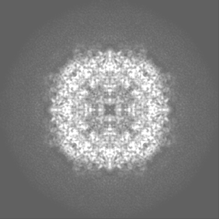
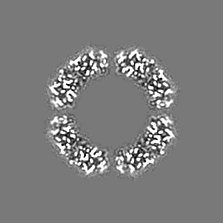
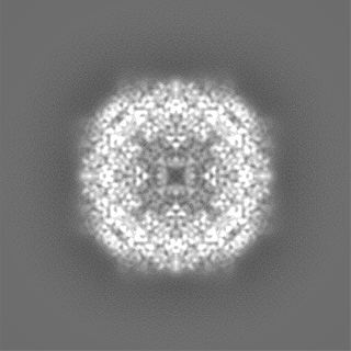
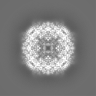
 Map data Map data | Sharpened map with Locspiral. | |||||||||
 Sample Sample |
| |||||||||
 Keywords Keywords | Iron storage / Ferroxidase / Bacterial Ferritin / Octahedral symmetry. / METAL TRANSPORT | |||||||||
| Function / homology |  Function and homology information Function and homology informationMtb iron assimilation by chelation / response to nitrosative stress / encapsulin nanocompartment / iron ion sequestering activity / ferroxidase / ferroxidase activity / ferric iron binding / peptidoglycan-based cell wall / iron ion transport / ferrous iron binding ...Mtb iron assimilation by chelation / response to nitrosative stress / encapsulin nanocompartment / iron ion sequestering activity / ferroxidase / ferroxidase activity / ferric iron binding / peptidoglycan-based cell wall / iron ion transport / ferrous iron binding / intracellular iron ion homeostasis / response to hypoxia / extracellular region / plasma membrane / cytoplasm / cytosol Similarity search - Function | |||||||||
| Biological species |  Mycobacterium tuberculosis H37Rv (bacteria) Mycobacterium tuberculosis H37Rv (bacteria) | |||||||||
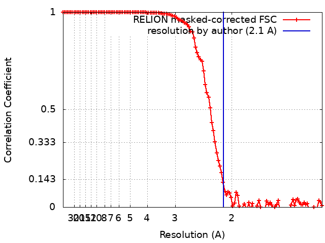
| Method | single particle reconstruction / cryo EM / Resolution: 2.1 Å | |||||||||
 Authors Authors | Gijsbers A / Zhang Y | |||||||||
| Funding support | European Union,  Netherlands, 2 items Netherlands, 2 items
| |||||||||
 Citation Citation | Journal: Acta Crystallogr D Struct Biol / Year: 2021 Title: Mycobacterium tuberculosis ferritin: a suitable workhorse protein for cryo-EM development. Authors: Abril Gijsbers / Yue Zhang / Ye Gao / Peter J Peters / Raimond B G Ravelli / Abstract: The use of cryo-EM continues to expand worldwide and calls for good-quality standard proteins with simple protocols for their production. Here, a straightforward expression and purification protocol ...The use of cryo-EM continues to expand worldwide and calls for good-quality standard proteins with simple protocols for their production. Here, a straightforward expression and purification protocol is presented that provides an apoferritin, bacterioferritin B (BfrB), from Mycobacterium tuberculosis with high yield and purity. A 2.12 Å resolution cryo-EM structure of BfrB is reported, showing the typical cage-like oligomer constituting of 24 monomers related by 432 symmetry. However, it also contains a unique C-terminal extension (164-181), which loops into the cage region of the shell and provides extra stability to the protein. Part of this region was ambiguous in previous crystal structures but could be built within the cryo-EM map. These findings and this protocol could serve the growing cryo-EM community in characterizing and pushing the limits of their electron microscopes and workflows. | |||||||||
| History |
|
- Structure visualization
| Movie |
Movie viewer |
|---|---|
| Structure viewer | EM map: SurfViewMolmilJmol/JSmol |
| Supplemental images |
- Downloads & links
-EMDB archive
| Map data | emd_12738.map.gz | 22.7 MB | EMDB map data format | |
|---|---|---|---|---|
| Header (meta data) | emd-12738-v30.xmlemd-12738.xml | 22.3 KB 22.3 KB | Display Display | EMDB header |
| FSC (resolution estimation) | emd_12738_fsc.xml | 11.3 KB | Display | FSC data file |
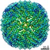
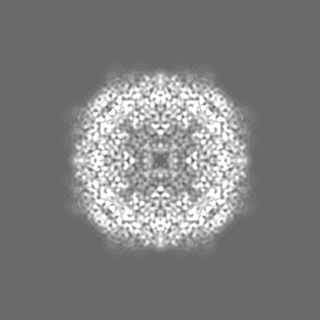
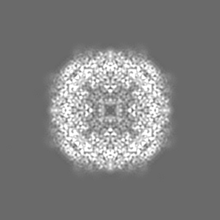
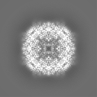
| Images |  emd_12738.png emd_12738.png | 70 KB | ||
| Masks | emd_12738_msk_1.map | 125 MB | Mask map | |
| Filedesc metadata | emd-12738.cif.gz | 6.4 KB | ||
| Others | emd_12738_additional_1.map.gzemd_12738_half_map_1.map.gzemd_12738_half_map_2.map.gz | 20.6 MB 94.1 MB 94.1 MB | ||
| Archive directory |  http://ftp.pdbj.org/pub/emdb/structures/EMD-12738ftp://ftp.pdbj.org/pub/emdb/structures/EMD-12738 http://ftp.pdbj.org/pub/emdb/structures/EMD-12738ftp://ftp.pdbj.org/pub/emdb/structures/EMD-12738 | HTTPS FTP |
-Related structure data
| Related structure data |  7o6eMC M: atomic model generated by this map C: citing same article ( |
|---|---|
| Similar structure data |


-Links
| EMDB pages | EMDB (EBI/PDBe) / EMDataResource |
|---|---|
| Related items in Molecule of the Month |


-Map
| File | Download / File: emd_12738.map.gz / Format: CCP4 / Size: 125 MB / Type: IMAGE STORED AS FLOATING POINT NUMBER (4 BYTES) | ||||||||||||||||||||||||||||||||||||||||||||||||||||||||||||
|---|---|---|---|---|---|---|---|---|---|---|---|---|---|---|---|---|---|---|---|---|---|---|---|---|---|---|---|---|---|---|---|---|---|---|---|---|---|---|---|---|---|---|---|---|---|---|---|---|---|---|---|---|---|---|---|---|---|---|---|---|---|
| Annotation | Sharpened map with Locspiral. | ||||||||||||||||||||||||||||||||||||||||||||||||||||||||||||
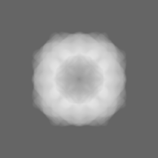
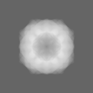
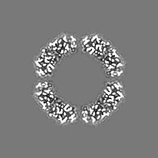
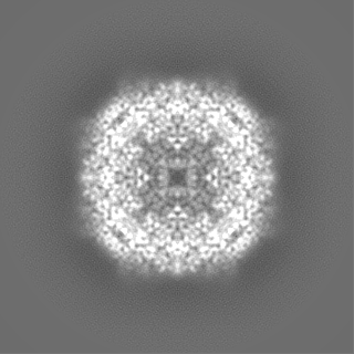
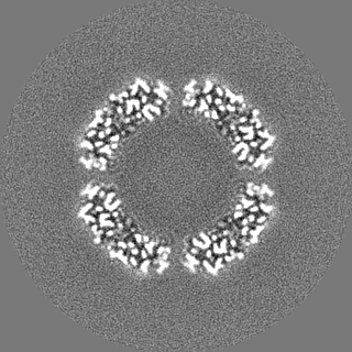
| Projections & slices | Image control
Images are generated by Spider. | ||||||||||||||||||||||||||||||||||||||||||||||||||||||||||||
| Voxel size | X=Y=Z: 0.6514 Å | ||||||||||||||||||||||||||||||||||||||||||||||||||||||||||||
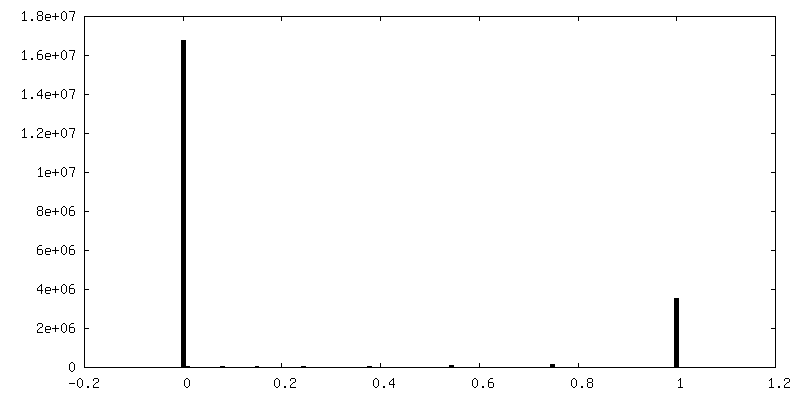
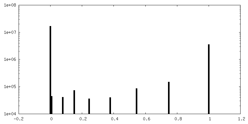
| Density |
| ||||||||||||||||||||||||||||||||||||||||||||||||||||||||||||
| Symmetry | Space group: 1 | ||||||||||||||||||||||||||||||||||||||||||||||||||||||||||||
| Details | EMDB XML:
CCP4 map header:
| ||||||||||||||||||||||||||||||||||||||||||||||||||||||||||||
 Z (Sec.)
 Y (Row.)
 X (Col.)


-Supplemental data
-Mask #1
| File | emd_12738_msk_1.map | ||||||||||||
|---|---|---|---|---|---|---|---|---|---|---|---|---|---|
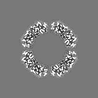
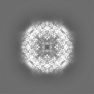
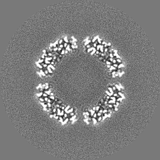
| Projections & Slices |
| ||||||||||||
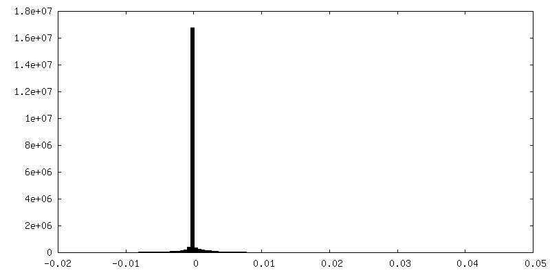
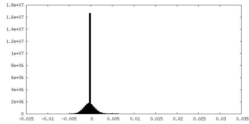
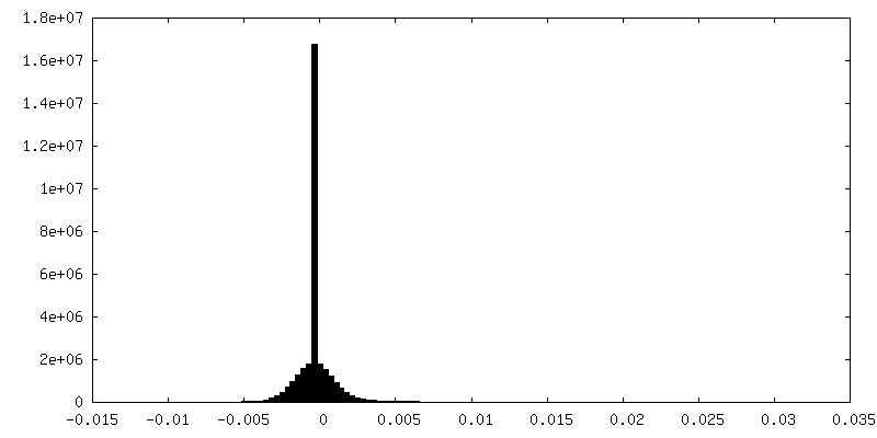
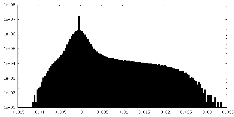
| Density Histograms |


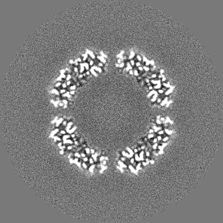
-Additional map: Relion postprocessing sharpened map
| File | emd_12738_additional_1.map | ||||||||||||
|---|---|---|---|---|---|---|---|---|---|---|---|---|---|
| Annotation | Relion postprocessing sharpened map | ||||||||||||
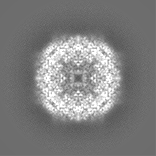
| Projections & Slices |
| ||||||||||||
| Density Histograms |

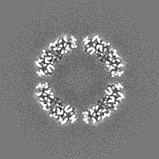
-Half map: #2
| File | emd_12738_half_map_1.map | ||||||||||||
|---|---|---|---|---|---|---|---|---|---|---|---|---|---|
| Projections & Slices |
| ||||||||||||
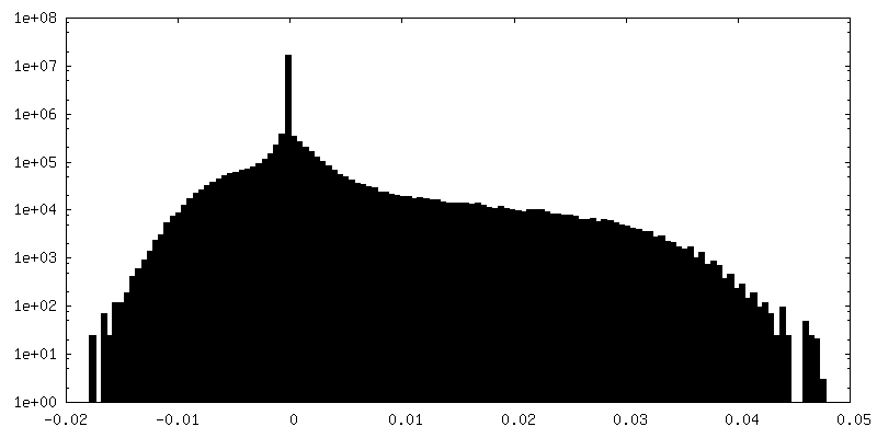
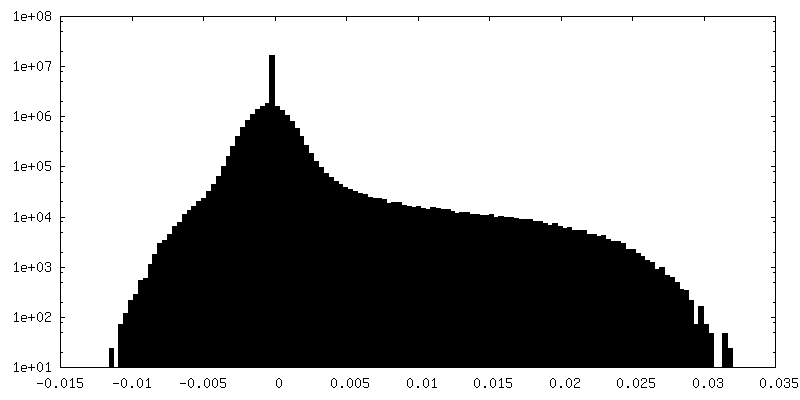
| Density Histograms |

-Half map: #1
| File | emd_12738_half_map_2.map | ||||||||||||
|---|---|---|---|---|---|---|---|---|---|---|---|---|---|
| Projections & Slices |
| ||||||||||||
| Density Histograms |

- Sample components
-Entire : Mycobacterium tuberculosis ferritin
| Entire | Name: Mycobacterium tuberculosis ferritin |
|---|---|
| Components |
|
-Supramolecule #1: Mycobacterium tuberculosis ferritin
| Supramolecule | Name: Mycobacterium tuberculosis ferritin / type: organelle_or_cellular_component / ID: 1 / Parent: 0 / Macromolecule list: #1 / Details: Mycobacterium tuberculosis ferritin |
|---|---|
| Source (natural) | Organism: Mycobacterium tuberculosis H37Rv (bacteria) / Strain: H37Rv |
| Molecular weight | Theoretical: 490 KDa |
-Macromolecule #1: Ferritin BfrB
| Macromolecule | Name: Ferritin BfrB / type: protein_or_peptide / ID: 1 / Number of copies: 24 / Enantiomer: LEVO / EC number: ferroxidase |
|---|---|
| Source (natural) | Organism: Mycobacterium tuberculosis H37Rv (bacteria) |
| Molecular weight | Theoretical: 20.463936 KDa |
| Recombinant expression | Organism: |
| Sequence | String: MTEYEGPKTK FHALMQEQIH NEFTAAQQYV AIAVYFDSED LPQLAKHFYS QAVEERNHAM MLVQHLLDRD LRVEIPGVDT VRNQFDRPR EALALALDQE RTVTDQVGRL TAVARDEGDF LGEQFMQWFL QEQIEEVALM ATLVRVADRA GANLFELENF V AREVDVAP AASGAPHAAG GRL UniProtKB: Bacterioferritin BfrB |
-Macromolecule #2: water
| Macromolecule | Name: water / type: ligand / ID: 2 / Number of copies: 1392 / Formula: HOH |
|---|---|
| Molecular weight | Theoretical: 18.015 Da |
| Chemical component information |  ChemComp-HOH: |
-Experimental details
-Structure determination
| Method | cryo EM |
|---|---|
 Processing Processing | single particle reconstruction |
| Aggregation state | particle |
-Sample preparation
| Concentration | 11 mg/mL | |||||||||
|---|---|---|---|---|---|---|---|---|---|---|
| Buffer | pH: 8 Component:
| |||||||||
| Grid | Model: UltrAuFoil / Material: GOLD / Mesh: 300 / Support film - Material: GOLD / Support film - topology: HOLEY ARRAY / Support film - Film thickness: 2000 / Pretreatment - Type: GLOW DISCHARGE / Pretreatment - Time: 40 sec. / Pretreatment - Atmosphere: AIR / Pretreatment - Pressure: 0.01 kPa | |||||||||
| Vitrification | Cryogen name: ETHANE / Chamber humidity: 100 % / Chamber temperature: 277 K / Instrument: FEI VITROBOT MARK IV | |||||||||
| Details | This sample was monodisperse. |
- Electron microscopy
| Microscope | FEI TITAN KRIOS |
|---|---|
| Temperature | Min: 93.0 K / Max: 105.0 K |
| Specialist optics | Energy filter - Name: GIF Bioquantum / Energy filter - Slit width: 20 eV |
| Details | Basic direct alignments were done as well as astigmatism and coma alignment using AutoCTF |
| Image recording | Film or detector model: GATAN K3 BIOQUANTUM (6k x 4k) / Digitization - Dimensions - Width: 5760 pixel / Digitization - Dimensions - Height: 4092 pixel / Number grids imaged: 1 / Number real images: 2518 / Average exposure time: 1.3 sec. / Average electron dose: 40.0 e/Å2 Details: Images were acquired in superresolution counting mode at a speed of 64 movies/hour and stored as tiff lzw non-gain normalised |
| Electron beam | Acceleration voltage: 300 kV / Electron source:  FIELD EMISSION GUN FIELD EMISSION GUN |
| Electron optics | C2 aperture diameter: 50.0 µm / Calibrated defocus max: 2.0 µm / Calibrated defocus min: 0.2 µm / Calibrated magnification: 76757 / Illumination mode: FLOOD BEAM / Imaging mode: BRIGHT FIELD / Cs: 2.7 mm / Nominal defocus max: 1.4000000000000001 µm / Nominal defocus min: 0.4 µm / Nominal magnification: 130000 |
| Sample stage | Specimen holder model: FEI TITAN KRIOS AUTOGRID HOLDER / Cooling holder cryogen: NITROGEN |
| Experimental equipment |  Model: Titan Krios / Image courtesy: FEI Company |
+Image processing

-Atomic model buiding 1
| Initial model | PDB ID: Chain - Chain ID: A / Chain - Residue range: 10-181 / Chain - Source name: PDB / Chain - Initial model type: experimental model |
|---|---|
| Refinement | Space: REAL / Protocol: OTHER / Overall B value: 28.1 / Target criteria: Correlation coefficient |
| Output model | PDB-7o6e: |
