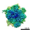
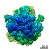
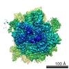
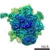
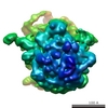
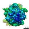
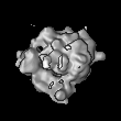
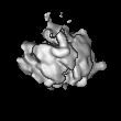
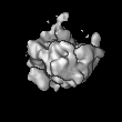
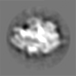
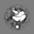
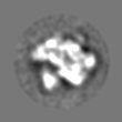
[日本語] English
- EMDB-1251: Structure of the E. coli signal recognition particle bound to a t... -

+
データを開く


IDまたはキーワード:

読み込み中...

-
基本情報

登録情報
データベース: EMDB / ID: EMD-1251
タイトルStructure of the E. coli signal recognition particle bound to a translating ribosome.
マップデータThis is the map file of the E. coli signal recognition particle bound to a non-translating ribosome.
試料
  • 試料: E. coli ribosome and signal recognition particle
  • 複合体: E.coli 70S ribosome
  • リガンド: E.coli SRP
機能・相同性
機能・相同性情報


signal recognition particle / endoplasmic reticulum signal peptide binding / signal-recognition-particle GTPase / 7S RNA binding / SRP-dependent cotranslational protein targeting to membrane / SRP-dependent cotranslational protein targeting to membrane, translocation / GTPase activity / GTP binding / ATP hydrolysis activity / cytosol
類似検索 - 分子機能
Signal recognition particle protein / SRP/SRP receptor, N-terminal / Signal recognition particle, SRP54 subunit / Signal recognition particle, SRP54 subunit, M-domain / Signal recognition particle, SRP54 subunit, M-domain superfamily / Signal peptide binding domain / SRP54-type proteins GTP-binding domain signature. / Signal recognition particle SRP54, helical bundle / Signal recognition particle SRP54, N-terminal domain superfamily / SRP54-type protein, helical bundle domain ...Signal recognition particle protein / SRP/SRP receptor, N-terminal / Signal recognition particle, SRP54 subunit / Signal recognition particle, SRP54 subunit, M-domain / Signal recognition particle, SRP54 subunit, M-domain superfamily / Signal peptide binding domain / SRP54-type proteins GTP-binding domain signature. / Signal recognition particle SRP54, helical bundle / Signal recognition particle SRP54, N-terminal domain superfamily / SRP54-type protein, helical bundle domain / SRP54-type protein, helical bundle domain / Signal recognition particle, SRP54 subunit, GTPase domain / SRP54-type protein, GTPase domain / SRP54-type protein, GTPase domain / ATPases associated with a variety of cellular activities / AAA+ ATPase domain / P-loop containing nucleoside triphosphate hydrolase
類似検索 - ドメイン・相同性
Signal recognition particle 54 kDa protein / Signal recognition particle protein
類似検索 - 構成要素
生物種Escherichia coli (大腸菌)
手法単粒子再構成法 / クライオ電子顕微鏡法 / 解像度: 20.0 Å
データ登録者Schaffitzel C / Oswald M / Berger I / Ishikawa T / Abrahams JP / Koerten HK / Koning RI / Ban N
引用ジャーナル: Nature / : 2006
タイトル: Structure of the E. coli signal recognition particle bound to a translating ribosome.
著者: Christiane Schaffitzel / Miro Oswald / Imre Berger / Takashi Ishikawa / Jan Pieter Abrahams / Henk K Koerten / Roman I Koning / Nenad Ban /
要旨: The prokaryotic signal recognition particle (SRP) targets membrane proteins into the inner membrane. It binds translating ribosomes and screens the emerging nascent chain for a hydrophobic signal ...The prokaryotic signal recognition particle (SRP) targets membrane proteins into the inner membrane. It binds translating ribosomes and screens the emerging nascent chain for a hydrophobic signal sequence, such as the transmembrane helix of inner membrane proteins. If such a sequence emerges, the SRP binds tightly, allowing the SRP receptor to lock on. This assembly delivers the ribosome-nascent chain complex to the protein translocation machinery in the membrane. Using cryo-electron microscopy and single-particle reconstruction, we obtained a 16 A structure of the Escherichia coli SRP in complex with a translating E. coli ribosome containing a nascent chain with a transmembrane helix anchor. We also obtained structural information on the SRP bound to an empty E. coli ribosome. The latter might share characteristics with a scanning SRP complex, whereas the former represents the next step: the targeting complex ready for receptor binding. High-resolution structures of the bacterial ribosome and of the bacterial SRP components are available, and their fitting explains our electron microscopic density. The structures reveal the regions that are involved in complex formation, provide insight into the conformation of the SRP on the ribosome and indicate the conformational changes that accompany high-affinity SRP binding to ribosome nascent chain complexes upon recognition of the signal sequence.
履歴
登録2006年7月29日-
ヘッダ(付随情報) 公開2006年7月29日-
マップ公開2008年7月29日-
更新2012年10月24日-
現状2012年10月24日処理サイト: PDBe / 状態: 公開

-
構造の表示

ムービー
  • 表面図(断面を密度値に従い着色)
  • 表面レベル: 0.25
  • UCSF Chimeraによる作画
  • ダウンロード
  • 表面図(高さに従い着色)
  • 表面レベル: 0.25
  • UCSF Chimeraによる作画
  • ダウンロード
  • あてはめたモデルとの重ね合わせ
  • 原子モデル: PDB-2iy3
  • 表面レベル: 0.25
  • UCSF Chimeraによる作画
  • ダウンロード
  • 単純化した表面モデル + あてはめた原子モデル
  • 原子モデルPDB-2iy3
  • Jmolによる作画
  • ダウンロード
ムービービューア
構造ビューアEMマップ:
SurfViewMolmilJmol/JSmol
添付画像

ダウンロードとリンク

-
マップ

ファイルダウンロード / ファイル: emd_1251.map.gz / 形式: CCP4 / 大きさ: 5 MB / タイプ: IMAGE STORED AS FLOATING POINT NUMBER (4 BYTES)
注釈This is the map file of the E. coli signal recognition particle bound to a non-translating ribosome.
投影像・断面図

画像のコントロール

大きさ
明度
コントラスト
その他
Z (Sec.)Y (Row.)X (Col.)
3.81 Å/pix.
x 110 pix.
= 419.1 Å
3.81 Å/pix.
x 110 pix.
= 419.1 Å
3.81 Å/pix.
x 110 pix.
= 419.1 Å

表面

投影像

断面 (1/3)

断面 (1/2)

断面 (2/3)

画像は Spider により作成

ボクセルのサイズX=Y=Z: 3.81 Å
密度
表面レベル1: 0.926 / ムービー #1: 0.25
最小 - 最大-2.74426 - 4.88095
平均 (標準偏差)-0.000000000234658 (±0.48842)
対称性空間群: 1
詳細

EMDB XML:

マップ形状
Axis orderXYZ
Origin000
サイズ110110110
Spacing110110110
セルA=B=C: 419.1 Å
α=β=γ: 90 °

CCP4マップ ヘッダ情報:

modeImage stored as Reals
Å/pix. X/Y/Z3.813.813.81
M x/y/z110110110
origin x/y/z0.0000.0000.000
length x/y/z419.100419.100419.100
α/β/γ90.00090.00090.000
start NX/NY/NZ-64-64-64
NX/NY/NZ128128128
MAP C/R/S123
start NC/NR/NS000
NC/NR/NS110110110
D min/max/mean-2.7444.881-0.000

-
添付データ

-
試料の構成要素

-
全体 : E. coli ribosome and signal recognition particle

全体名称: E. coli ribosome and signal recognition particle
要素
  • 試料: E. coli ribosome and signal recognition particle
  • 複合体: E.coli 70S ribosome
  • リガンド: E.coli SRP

-
超分子 #1000: E. coli ribosome and signal recognition particle

超分子名称: E. coli ribosome and signal recognition particle / タイプ: sample / ID: 1000 / 集合状態: One SRP binds to one ribosome. / Number unique components: 2

-
超分子 #1: E.coli 70S ribosome

超分子名称: E.coli 70S ribosome / タイプ: complex / ID: 1 / 組換発現: No / Ribosome-details: ribosome-prokaryote: ALL
由来(天然)生物種: Escherichia coli (大腸菌)
分子量理論値: 2.5 MDa

-
分子 #1: E.coli SRP

分子名称: E.coli SRP / タイプ: ligand / ID: 1 / Name.synonym: Signal rocognition particle / コピー数: 1 / 集合状態: monomer / 組換発現: Yes
由来(天然)生物種: Escherichia coli (大腸菌) / 細胞中の位置: cytosol
分子量理論値: 90 KDa
組換発現生物種: Escherichia coli BL21(DE3) (大腸菌) / 組換プラスミド: pET24a_Ffh and pUC19_Ffs

-
実験情報

-
構造解析

手法クライオ電子顕微鏡法
解析単粒子再構成法
試料の集合状態particle

-
試料調製

濃度0.375 mg/mL
緩衝液pH: 7.5
詳細: 50mM Hepes-KOH pH7.5,100mM KCl,25mM MgCl2,1mM DTT,1mM GTP
グリッド詳細: carbon-coated lacey formvar grids
凍結凍結剤: ETHANE / チャンバー内湿度: 100 % / チャンバー内温度: 298 K / 装置: HOMEMADE PLUNGER
詳細: Vitrification instrument: home-built environmental chamber and vitrification device
手法: blot for 1.5 sec before plunging

-
電子顕微鏡法

顕微鏡FEI TECNAI F20
撮影カテゴリ: FILM / フィルム・検出器のモデル: KODAK SO-163 FILM / デジタル化 - スキャナー: OTHER / 実像数: 212 / 平均電子線量: 10 e/Å2
電子線加速電圧: 200 kV / 電子線源: FIELD EMISSION GUN
電子光学系倍率(補正後): 51000 / 照射モード: FLOOD BEAM / 撮影モード: BRIGHT FIELD / Cs: 2 mm / 最大 デフォーカス(公称値): 4.5 µm / 最小 デフォーカス(公称値): 1.0 µm / 倍率(公称値): 50000
試料ステージ試料ホルダー: cryo stage / 試料ホルダーモデル: GATAN LIQUID NITROGEN
実験機器
モデル: Tecnai F20 / 画像提供: FEI Company

-
画像解析

最終 再構成想定した対称性 - 点群: C1 (非対称) / 解像度のタイプ: BY AUTHOR / 解像度: 20.0 Å / 解像度の算出法: FSC 0.5 CUT-OFF / ソフトウェア - 名称: spider / 使用した粒子像数: 9300

+
万見について

-
お知らせ

-
2022年2月9日: EMDBエントリの付随情報ファイルのフォーマットが新しくなりました

EMDBエントリの付随情報ファイルのフォーマットが新しくなりました

  • EMDBのヘッダファイルのバージョン3が、公式のフォーマットとなりました。
  • これまでは公式だったバージョン1.9は、アーカイブから削除されます。

関連情報:EMDBヘッダ

外部リンク:wwPDBはEMDBデータモデルのバージョン3へ移行します

-
2020年8月12日: 新型コロナ情報

新型コロナ情報

URL: https://pdbjlvh1.pdbj.org/emnavi/covid19.php

新ページ: EM Navigatorに新型コロナウイルスの特設ページを開設しました。

関連情報:Covid-19情報 / 2020年3月5日: 新型コロナウイルスの構造データ

+
2020年3月5日: 新型コロナウイルスの構造データ

新型コロナウイルスの構造データ

関連情報:万見生物種 / 2020年8月12日: 新型コロナ情報

外部リンク:COVID-19特集ページ - PDBj / 今月の分子2020年2月:コロナウイルスプロテーアーゼ

+
2019年1月31日: EMDBのIDの桁数の変更

EMDBのIDの桁数の変更

  • EMDBエントリに付与されているアクセスコード(EMDB-ID)は4桁の数字(例、EMD-1234)でしたが、間もなく枯渇します。これまでの4桁のID番号は4桁のまま変更されませんが、4桁の数字を使い切った後に発行されるIDは5桁以上の数字(例、EMD-12345)になります。5桁のIDは2019年の春頃から発行される見通しです。
  • EM Navigator/万見では、接頭語「EMD-」は省略されています。

関連情報:Q: 「EMD」とは何ですか? / 万見/EM NavigatorにおけるID/アクセスコードの表記

外部リンク:EMDB Accession Codes are Changing Soon! / PDBjへお問い合わせ

+
2017年7月12日: PDB大規模アップデート

PDB大規模アップデート

  • 新バージョンのPDBx/mmCIF辞書形式に基づくデータがリリースされました。
  • 今回の更新はバージョン番号が4から5になる大規模なもので、全エントリデータの書き換えが行われる「Remediation」というアップデートに該当します。
  • このバージョンアップで、電子顕微鏡の実験手法に関する多くの項目の書式が改定されました(例:em_softwareなど)。
  • EM NavigatorとYorodumiでも、この改定に基づいた表示内容になります。

外部リンク:wwPDB Remediation / OneDepデータ基準に準拠した、より強化された内容のモデル構造ファイルが、PDBアーカイブで公開されました。

-
万見 (Yorodumi)

幾万の構造データを、幾万の視点から

  • 万見(Yorodumi)は、EMDB/PDB/SASBDBなどの構造データを閲覧するためのページです。
  • EM Navigatorの詳細ページの後継、Omokage検索のフロントエンドも兼ねています。

関連情報:EMDB / PDB / SASBDB / 3つのデータバンクの比較 / 万見検索 / 2016年8月31日: 新しいEM Navigatorと万見 / 万見文献 / Jmol/JSmol / 機能・相同性情報 / 新しいEM Navigatorと万見の変更点

他の情報も見る