Journal: Sci Adv / Year: 2021 Title: Cryo-electron microscopy structure of the antidiuretic hormone arginine-vasopressin V2 receptor signaling complex. Authors: Julien Bous / Hélène Orcel / Nicolas Floquet / Cédric Leyrat / Joséphine Lai-Kee-Him / Gérald Gaibelet / Aurélie Ancelin / Julie Saint-Paul / Stefano Trapani / Maxime Louet / Rémy ...Authors: Julien Bous / Hélène Orcel / Nicolas Floquet / Cédric Leyrat / Joséphine Lai-Kee-Him / Gérald Gaibelet / Aurélie Ancelin / Julie Saint-Paul / Stefano Trapani / Maxime Louet / Rémy Sounier / Hélène Déméné / Sébastien Granier / Patrick Bron / Bernard Mouillac / Abstract: The antidiuretic hormone arginine-vasopressin (AVP) forms a signaling complex with the V2 receptor (V2R) and the G protein, promoting kidney water reabsorption. Molecular mechanisms underlying ...The antidiuretic hormone arginine-vasopressin (AVP) forms a signaling complex with the V2 receptor (V2R) and the G protein, promoting kidney water reabsorption. Molecular mechanisms underlying activation of this critical G protein-coupled receptor (GPCR) signaling system are still unknown. To fill this gap of knowledge, we report here the cryo-electron microscopy structure of the AVP-V2R-G complex. Single-particle analysis revealed the presence of three different states. The two best maps were combined with computational and nuclear magnetic resonance spectroscopy constraints to reconstruct two structures of the ternary complex. These structures differ in AVP and G binding modes. They reveal an original receptor-G interface in which the Gα subunit penetrates deep into the active V2R. The structures help to explain how V2R R137H or R137L/C variants can lead to two severe genetic diseases. Our study provides important structural insights into the function of this clinically relevant GPCR signaling complex.
History
Deposition
Dec 17, 2020
-
Header (metadata) release
Jun 2, 2021
-
Map release
Jun 2, 2021
-
Update
Oct 23, 2024
-
Current status
Oct 23, 2024
Processing site: PDBe / Status: Released
-
Structure visualization
Movie
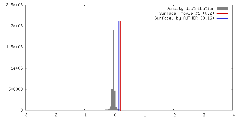
Surface view with section colored by density value
In the structure databanks used in Yorodumi, some data are registered as the other names, "COVID-19 virus" and "2019-nCoV". Here are the details of the virus and the list of structure data.
Jan 31, 2019. EMDB accession codes are about to change! (news from PDBe EMDB page)
EMDB accession codes are about to change! (news from PDBe EMDB page)
The allocation of 4 digits for EMDB accession codes will soon come to an end. Whilst these codes will remain in use, new EMDB accession codes will include an additional digit and will expand incrementally as the available range of codes is exhausted. The current 4-digit format prefixed with “EMD-” (i.e. EMD-XXXX) will advance to a 5-digit format (i.e. EMD-XXXXX), and so on. It is currently estimated that the 4-digit codes will be depleted around Spring 2019, at which point the 5-digit format will come into force.
The EM Navigator/Yorodumi systems omit the EMD- prefix.
Related info.:Q: What is EMD? / ID/Accession-code notation in Yorodumi/EM Navigator
Yorodumi is a browser for structure data from EMDB, PDB, SASBDB, etc.
This page is also the successor to EM Navigator detail page, and also detail information page/front-end page for Omokage search.
The word "yorodu" (or yorozu) is an old Japanese word meaning "ten thousand". "mi" (miru) is to see.
Related info.:EMDB / PDB / SASBDB / Comparison of 3 databanks / Yorodumi Search / Aug 31, 2016. New EM Navigator & Yorodumi / Yorodumi Papers / Jmol/JSmol / Function and homology information / Changes in new EM Navigator and Yorodumi
 Movie
Movie Controller


 Open data
 Basic information
 Map data
 Sample
 Keywords
 Function and homology information
 Homo sapiens (human) /
 Authors
 France, 2 items
  Citation
 Structure visualization
 Downloads & links
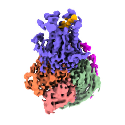 emd_12128.png
 http://ftp.pdbj.org/pub/emdb/structures/EMD-12128


 X (Sec.)
 Y (Row.)
 Z (Col.)


 Sample components
 Spodoptera frugiperda (fall armyworm)
 Processing
 Electron microscopy
 FIELD EMISSION GUN

