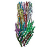
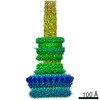
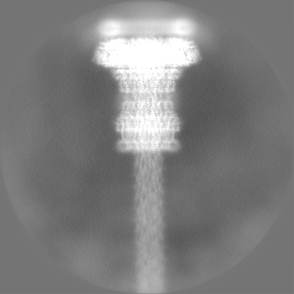
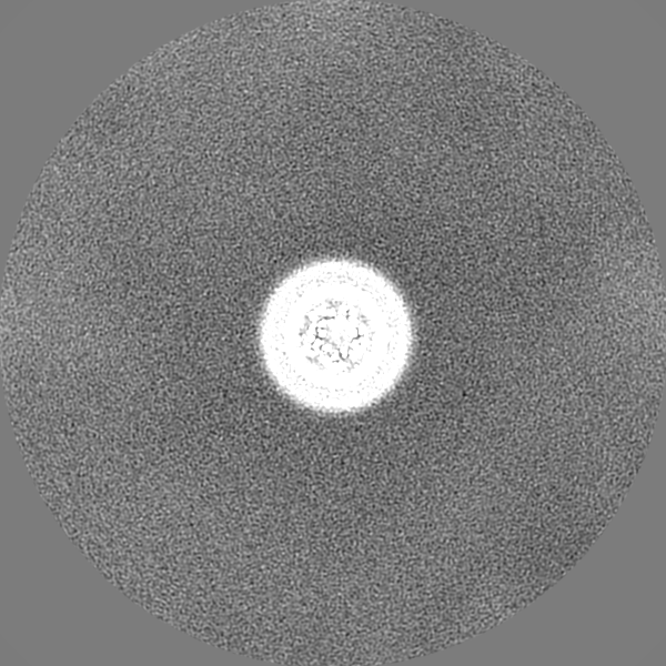
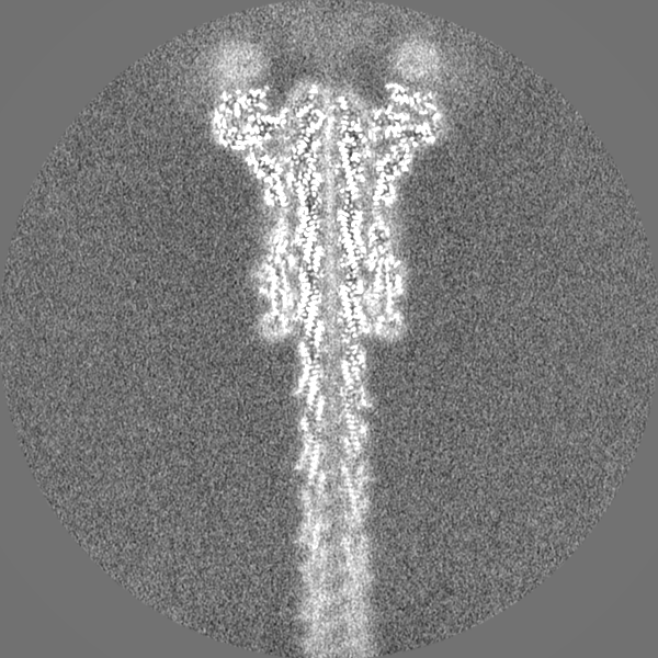
- EMDB-11781: Substrate-engaged type 3 secretion system needle complex from Sal... -
+
データを開く
IDまたはキーワード:
読み込み中...
-
基本情報
登録情報
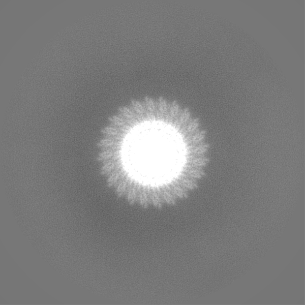
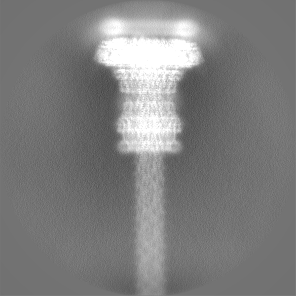
データベース: EMDB / ID: EMD-11781
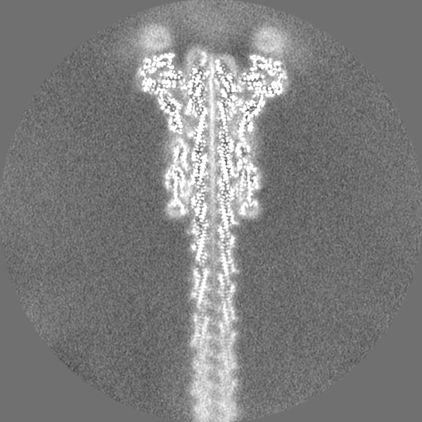
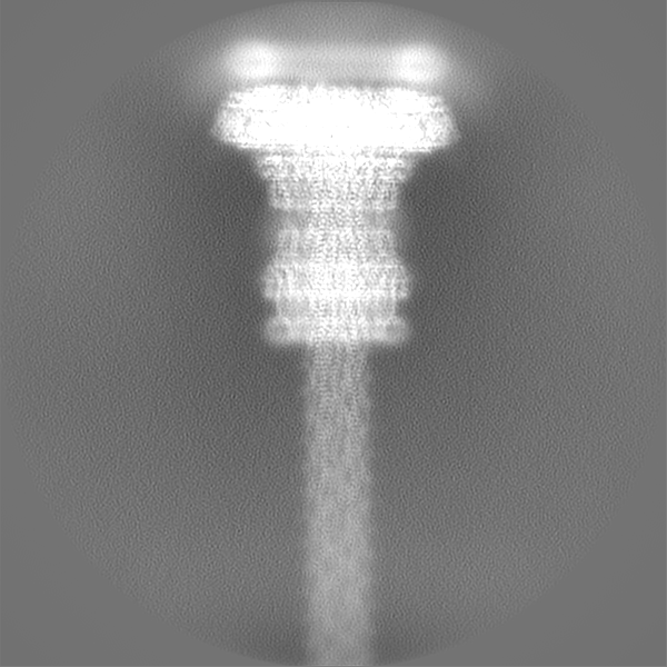
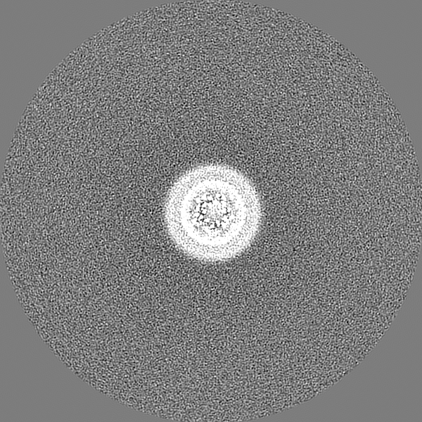
タイトル
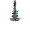
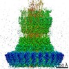
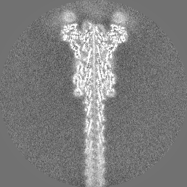
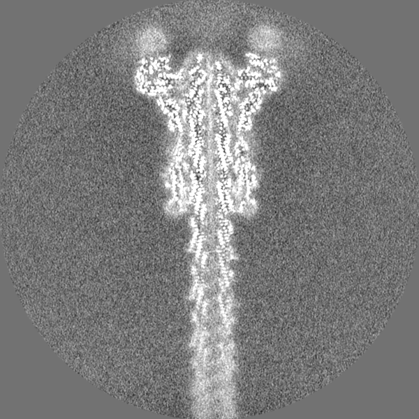
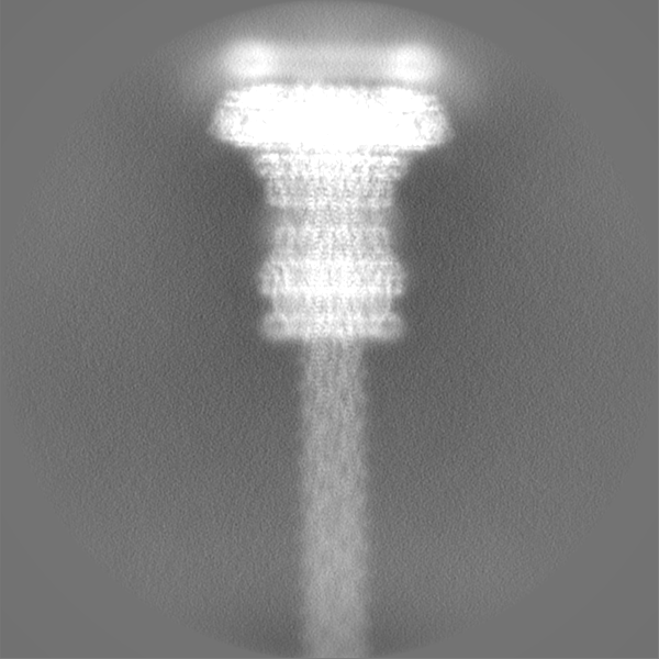
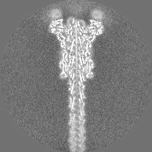
Substrate-engaged type 3 secretion system needle complex from Salmonella enterica typhimurium - SpaR state 1
マップデータ
試料
複合体: Substrate-engaged T3SS needle complex
タンパク質・ペプチド: x 9種
リガンド: x 2種
キーワード
T3SS / Export Apparatus / Injectisome / Needle Complex / PROTEIN TRANSPORT / Substrate
機能・相同性
機能・相同性情報
The IPAF inflammasome / type III protein secretion system complex / type II protein secretion system complex / protein secretion by the type III secretion system / protein secretion / protein targeting / cell outer membrane / protein transport / cell surface / extracellular region ...The IPAF inflammasome / type III protein secretion system complex / type II protein secretion system complex / protein secretion by the type III secretion system / protein secretion / protein targeting / cell outer membrane / protein transport / cell surface / extracellular region / identical protein binding / plasma membrane 類似検索 - 分子機能
: / Type III secretion protein SpaR/YscT / Type III secretion protein HrpO / Yop virulence translocation protein R / Type III secretion system, PrgH/EprH / Type III secretion system lipoprotein HrcJ/YscJ / Type III secretion system inner membrane R protein / Bacterial export protein family 3 / Bacterial export proteins, family 1 / Bacterial export proteins, family 3 ...: / Type III secretion protein SpaR/YscT / Type III secretion protein HrpO / Yop virulence translocation protein R / Type III secretion system, PrgH/EprH / Type III secretion system lipoprotein HrcJ/YscJ / Type III secretion system inner membrane R protein / Bacterial export protein family 3 / Bacterial export proteins, family 1 / Bacterial export proteins, family 3 / Flagella transport protein fliP family signature 1. / Type III secretion system inner membrane P protein / Type III secretion system, PrgH/EprH-like / FliP family / Type III secretion system protein PrgH-EprH (PrgH) / Flagella transport protein fliP family signature 2. / : / SPI-1 type 3 secretion system secretin, N0 domain / Type III secretion system outer membrane pore YscC/HrcC / Type III secretion, needle-protein-like / Type III secretion, needle-protein-like superfamily / Type III secretion needle MxiH, YscF, SsaG, EprI, PscF, EscF / Type III secretion system, needle protein / : / Bacterial type II secretion system protein D signature. / Type II secretion system protein GspD, conserved site / NolW-like / Bacterial type II/III secretion system short domain / NolW-like superfamily / Type II/III secretion system / Bacterial type II and III secretion system protein / Lipoprotein YscJ/Flagellar M-ring protein / Secretory protein of YscJ/FliF family / Flagellar M-ring , N-terminal / AMP-binding enzyme, C-terminal domain superfamily / Prokaryotic membrane lipoprotein lipid attachment site profile. 類似検索 - ドメイン・相同性
Surface presentation of antigens protein SpaQ / SPI-1 type 3 secretion system secretin / Surface presentation of antigens protein SpaP / Surface presentation of antigens protein SpaR / Protein PrgH / SPI-1 type 3 secretion system needle filament protein / Protein PrgJ / Lipoprotein PrgK 類似検索 - 構成要素
生物種
Salmonella enterica subsp. enterica serovar Typhimurium str. LT2 (サルモネラ菌)
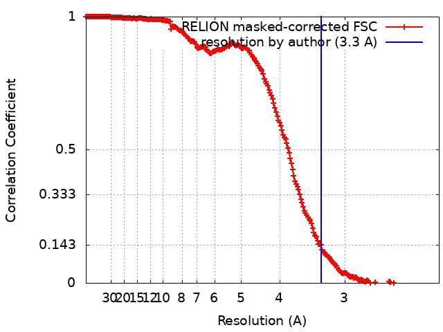
ジャーナル: Nat Commun / 年: 2021 タイトル: Substrate-engaged type III secretion system structures reveal gating mechanism for unfolded protein translocation. 著者: Sean Miletic / Dirk Fahrenkamp / Nikolaus Goessweiner-Mohr / Jiri Wald / Maurice Pantel / Oliver Vesper / Vadim Kotov / Thomas C Marlovits / 要旨: Many bacterial pathogens rely on virulent type III secretion systems (T3SSs) or injectisomes to translocate effector proteins in order to establish infection. The central component of the injectisome ...Many bacterial pathogens rely on virulent type III secretion systems (T3SSs) or injectisomes to translocate effector proteins in order to establish infection. The central component of the injectisome is the needle complex which assembles a continuous conduit crossing the bacterial envelope and the host cell membrane to mediate effector protein translocation. However, the molecular principles underlying type III secretion remain elusive. Here, we report a structure of an active Salmonella enterica serovar Typhimurium needle complex engaged with the effector protein SptP in two functional states, revealing the complete 800Å-long secretion conduit and unraveling the critical role of the export apparatus (EA) subcomplex in type III secretion. Unfolded substrates enter the EA through a hydrophilic constriction formed by SpaQ proteins, which enables side chain-independent substrate transport. Above, a methionine gasket formed by SpaP proteins functions as a gate that dilates to accommodate substrates while preventing leaky pore formation. Following gate penetration, a moveable SpaR loop first folds up to then support substrate transport. Together, these findings establish the molecular basis for substrate translocation through T3SSs and improve our understanding of bacterial pathogenicity and motility.
名称: SptP3x-GFP-FLAG / タイプ: protein_or_peptide / ID: 5 詳細: SptP3xGFP sequence modeled as poly-alanine and named as unknown (UNK) in the coordinate file コピー数: 1 / 光学異性体: LEVO
由来(天然)
生物種: Salmonella enterica subsp. enterica serovar Typhimurium str. LT2 (サルモネラ菌)
分子量
理論値: 12.017806 KDa
組換発現
生物種: Salmonella enterica subsp. enterica serovar Typhimurium str. LT2 (サルモネラ菌)
 ムービー
ムービー コントローラー


 データを開く
 基本情報
基本情報 マップデータ
 試料
試料 キーワード
キーワード 機能・相同性情報
 Salmonella enterica subsp. enterica serovar Typhimurium str. LT2 (サルモネラ菌)
 データ登録者
 オーストリア,
  ドイツ, 2件
  引用
引用 構造の表示
構造の表示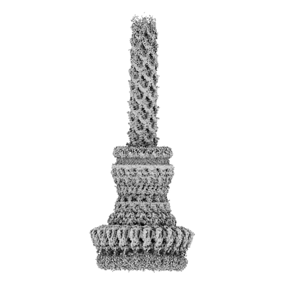
 ダウンロードとリンク
 emd_11781.png
 http://ftp.pdbj.org/pub/emdb/structures/EMD-11781


 Z
Z Y
Y X
X


 試料の構成要素

 解析
解析 電子顕微鏡法
 FIELD EMISSION GUN