 Movie
Movie Controller

[English] 日本語
 Yorodumi
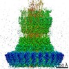
- PDB-7ahi: Substrate-engaged type 3 secretion system needle complex from Sal... -
+ Open data

- Basic information
| Entry | Database: PDB / ID: 7ahi | |||||||||
|---|---|---|---|---|---|---|---|---|---|---|
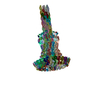
| Title | Substrate-engaged type 3 secretion system needle complex from Salmonella enterica typhimurium - SpaR state 2 | |||||||||
 Components Components |
| |||||||||
 Keywords Keywords | PROTEIN TRANSPORT / T3SS / Export Apparatus / Injectisome / Needle Complex / Substrate | |||||||||
| Function / homology |  Function and homology information Function and homology informationThe IPAF inflammasome / type III protein secretion system complex / type II protein secretion system complex / protein secretion by the type III secretion system / bacterial-type flagellum-dependent swarming motility / bacterial-type flagellum assembly / protein secretion / protein targeting / cell outer membrane / protein transport ...The IPAF inflammasome / type III protein secretion system complex / type II protein secretion system complex / protein secretion by the type III secretion system / bacterial-type flagellum-dependent swarming motility / bacterial-type flagellum assembly / protein secretion / protein targeting / cell outer membrane / protein transport / cell surface / extracellular region / identical protein binding / plasma membrane Similarity search - Function | |||||||||
| Biological species |  Salmonella enterica subsp. enterica serovar Typhimurium str. LT2 (bacteria) Salmonella enterica subsp. enterica serovar Typhimurium str. LT2 (bacteria) | |||||||||
| Method | ELECTRON MICROSCOPY / single particle reconstruction / cryo EM / Resolution: 3.3 Å | |||||||||
 Authors Authors | Fahrenkamp, D. / Goessweiner-Mohr, N. / Miletic, S. / Wald, J. / Marlovits, T. | |||||||||
| Funding support |  Austria, Austria,  Germany, 2items Germany, 2items
| |||||||||
 Citation Citation | Journal: Nat Commun / Year: 2021 Title: Substrate-engaged type III secretion system structures reveal gating mechanism for unfolded protein translocation. Authors: Sean Miletic / Dirk Fahrenkamp / Nikolaus Goessweiner-Mohr / Jiri Wald / Maurice Pantel / Oliver Vesper / Vadim Kotov / Thomas C Marlovits / Abstract: Many bacterial pathogens rely on virulent type III secretion systems (T3SSs) or injectisomes to translocate effector proteins in order to establish infection. The central component of the injectisome ...Many bacterial pathogens rely on virulent type III secretion systems (T3SSs) or injectisomes to translocate effector proteins in order to establish infection. The central component of the injectisome is the needle complex which assembles a continuous conduit crossing the bacterial envelope and the host cell membrane to mediate effector protein translocation. However, the molecular principles underlying type III secretion remain elusive. Here, we report a structure of an active Salmonella enterica serovar Typhimurium needle complex engaged with the effector protein SptP in two functional states, revealing the complete 800Å-long secretion conduit and unraveling the critical role of the export apparatus (EA) subcomplex in type III secretion. Unfolded substrates enter the EA through a hydrophilic constriction formed by SpaQ proteins, which enables side chain-independent substrate transport. Above, a methionine gasket formed by SpaP proteins functions as a gate that dilates to accommodate substrates while preventing leaky pore formation. Following gate penetration, a moveable SpaR loop first folds up to then support substrate transport. Together, these findings establish the molecular basis for substrate translocation through T3SSs and improve our understanding of bacterial pathogenicity and motility. | |||||||||
| History |
|
- Structure visualization
| Movie |
Movie viewer |
|---|---|
| Structure viewer | Molecule: MolmilJmol/JSmol |
- Downloads & links
-Download
| PDBx/mmCIF format | 7ahi.cif.gz | 7.8 MB | Display | PDBx/mmCIF format |
|---|---|---|---|---|
| PDB format | pdb7ahi.ent.gz | Display | PDB format | |
| PDBx/mmJSON format | 7ahi.json.gz | Tree view | PDBx/mmJSON format | |
| Others |  Other downloads Other downloads |
-Validation report
| Arichive directory | https://data.pdbj.org/pub/pdb/validation_reports/ah/7ahiftp://data.pdbj.org/pub/pdb/validation_reports/ah/7ahi | HTTPS FTP |
|---|
-Related structure data
| Related structure data | 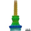 11781MC  7agxC 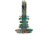 7ah9C C: citing same article ( M: map data used to model this data |
|---|---|
| Similar structure data |
-Links
 PDBj
PDBj


- Assembly
| Deposited unit | 
|
|---|---|
| 1 |
|
-Components
-Surface presentation of antigens protein ... , 3 types, 10 molecules 1A1B1C1D1E1F1G1H1I1J
| #1: Protein | Mass: 25249.596 Da / Num. of mol.: 5 / Source method: isolated from a natural source Source: (natural) Salmonella enterica subsp. enterica serovar Typhimurium str. LT2 (bacteria)References: UniProt: P40700 #2: Protein | | Mass: 28499.533 Da / Num. of mol.: 1 / Source method: isolated from a natural source Source: (natural) Salmonella enterica subsp. enterica serovar Typhimurium str. LT2 (bacteria)References: UniProt: P40701 #3: Protein | Mass: 9363.229 Da / Num. of mol.: 4 / Source method: isolated from a natural source Source: (natural) Salmonella enterica subsp. enterica serovar Typhimurium str. LT2 (bacteria)References: UniProt: P0A1L7 |
|---|
-Protein , 6 types, 143 molecules 1K1L1M1N1O1P1Z2A2B2C2D2E2F2G2H2I2J2K2L2M2N2O2P2Q2R2S2T2U2V2W...
| #4: Protein | Mass: 10934.425 Da / Num. of mol.: 6 / Source method: isolated from a natural source Source: (natural) Salmonella enterica subsp. enterica serovar Typhimurium str. LT2 (bacteria)References: UniProt: P41785 #5: Protein | | Mass: 12017.806 Da / Num. of mol.: 1 Source method: isolated from a genetically manipulated source Source: (gene. exp.) Salmonella enterica subsp. enterica serovar Typhimurium str. LT2 (bacteria)Production host: Salmonella enterica subsp. enterica serovar Typhimurium str. LT2 (bacteria)#6: Protein | Mass: 8864.868 Da / Num. of mol.: 72 / Source method: isolated from a natural source Source: (natural) Salmonella enterica subsp. enterica serovar Typhimurium str. LT2 (bacteria)References: UniProt: P41784 #7: Protein | Mass: 61835.559 Da / Num. of mol.: 16 / Source method: isolated from a natural source Source: (natural) Salmonella enterica subsp. enterica serovar Typhimurium str. LT2 (bacteria)References: UniProt: P35672 #8: Protein | Mass: 28245.287 Da / Num. of mol.: 24 / Source method: isolated from a natural source Source: (natural) Salmonella enterica subsp. enterica serovar Typhimurium str. LT2 (bacteria)References: UniProt: P41786 #9: Protein | Mass: 44509.367 Da / Num. of mol.: 24 / Source method: isolated from a natural source Source: (natural) Salmonella enterica subsp. enterica serovar Typhimurium str. LT2 (bacteria)References: UniProt: P41783 |
|---|
-Non-polymers , 2 types, 14 molecules 

| #10: Chemical | ChemComp-3PH /  Mass: 704.998 Da / Num. of mol.: 5 / Source method: obtained synthetically / Formula: C39H77O8P Mass: 704.998 Da / Num. of mol.: 5 / Source method: obtained synthetically / Formula: C39H77O8P#11: Chemical | ChemComp-LDA /  Mass: 229.402 Da / Num. of mol.: 9 / Source method: obtained synthetically / Formula: C14H31NO / Comment: LDAO, detergent*YM Mass: 229.402 Da / Num. of mol.: 9 / Source method: obtained synthetically / Formula: C14H31NO / Comment: LDAO, detergent*YM |
|---|
-Details
| Has ligand of interest | N |
|---|
-Experimental details
-Experiment
| Experiment | Method: ELECTRON MICROSCOPY |
|---|---|
| EM experiment | Aggregation state: PARTICLE / 3D reconstruction method: single particle reconstruction |
- Sample preparation
| Component | Name: Substrate-engaged T3SS needle complex / Type: COMPLEX / Entity ID: #1-#9 / Source: NATURAL |
|---|---|
| Molecular weight | Value: 2.84 MDa / Experimental value: NO |
| Source (natural) | Organism: Salmonella enterica subsp. enterica serovar Typhimurium str. LT2 (bacteria) |
| Buffer solution | pH: 8 |
| Specimen | Embedding applied: NO / Shadowing applied: NO / Staining applied: NO / Vitrification applied: YES |
| Vitrification | Cryogen name: ETHANE-PROPANE |
- Electron microscopy imaging
| Experimental equipment |  Model: Titan Krios / Image courtesy: FEI Company |
|---|---|
| Microscopy | Model: TFS KRIOS |
| Electron gun | Electron source:  FIELD EMISSION GUN / Accelerating voltage: 300 kV / Illumination mode: SPOT SCAN FIELD EMISSION GUN / Accelerating voltage: 300 kV / Illumination mode: SPOT SCAN |
| Electron lens | Mode: BRIGHT FIELD |
| Image recording | Electron dose: 53 e/Å2 / Film or detector model: GATAN K3 (6k x 4k) |
- Processing
| Software |
| ||||||||||||||||||||
|---|---|---|---|---|---|---|---|---|---|---|---|---|---|---|---|---|---|---|---|---|---|
| EM software |
| ||||||||||||||||||||
| CTF correction | Type: PHASE FLIPPING AND AMPLITUDE CORRECTION | ||||||||||||||||||||
| 3D reconstruction | Resolution: 3.3 Å / Resolution method: FSC 0.143 CUT-OFF / Num. of particles: 77411 / Symmetry type: POINT |
