Movie
Movie Controller

[English] 日本語
 Yorodumi
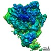
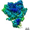
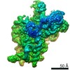
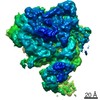
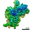
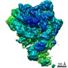
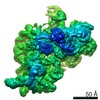
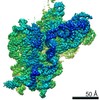
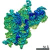
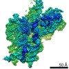
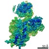
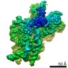
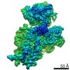
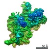
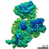
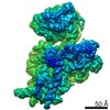
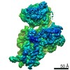
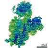
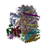
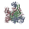
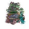
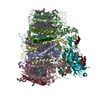
- EMDB-11751: Bacterial 30S ribosomal subunit assembly complex state M (head domain) -
+ Open data

- Basic information
| Entry | Database: EMDB / ID: EMD-11751 | |||||||||
|---|---|---|---|---|---|---|---|---|---|---|
| Title | Bacterial 30S ribosomal subunit assembly complex state M (head domain) | |||||||||
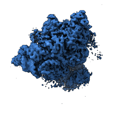
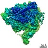
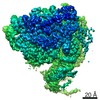
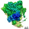
 Map data Map data | Full map | |||||||||
 Sample Sample |
| |||||||||
 Keywords Keywords | Cryo-EM / 30S biogenesis / ribosome assembly / RbfA / RsgA / YjeQ / RimP / KsgA / RsmA / RIBOSOME | |||||||||
| Function / homology |  Function and homology information Function and homology informationribosomal small subunit assembly / small ribosomal subunit / cytosolic small ribosomal subunit / tRNA binding / rRNA binding / structural constituent of ribosome / translation / mRNA binding / RNA binding Similarity search - Function | |||||||||
| Biological species |  | |||||||||
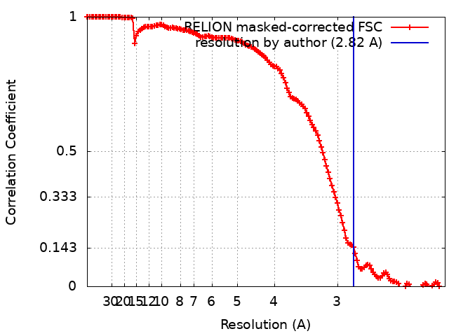
| Method | single particle reconstruction / cryo EM / Resolution: 2.82 Å | |||||||||
 Authors Authors | Schedlbauer A / Iturrioz I | |||||||||
| Funding support |  Spain, 2 items Spain, 2 items
| |||||||||
 Citation Citation | Journal: Sci Adv / Year: 2021 Title: A conserved rRNA switch is central to decoding site maturation on the small ribosomal subunit. Authors: Andreas Schedlbauer / Idoia Iturrioz / Borja Ochoa-Lizarralde / Tammo Diercks / Jorge Pedro López-Alonso / José Luis Lavin / Tatsuya Kaminishi / Retina Çapuni / Neha Dhimole / Elisa de ...Authors: Andreas Schedlbauer / Idoia Iturrioz / Borja Ochoa-Lizarralde / Tammo Diercks / Jorge Pedro López-Alonso / José Luis Lavin / Tatsuya Kaminishi / Retina Çapuni / Neha Dhimole / Elisa de Astigarraga / David Gil-Carton / Paola Fucini / Sean R Connell /  Abstract: While a structural description of the molecular mechanisms guiding ribosome assembly in eukaryotic systems is emerging, bacteria use an unrelated core set of assembly factors for which high- ...While a structural description of the molecular mechanisms guiding ribosome assembly in eukaryotic systems is emerging, bacteria use an unrelated core set of assembly factors for which high-resolution structural information is still missing. To address this, we used single-particle cryo-electron microscopy to visualize the effects of bacterial ribosome assembly factors RimP, RbfA, RsmA, and RsgA on the conformational landscape of the 30 ribosomal subunit and obtained eight snapshots representing late steps in the folding of the decoding center. Analysis of these structures identifies a conserved secondary structure switch in the 16 ribosomal RNA central to decoding site maturation and suggests both a sequential order of action and molecular mechanisms for the assembly factors in coordinating and controlling this switch. Structural and mechanistic parallels between bacterial and eukaryotic systems indicate common folding features inherent to all ribosomes. | |||||||||
| History |
|
- Structure visualization
| Movie |
Movie viewer |
|---|---|
| Structure viewer | EM map: SurfViewMolmilJmol/JSmol |
| Supplemental images |
- Downloads & links
-EMDB archive
| Map data | emd_11751.map.gz | 163.2 MB | EMDB map data format | |
|---|---|---|---|---|
| Header (meta data) | emd-11751-v30.xmlemd-11751.xml | 26.2 KB 26.2 KB | Display Display | EMDB header |
| FSC (resolution estimation) | emd_11751_fsc.xml | 14.1 KB | Display | FSC data file |
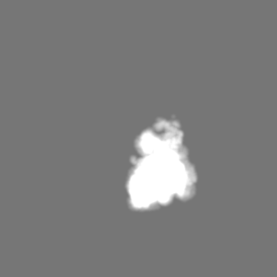
| Images |  emd_11751.png emd_11751.png | 107.5 KB | ||
| Masks | emd_11751_msk_1.map | 244.1 MB | Mask map | |
| Filedesc metadata | emd-11751.cif.gz | 8.1 KB | ||
| Others | emd_11751_half_map_1.map.gzemd_11751_half_map_2.map.gz | 163.6 MB 163.7 MB | ||
| Archive directory |  http://ftp.pdbj.org/pub/emdb/structures/EMD-11751ftp://ftp.pdbj.org/pub/emdb/structures/EMD-11751 http://ftp.pdbj.org/pub/emdb/structures/EMD-11751ftp://ftp.pdbj.org/pub/emdb/structures/EMD-11751 | HTTPS FTP |
-Related structure data
| Related structure data |  7af3MC  7af5C 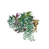 7af8C  7afaC 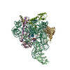 7afdC 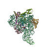 7afhC 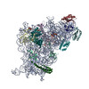 7afiC  7afkC 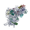 7aflC 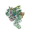 7afnC 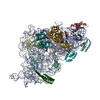 7afoC 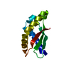 7afqC 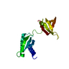 7afrC 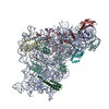 7bodC 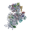 7boeC 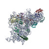 7bofC 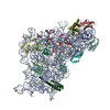 7bogC 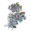 7bohC 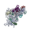 7boiC 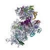 7narC 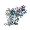 7nasC 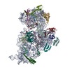 7natC 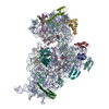 7nauC 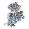 7navC  7nawC 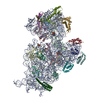 7naxC M: atomic model generated by this map C: citing same article ( |
|---|---|
| Similar structure data |


-Links
| EMDB pages | EMDB (EBI/PDBe) / EMDataResource |
|---|---|
| Related items in Molecule of the Month |


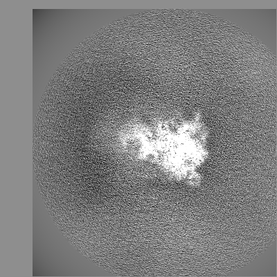
-Map
| File | Download / File: emd_11751.map.gz / Format: CCP4 / Size: 244.1 MB / Type: IMAGE STORED AS FLOATING POINT NUMBER (4 BYTES) | ||||||||||||||||||||||||||||||||||||||||||||||||||||||||||||
|---|---|---|---|---|---|---|---|---|---|---|---|---|---|---|---|---|---|---|---|---|---|---|---|---|---|---|---|---|---|---|---|---|---|---|---|---|---|---|---|---|---|---|---|---|---|---|---|---|---|---|---|---|---|---|---|---|---|---|---|---|---|
| Annotation | Full map | ||||||||||||||||||||||||||||||||||||||||||||||||||||||||||||
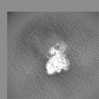
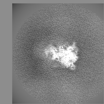
| Projections & slices | Image control
Images are generated by Spider. | ||||||||||||||||||||||||||||||||||||||||||||||||||||||||||||
| Voxel size | X=Y=Z: 1.05 Å | ||||||||||||||||||||||||||||||||||||||||||||||||||||||||||||
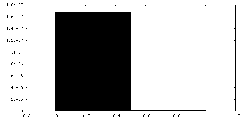
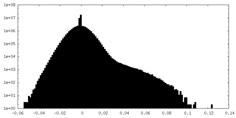
| Density |
| ||||||||||||||||||||||||||||||||||||||||||||||||||||||||||||
| Symmetry | Space group: 1 | ||||||||||||||||||||||||||||||||||||||||||||||||||||||||||||
| Details | EMDB XML:
CCP4 map header:
| ||||||||||||||||||||||||||||||||||||||||||||||||||||||||||||
 Z (Sec.)
 Y (Row.)
 X (Col.)


-Supplemental data
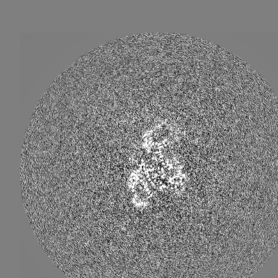
-Mask #1
| File | emd_11751_msk_1.map | ||||||||||||
|---|---|---|---|---|---|---|---|---|---|---|---|---|---|
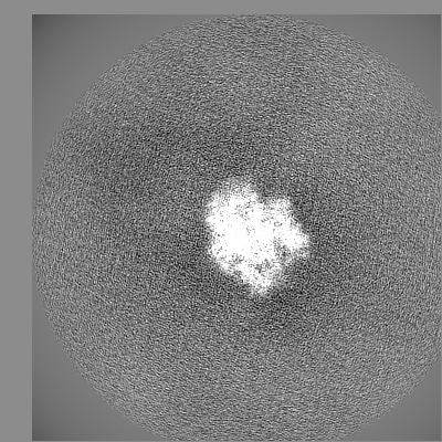
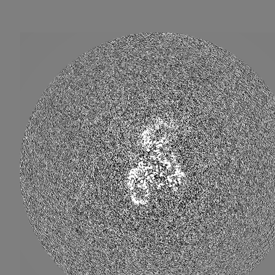
| Projections & Slices |
| ||||||||||||
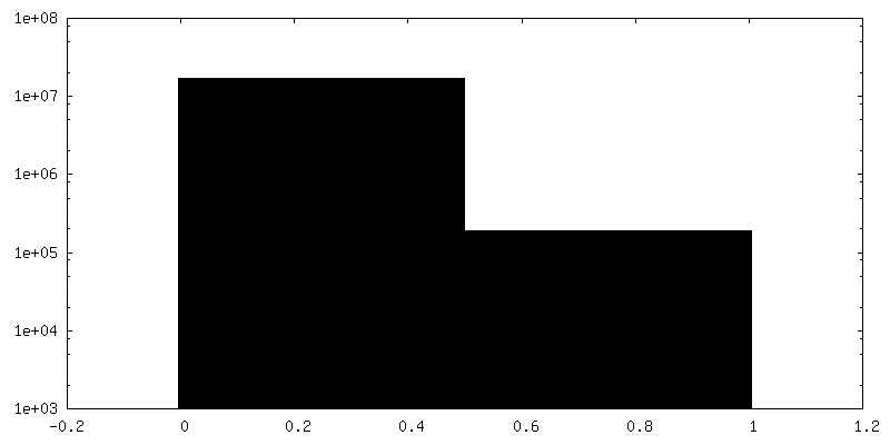
| Density Histograms |


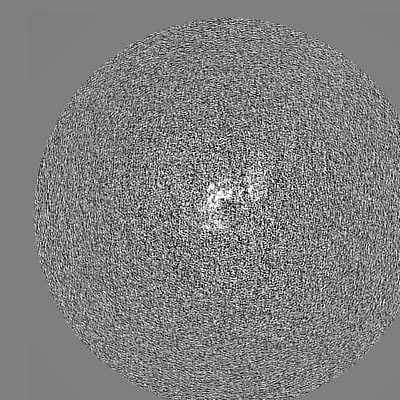
-Half map: half map 1
| File | emd_11751_half_map_1.map | ||||||||||||
|---|---|---|---|---|---|---|---|---|---|---|---|---|---|
| Annotation | half map 1 | ||||||||||||
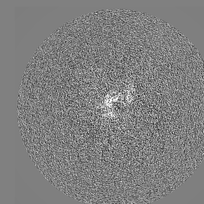
| Projections & Slices |
| ||||||||||||
| Density Histograms |


-Half map: half map 2
| File | emd_11751_half_map_2.map | ||||||||||||
|---|---|---|---|---|---|---|---|---|---|---|---|---|---|
| Annotation | half map 2 | ||||||||||||
| Projections & Slices |
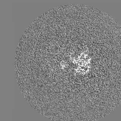
| ||||||||||||
| Density Histograms |


- Sample components
+Entire : Bacterial 30S ribosomal subunit assembly complex state M (head domain)
+Supramolecule #1: Bacterial 30S ribosomal subunit assembly complex state M (head domain)
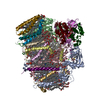
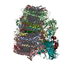
+Macromolecule #1: 16S rRNA (head)
+Macromolecule #2: 30S ribosomal protein S3
+Macromolecule #3: 30S ribosomal protein S7
+Macromolecule #4: 30S ribosomal protein S9
+Macromolecule #5: 30S ribosomal protein S10
+Macromolecule #6: 30S ribosomal protein S13
+Macromolecule #7: 30S ribosomal protein S14
+Macromolecule #8: 30S ribosomal protein S19
+Macromolecule #9: 30S ribosomal protein S2
+Macromolecule #10: MAGNESIUM ION
+Macromolecule #11: ZINC ION
-Experimental details
-Structure determination
| Method | cryo EM |
|---|---|
 Processing Processing | single particle reconstruction |
| Aggregation state | particle |
-Sample preparation
| Buffer | pH: 7.8 |
|---|---|
| Grid | Model: Quantifoil R2/2 / Support film - Material: CARBON / Support film - topology: CONTINUOUS |
| Vitrification | Cryogen name: ETHANE / Chamber humidity: 100 % / Chamber temperature: 277 K / Instrument: FEI VITROBOT MARK III |
- Electron microscopy
| Microscope | FEI TITAN KRIOS |
|---|---|
| Image recording | Film or detector model: GATAN K2 SUMMIT (4k x 4k) / Detector mode: COUNTING / Average electron dose: 38.8 e/Å2 |
| Electron beam | Acceleration voltage: 300 kV / Electron source:  FIELD EMISSION GUN FIELD EMISSION GUN |
| Electron optics | Illumination mode: SPOT SCAN / Imaging mode: BRIGHT FIELD |
| Sample stage | Specimen holder model: FEI TITAN KRIOS AUTOGRID HOLDER / Cooling holder cryogen: NITROGEN |
| Experimental equipment |  Model: Titan Krios / Image courtesy: FEI Company |