COPI-independent Golgi-to-ER retrograde traffic / HSP90 chaperone cycle for steroid hormone receptors (SHR) in the presence of ligand / Amplification of signal from unattached kinetochores via a MAD2 inhibitory signal / COPI-mediated anterograde transport / Aggrephagy / positive regulation of intracellular transport / Mitotic Prometaphase / EML4 and NUDC in mitotic spindle formation / cilium movement / regulation of metaphase plate congression ...COPI-independent Golgi-to-ER retrograde traffic / HSP90 chaperone cycle for steroid hormone receptors (SHR) in the presence of ligand / Amplification of signal from unattached kinetochores via a MAD2 inhibitory signal / COPI-mediated anterograde transport / Aggrephagy / positive regulation of intracellular transport / Mitotic Prometaphase / EML4 and NUDC in mitotic spindle formation / cilium movement / regulation of metaphase plate congression / Resolution of Sister Chromatid Cohesion / establishment of spindle localization / axonemal dynein complex / positive regulation of spindle assembly / RHO GTPases Activate Formins / Separation of Sister Chromatids / Loss of Nlp from mitotic centrosomes / Recruitment of mitotic centrosome proteins and complexes / Loss of proteins required for interphase microtubule organization from the centrosome / Recruitment of NuMA to mitotic centrosomes / Anchoring of the basal body to the plasma membrane / AURKA Activation by TPX2 / Regulation of PLK1 Activity at G2/M Transition / manchette / dynein complex / MHC class II antigen presentation / P-body assembly / minus-end-directed microtubule motor activity / dynein light intermediate chain binding / cytoplasmic dynein complex / Microtubule-dependent trafficking of connexons from Golgi to the plasma membrane / Hedgehog 'off' state / Cilium Assembly / Intraflagellar transport / COPI-dependent Golgi-to-ER retrograde traffic / Carboxyterminal post-translational modifications of tubulin / RHOH GTPase cycle / Sealing of the nuclear envelope (NE) by ESCRT-III / Kinesins / PKR-mediated signaling / The role of GTSE1 in G2/M progression after G2 checkpoint / Aggrephagy / Resolution of Sister Chromatid Cohesion / Mitotic Prometaphase / EML4 and NUDC in mitotic spindle formation / Separation of Sister Chromatids / retrograde axonal transport / RHO GTPases activate IQGAPs / RHO GTPases Activate Formins / nuclear migration / Recruitment of NuMA to mitotic centrosomes / HSP90 chaperone cycle for steroid hormone receptors (SHR) in the presence of ligand / MHC class II antigen presentation / COPI-mediated anterograde transport / dynein intermediate chain binding / microtubule-based movement / cytoplasmic microtubule / stress granule assembly / cytoplasmic microtubule organization / regulation of mitotic spindle organization / axon cytoplasm / Neutrophil degranulation / mitotic spindle organization / filopodium / 加水分解酵素; 酸無水物に作用; GTPに作用・細胞または細胞小器官の運動に関与 / structural constituent of cytoskeleton / microtubule cytoskeleton organization / microtubule cytoskeleton / nuclear envelope / positive regulation of cold-induced thermogenesis / mitotic cell cycle / cell cortex / microtubule / cell division / axon / GTPase activity / centrosome / neuronal cell body / GTP binding / ATP hydrolysis activity / ATP binding / metal ion binding / cytoplasm 類似検索 - 分子機能
Dynein heavy chain, AAA 5 extension domain / Dynein heavy chain AAA lid domain / Dynein heavy chain, C-terminal domain / Dynein heavy chain, C-terminal domain, barrel region / Dynein heavy chain C-terminal domain / P-loop containing dynein motor region / Dynein heavy chain, tail / Dynein heavy chain, N-terminal region 1 / Dynein heavy chain / Dynein heavy chain region D6 P-loop domain ...Dynein heavy chain, AAA 5 extension domain / Dynein heavy chain AAA lid domain / Dynein heavy chain, C-terminal domain / Dynein heavy chain, C-terminal domain, barrel region / Dynein heavy chain C-terminal domain / P-loop containing dynein motor region / Dynein heavy chain, tail / Dynein heavy chain, N-terminal region 1 / Dynein heavy chain / Dynein heavy chain region D6 P-loop domain / Dynein heavy chain, linker / Dynein heavy chain, AAA module D4 / Dynein heavy chain, coiled coil stalk / Dynein heavy chain, hydrolytic ATP-binding dynein motor region / Dynein heavy chain, ATP-binding dynein motor region / Dynein heavy chain AAA lid domain / Dynein heavy chain AAA lid domain superfamily / Dynein heavy chain, domain 2, N-terminal / Dynein heavy chain, linker, subdomain 3 / Dynein heavy chain, AAA1 domain, small subdomain / Dynein heavy chain region D6 P-loop domain / Dynein heavy chain, N-terminal region 2 / Hydrolytic ATP binding site of dynein motor region / Microtubule-binding stalk of dynein motor / P-loop containing dynein motor region D4 / ATP-binding dynein motor region / Dynein heavy chain AAA lid domain / Tubulin-beta mRNA autoregulation signal. / Alpha tubulin / Beta tubulin, autoregulation binding site / Beta tubulin / Tubulin / Tubulin, C-terminal / Tubulin C-terminal domain / Tubulin, conserved site / Tubulin subunits alpha, beta, and gamma signature. / Tubulin/FtsZ family, C-terminal domain / Tubulin/FtsZ-like, C-terminal domain / Tubulin/FtsZ, C-terminal / Tubulin/FtsZ, 2-layer sandwich domain / Tubulin/FtsZ family, GTPase domain / Tubulin/FtsZ family, GTPase domain / Tubulin/FtsZ, GTPase domain / Tubulin/FtsZ, GTPase domain superfamily / ATPases associated with a variety of cellular activities / AAA+ ATPase domain / P-loop containing nucleoside triphosphate hydrolase 類似検索 - ドメイン・相同性
Tubulin beta chain / Tubulin alpha-1B chain / MKIAA0325 protein / Cytoplasmic dynein 1 heavy chain 1 類似検索 - 構成要素
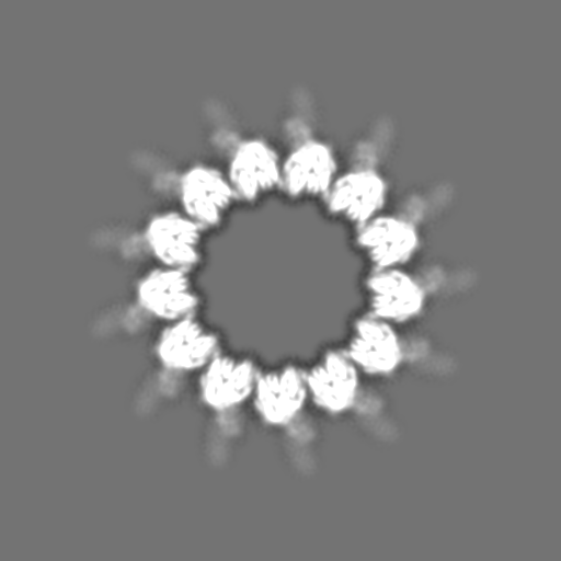
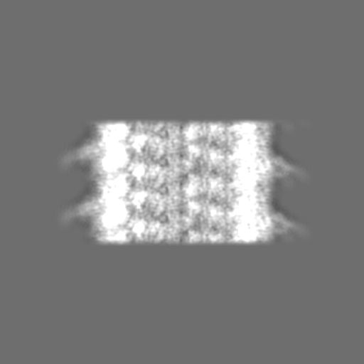
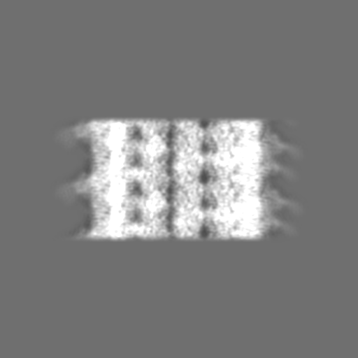
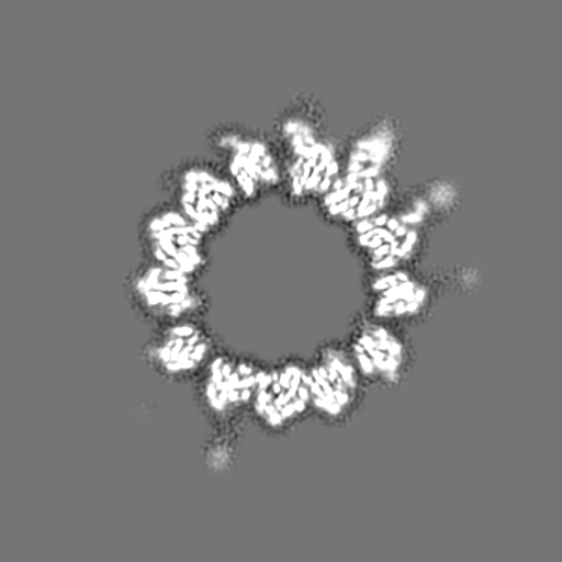
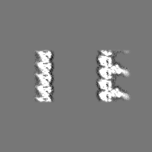
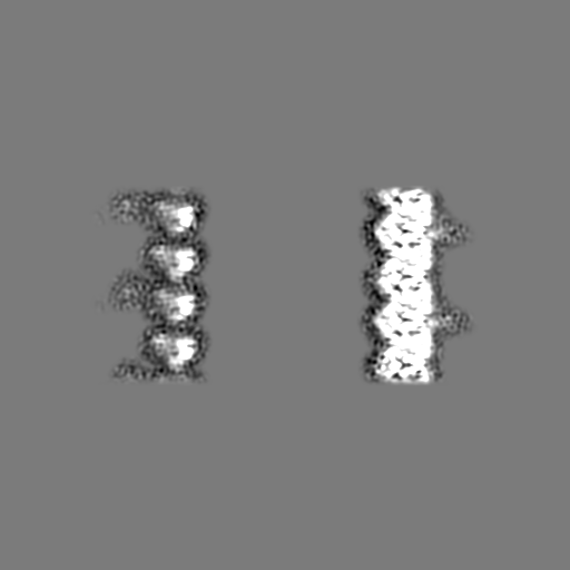
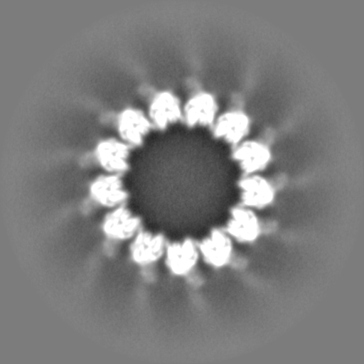
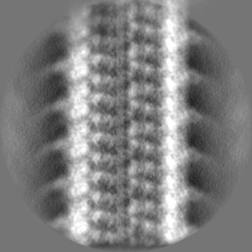
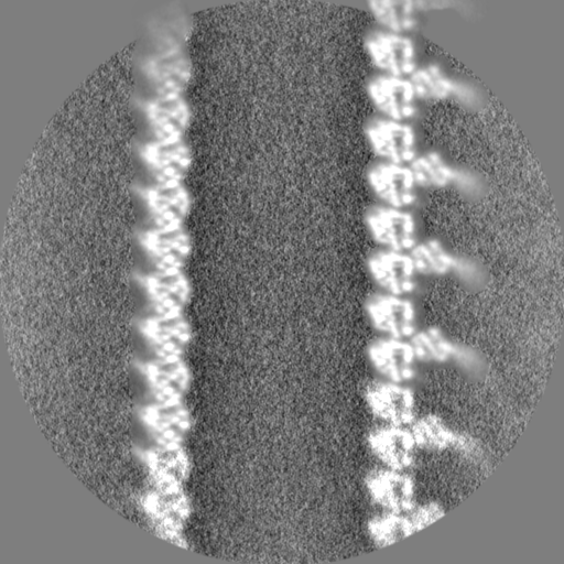
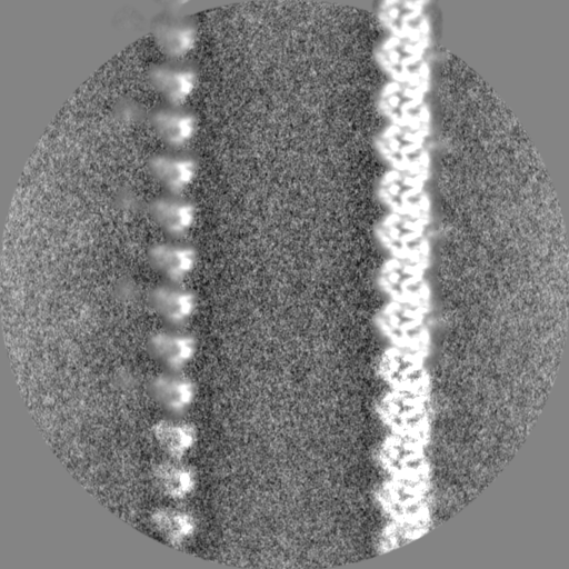
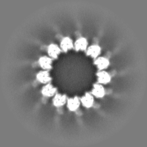
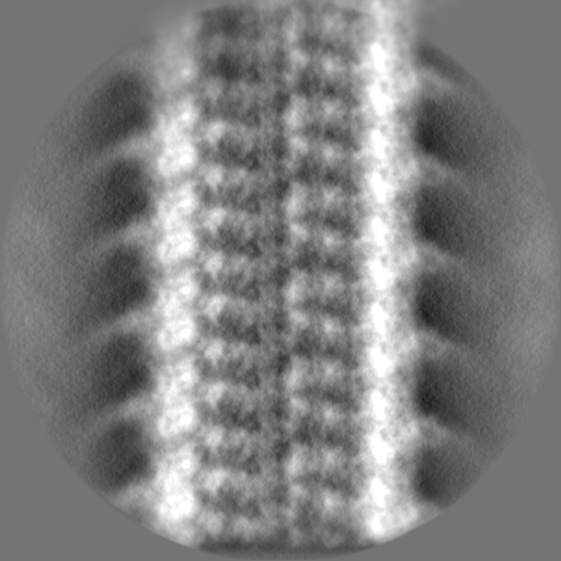
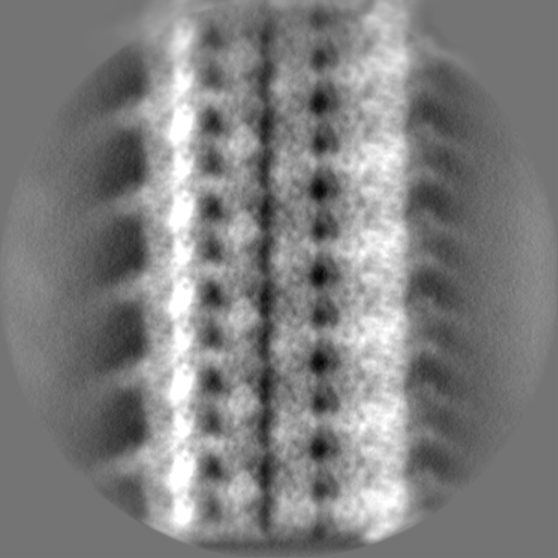
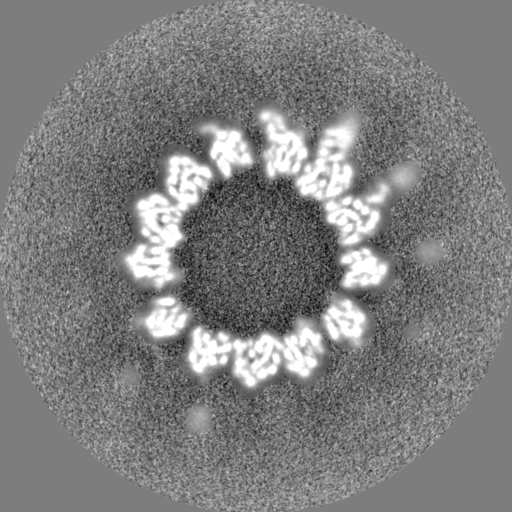
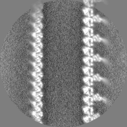
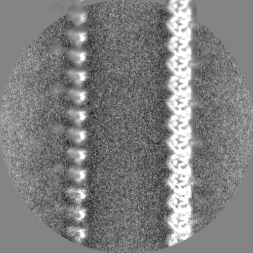
ジャーナル: Elife / 年: 2019 タイトル: Cryo-EM of dynein microtubule-binding domains shows how an axonemal dynein distorts the microtubule. 著者: Samuel E Lacey / Shaoda He / Sjors Hw Scheres / Andrew P Carter / 要旨: Dyneins are motor proteins responsible for transport in the cytoplasm and the beating of axonemes in cilia and flagella. They bind and release microtubules via a compact microtubule-binding domain ...Dyneins are motor proteins responsible for transport in the cytoplasm and the beating of axonemes in cilia and flagella. They bind and release microtubules via a compact microtubule-binding domain (MTBD) at the end of a coiled-coil stalk. We address how cytoplasmic and axonemal dynein MTBDs bind microtubules at near atomic resolution. We decorated microtubules with MTBDs of cytoplasmic dynein-1 and axonemal dynein DNAH7 and determined their cryo-EM structures using helical Relion. The majority of the MTBD is rigid upon binding, with the transition to the high-affinity state controlled by the movement of a single helix at the MTBD interface. DNAH7 contains an 18-residue insertion, found in many axonemal dyneins, that contacts the adjacent protofilament. Unexpectedly, we observe that DNAH7, but not dynein-1, induces large distortions in the microtubule cross-sectional curvature. This raises the possibility that dynein coordination in axonemes is mediated via conformational changes in the microtubule.
 ムービー
ムービー コントローラー


 データを開く
 基本情報
基本情報 マップデータ
 試料
試料 キーワード
キーワード 機能・相同性情報
 データ登録者
 英国, 2件
  引用
引用 構造の表示
構造の表示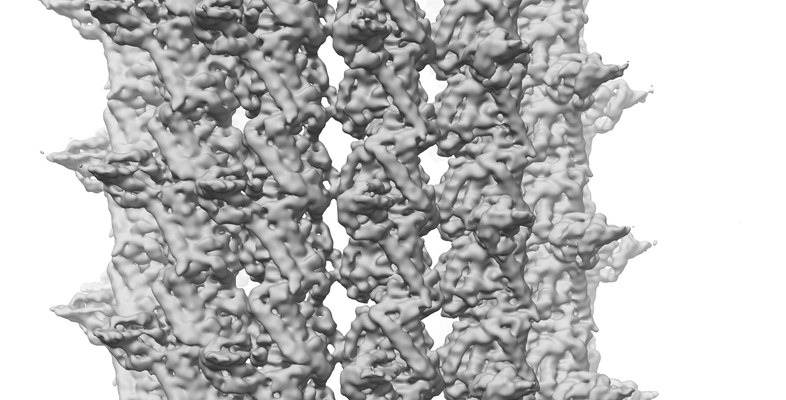
 ダウンロードとリンク
 emd_10061.png
 http://ftp.pdbj.org/pub/emdb/structures/EMD-10061


 Z
Z Y
Y X
X


 試料の構成要素


 解析
解析 電子顕微鏡法
 FIELD EMISSION GUN

