 ムービー
ムービー コントローラー


+ データを開く

- 基本情報
基本情報
| 登録情報 | データベース: EMDB / ID: EMD-1005 | |||||||||
|---|---|---|---|---|---|---|---|---|---|---|
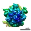
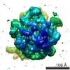
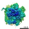
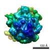
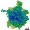
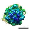
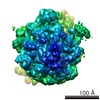
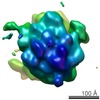
| タイトル | Structure of the Escherichia coli ribosomal termination complex with release factor 2. | |||||||||
 マップデータ マップデータ | E. coli ribosomal termination complex with release factor RF2. | |||||||||
 試料 試料 |
| |||||||||
| 機能・相同性 |  機能・相同性情報 機能・相同性情報translation release factor activity, codon specific / translational termination / ribosomal large subunit biogenesis / response to radiation / ribosomal large subunit assembly / regulation of translation / ribosomal small subunit biogenesis / ribosomal small subunit assembly / small ribosomal subunit / small ribosomal subunit rRNA binding ...translation release factor activity, codon specific / translational termination / ribosomal large subunit biogenesis / response to radiation / ribosomal large subunit assembly / regulation of translation / ribosomal small subunit biogenesis / ribosomal small subunit assembly / small ribosomal subunit / small ribosomal subunit rRNA binding / transferase activity / 5S rRNA binding / large ribosomal subunit rRNA binding / cytosolic small ribosomal subunit / cytosolic large ribosomal subunit / tRNA binding / cytoplasmic translation / rRNA binding / negative regulation of translation / ribosome / structural constituent of ribosome / ribonucleoprotein complex / translation / mRNA binding / zinc ion binding / metal ion binding / cytoplasm / cytosol 類似検索 - 分子機能 | |||||||||
| 生物種 |  | |||||||||
| 手法 | 単粒子再構成法 / クライオ電子顕微鏡法 / ネガティブ染色法 / 解像度: 14.0 Å | |||||||||
 データ登録者 データ登録者 | Klaholz BP / Pape T / Zavialov AV / Myasnikov AG / Orlova EV / Vestergaard B / Ehrenberg M / van Heel M | |||||||||
 引用 引用 | ジャーナル: Nature / 年: 2003 タイトル: Structure of the Escherichia coli ribosomal termination complex with release factor 2. 著者: Bruno P Klaholz / Tillmann Pape / Andrey V Zavialov / Alexander G Myasnikov / Elena V Orlova / Bente Vestergaard / Måns Ehrenberg / Marin van Heel /  要旨: Termination of protein synthesis occurs when the messenger RNA presents a stop codon in the ribosomal aminoacyl (A) site. Class I release factor proteins (RF1 or RF2) are believed to recognize stop ...Termination of protein synthesis occurs when the messenger RNA presents a stop codon in the ribosomal aminoacyl (A) site. Class I release factor proteins (RF1 or RF2) are believed to recognize stop codons via tripeptide motifs, leading to release of the completed polypeptide chain from its covalent attachment to transfer RNA in the ribosomal peptidyl (P) site. Class I RFs possess a conserved GGQ amino-acid motif that is thought to be involved directly in protein-transfer-RNA bond hydrolysis. Crystal structures of bacterial and eukaryotic class I RFs have been determined, but the mechanism of stop codon recognition and peptidyl-tRNA hydrolysis remains unclear. Here we present the structure of the Escherichia coli ribosome in a post-termination complex with RF2, obtained by single-particle cryo-electron microscopy (cryo-EM). Fitting the known 70S and RF2 structures into the electron density map reveals that RF2 adopts a different conformation on the ribosome when compared with the crystal structure of the isolated protein. The amino-terminal helical domain of RF2 contacts the factor-binding site of the ribosome, the 'SPF' loop of the protein is situated close to the mRNA, and the GGQ-containing domain of RF2 interacts with the peptidyl-transferase centre (PTC). By connecting the ribosomal decoding centre with the PTC, RF2 functionally mimics a tRNA molecule in the A site. Translational termination in eukaryotes is likely to be based on a similar mechanism. | |||||||||
| 履歴 |
|
- 構造の表示
構造の表示
| ムービー |
ムービービューア |
|---|---|
| 構造ビューア | EMマップ: SurfViewMolmilJmol/JSmol |
| 添付画像 |

- ダウンロードとリンク
-EMDBアーカイブ
| マップデータ | emd_1005.map.gz | 2.8 MB | EMDBマップデータ形式 | |
|---|---|---|---|---|
| ヘッダ (付随情報) | emd-1005-v30.xmlemd-1005.xml | 12.5 KB 12.5 KB | 表示 表示 | EMDBヘッダ |
| 画像 |  1005.gif 1005.gif | 27.4 KB | ||
| アーカイブディレクトリ |  http://ftp.pdbj.org/pub/emdb/structures/EMD-1005ftp://ftp.pdbj.org/pub/emdb/structures/EMD-1005 http://ftp.pdbj.org/pub/emdb/structures/EMD-1005ftp://ftp.pdbj.org/pub/emdb/structures/EMD-1005 | HTTPS FTP |
-検証レポート
| 文書・要旨 | emd_1005_validation.pdf.gz | 334.3 KB | 表示 | EMDB検証レポート |
|---|---|---|---|---|
| 文書・詳細版 | emd_1005_full_validation.pdf.gz | 333.9 KB | 表示 | |
| XML形式データ | emd_1005_validation.xml.gz | 5.2 KB | 表示 | |
| アーカイブディレクトリ | https://ftp.pdbj.org/pub/emdb/validation_reports/EMD-1005ftp://ftp.pdbj.org/pub/emdb/validation_reports/EMD-1005 | HTTPS FTP |
-関連構造データ


-リンク
| EMDBのページ | EMDB (EBI/PDBe) / EMDataResource |
|---|---|
| 「今月の分子」の関連する項目 |


-マップ
| ファイル | ダウンロード / ファイル: emd_1005.map.gz / 形式: CCP4 / 大きさ: 6.4 MB / タイプ: IMAGE STORED AS FLOATING POINT NUMBER (4 BYTES) | ||||||||||||||||||||||||||||||||||||||||||||||||||||||||||||
|---|---|---|---|---|---|---|---|---|---|---|---|---|---|---|---|---|---|---|---|---|---|---|---|---|---|---|---|---|---|---|---|---|---|---|---|---|---|---|---|---|---|---|---|---|---|---|---|---|---|---|---|---|---|---|---|---|---|---|---|---|---|
| 注釈 | E. coli ribosomal termination complex with release factor RF2. | ||||||||||||||||||||||||||||||||||||||||||||||||||||||||||||
| ボクセルのサイズ | X=Y=Z: 2.419 Å | ||||||||||||||||||||||||||||||||||||||||||||||||||||||||||||
| 密度 |
| ||||||||||||||||||||||||||||||||||||||||||||||||||||||||||||
| 対称性 | 空間群: 1 | ||||||||||||||||||||||||||||||||||||||||||||||||||||||||||||
| 詳細 | EMDB XML:
CCP4マップ ヘッダ情報:
| ||||||||||||||||||||||||||||||||||||||||||||||||||||||||||||
-添付データ
- 試料の構成要素
-全体 : release factor RF2 bound to E. coli ribosomes
| 全体 | 名称: release factor RF2 bound to E. coli ribosomes |
|---|---|
| 要素 |
|
-超分子 #1000: release factor RF2 bound to E. coli ribosomes
| 超分子 | 名称: release factor RF2 bound to E. coli ribosomes / タイプ: sample / ID: 1000 / 集合状態: monomer / Number unique components: 2 |
|---|---|
| 分子量 | 実験値: 2.3 MDa / 手法: sedimentation |
-超分子 #1: ribosome
| 超分子 | 名称: ribosome / タイプ: complex / ID: 1 / Name.synonym: ribosome 詳細: Proteins L7/L12 of the LSU 50S subunit are not seen in the map 組換発現: No / Ribosome-details: ribosome-prokaryote: LSU 50S |
|---|---|
| 由来(天然) | 生物種: |
| 分子量 | 実験値: 2.3 MDa / 理論値: 2.3 MDa |
-分子 #1: release factor 2
| 分子 | 名称: release factor 2 / タイプ: protein_or_peptide / ID: 1 / Name.synonym: RF2 / コピー数: 1 / 集合状態: monomer / 組換発現: Yes |
|---|---|
| 由来(天然) | 生物種: |
| 分子量 | 実験値: 40 KDa / 理論値: 40 KDa |
| 組換発現 | 生物種: |
| 配列 | InterPro: Peptide chain release factor 2 |
-実験情報
-構造解析
| 手法 | ネガティブ染色法, クライオ電子顕微鏡法 |
|---|---|
 解析 解析 | 単粒子再構成法 |
| 試料の集合状態 | particle |
-試料調製
| 濃度 | 0.15 mg/mL |
|---|---|
| 緩衝液 | pH: 7.5 / 詳細: polymix buffer |
| 染色 | タイプ: NEGATIVE / 詳細: no staining, cryo-EM with holey carbon grids |
| 凍結 | 凍結剤: ETHANE / チャンバー内湿度: 45 % / チャンバー内温度: 20 K / 装置: HOMEMADE PLUNGER / 詳細: Vitrification instrument: home-made cryo-plunger / 手法: blot for 2 seconds before plunging |
- 電子顕微鏡法
| 顕微鏡 | FEI/PHILIPS CM200FEG/ST |
|---|---|
| 温度 | 最低: 100 K / 最高: 102 K / 平均: 100 K |
| 日付 | 2001年9月6日 |
| 撮影 | カテゴリ: FILM / フィルム・検出器のモデル: KODAK SO-163 FILM / デジタル化 - スキャナー: PATCHWORK DENSITOMETER / デジタル化 - サンプリング間隔: 3 µm / 実像数: 78 / 平均電子線量: 10 e/Å2 / ビット/ピクセル: 12 |
| 電子線 | 加速電圧: 200 kV / 電子線源:  FIELD EMISSION GUN FIELD EMISSION GUN |
| 電子光学系 | 倍率(補正後): 48000 / 照射モード: FLOOD BEAM / 撮影モード: BRIGHT FIELD / Cs: 2.1 mm / 最大 デフォーカス(公称値): 2.9 µm / 最小 デフォーカス(公称値): 0.5 µm / 倍率(公称値): 50000 |
| 試料ステージ | 試料ホルダー: Side entry liquid nitrogen-cooled cryo specimen holder. 試料ホルダーモデル: GATAN LIQUID NITROGEN |
-画像解析
| CTF補正 | 詳細: each particle |
|---|---|
| 最終 再構成 | 想定した対称性 - 点群: C1 (非対称) / アルゴリズム: OTHER / 解像度のタイプ: BY AUTHOR / 解像度: 14.0 Å / 解像度の算出法: OTHER / ソフトウェア - 名称: IMAGIC / 詳細: exact filtered back-projection / 使用した粒子像数: 15800 |
| 最終 角度割当 | 詳細: beta gamma |
| 最終 2次元分類 | クラス数: 103 |
-原子モデル構築 1
| 初期モデル | PDB ID:  1gix |
|---|---|
| ソフトウェア | 名称: eye |
| 詳細 | Protocol: rigid body. rigid body of three individual domains keeping the connectivity; linkers betweenn domains defined based on temperature factors of crystal structure, conserved Gly and Pro residues, proteolytic sites, and global domain architecture |
| 精密化 | 空間: REAL / プロトコル: RIGID BODY FIT 当てはまり具合の基準: best visual fit using the program O |
| 得られたモデル | 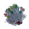 PDB-1ml5: |
-原子モデル構築 2
| 初期モデル | PDB ID: 1giy |
|---|---|
| ソフトウェア | 名称: eye |
| 詳細 | Protocol: rigid body. rigid body of three individual domains keeping the connectivity; linkers betweenn domains defined based on temperature factors of crystal structure, conserved Gly and Pro residues, proteolytic sites, and global domain architecture |
| 精密化 | 空間: REAL / プロトコル: RIGID BODY FIT 当てはまり具合の基準: best visual fit using the program O |
| 得られたモデル | PDB-1ml5: |
