 Movie
Movie Controller

[English] 日本語
 Yorodumi
- EMDB-1005: Structure of the Escherichia coli ribosomal termination complex w... -
+ Open data

- Basic information
| Entry | Database: EMDB / ID: EMD-1005 | |||||||||
|---|---|---|---|---|---|---|---|---|---|---|
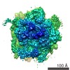
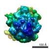
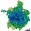
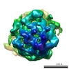
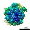
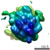
| Title | Structure of the Escherichia coli ribosomal termination complex with release factor 2. | |||||||||
 Map data Map data | E. coli ribosomal termination complex with release factor RF2. | |||||||||
 Sample Sample |
| |||||||||
| Function / homology |  Function and homology information Function and homology informationtranslation release factor activity, codon specific / translational termination / response to radiation / regulation of translation / large ribosomal subunit / transferase activity / ribosomal small subunit assembly / ribosomal small subunit biogenesis / 5S rRNA binding / ribosomal large subunit assembly ...translation release factor activity, codon specific / translational termination / response to radiation / regulation of translation / large ribosomal subunit / transferase activity / ribosomal small subunit assembly / ribosomal small subunit biogenesis / 5S rRNA binding / ribosomal large subunit assembly / small ribosomal subunit / small ribosomal subunit rRNA binding / cytosolic small ribosomal subunit / large ribosomal subunit rRNA binding / cytosolic large ribosomal subunit / cytoplasmic translation / tRNA binding / negative regulation of translation / rRNA binding / structural constituent of ribosome / ribosome / translation / ribonucleoprotein complex / viral translational frameshifting / mRNA binding / zinc ion binding / metal ion binding / cytoplasm / cytosol Similarity search - Function | |||||||||
| Biological species |  | |||||||||
| Method | single particle reconstruction / cryo EM / negative staining / Resolution: 14.0 Å | |||||||||
 Authors Authors | Klaholz BP / Pape T / Zavialov AV / Myasnikov AG / Orlova EV / Vestergaard B / Ehrenberg M / van Heel M | |||||||||
 Citation Citation | Journal: Nature / Year: 2003 Title: Structure of the Escherichia coli ribosomal termination complex with release factor 2. Authors: Bruno P Klaholz / Tillmann Pape / Andrey V Zavialov / Alexander G Myasnikov / Elena V Orlova / Bente Vestergaard / Måns Ehrenberg / Marin van Heel /  Abstract: Termination of protein synthesis occurs when the messenger RNA presents a stop codon in the ribosomal aminoacyl (A) site. Class I release factor proteins (RF1 or RF2) are believed to recognize stop ...Termination of protein synthesis occurs when the messenger RNA presents a stop codon in the ribosomal aminoacyl (A) site. Class I release factor proteins (RF1 or RF2) are believed to recognize stop codons via tripeptide motifs, leading to release of the completed polypeptide chain from its covalent attachment to transfer RNA in the ribosomal peptidyl (P) site. Class I RFs possess a conserved GGQ amino-acid motif that is thought to be involved directly in protein-transfer-RNA bond hydrolysis. Crystal structures of bacterial and eukaryotic class I RFs have been determined, but the mechanism of stop codon recognition and peptidyl-tRNA hydrolysis remains unclear. Here we present the structure of the Escherichia coli ribosome in a post-termination complex with RF2, obtained by single-particle cryo-electron microscopy (cryo-EM). Fitting the known 70S and RF2 structures into the electron density map reveals that RF2 adopts a different conformation on the ribosome when compared with the crystal structure of the isolated protein. The amino-terminal helical domain of RF2 contacts the factor-binding site of the ribosome, the 'SPF' loop of the protein is situated close to the mRNA, and the GGQ-containing domain of RF2 interacts with the peptidyl-transferase centre (PTC). By connecting the ribosomal decoding centre with the PTC, RF2 functionally mimics a tRNA molecule in the A site. Translational termination in eukaryotes is likely to be based on a similar mechanism. | |||||||||
| History |
|
- Structure visualization
| Movie |
Movie viewer |
|---|---|
| Structure viewer | EM map: SurfViewMolmilJmol/JSmol |
| Supplemental images |

- Downloads & links
-EMDB archive
| Map data | emd_1005.map.gz | 2.8 MB | EMDB map data format | |
|---|---|---|---|---|
| Header (meta data) | emd-1005-v30.xmlemd-1005.xml | 12.5 KB 12.5 KB | Display Display | EMDB header |
| Images | 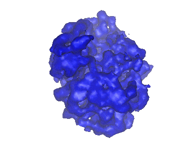 1005.gif 1005.gif | 27.4 KB | ||
| Archive directory |  http://ftp.pdbj.org/pub/emdb/structures/EMD-1005ftp://ftp.pdbj.org/pub/emdb/structures/EMD-1005 http://ftp.pdbj.org/pub/emdb/structures/EMD-1005ftp://ftp.pdbj.org/pub/emdb/structures/EMD-1005 | HTTPS FTP |
-Related structure data
| Related structure data |  1ml5MC M: atomic model generated by this map C: citing same article ( |
|---|---|
| Similar structure data |


-Links
| EMDB pages | EMDB (EBI/PDBe) / EMDataResource |
|---|---|
| Related items in Molecule of the Month |


-Map
| File | Download / File: emd_1005.map.gz / Format: CCP4 / Size: 6.4 MB / Type: IMAGE STORED AS FLOATING POINT NUMBER (4 BYTES) | ||||||||||||||||||||||||||||||||||||||||||||||||||||||||||||
|---|---|---|---|---|---|---|---|---|---|---|---|---|---|---|---|---|---|---|---|---|---|---|---|---|---|---|---|---|---|---|---|---|---|---|---|---|---|---|---|---|---|---|---|---|---|---|---|---|---|---|---|---|---|---|---|---|---|---|---|---|---|
| Annotation | E. coli ribosomal termination complex with release factor RF2. | ||||||||||||||||||||||||||||||||||||||||||||||||||||||||||||
| Projections & slices | Image control
Images are generated by Spider. | ||||||||||||||||||||||||||||||||||||||||||||||||||||||||||||
| Voxel size | X=Y=Z: 2.419 Å | ||||||||||||||||||||||||||||||||||||||||||||||||||||||||||||
| Density |
| ||||||||||||||||||||||||||||||||||||||||||||||||||||||||||||
| Symmetry | Space group: 1 | ||||||||||||||||||||||||||||||||||||||||||||||||||||||||||||
| Details | EMDB XML:
CCP4 map header:
| ||||||||||||||||||||||||||||||||||||||||||||||||||||||||||||
 Z (Sec.)
 Y (Row.)
 X (Col.)


-Supplemental data
- Sample components
-Entire : release factor RF2 bound to E. coli ribosomes
| Entire | Name: release factor RF2 bound to E. coli ribosomes |
|---|---|
| Components |
|
-Supramolecule #1000: release factor RF2 bound to E. coli ribosomes
| Supramolecule | Name: release factor RF2 bound to E. coli ribosomes / type: sample / ID: 1000 / Oligomeric state: monomer / Number unique components: 2 |
|---|---|
| Molecular weight | Experimental: 2.3 MDa / Method: sedimentation |
-Supramolecule #1: ribosome
| Supramolecule | Name: ribosome / type: complex / ID: 1 / Name.synonym: ribosome Details: Proteins L7/L12 of the LSU 50S subunit are not seen in the map Recombinant expression: No / Ribosome-details: ribosome-prokaryote: LSU 50S |
|---|---|
| Source (natural) | Organism: |
| Molecular weight | Experimental: 2.3 MDa / Theoretical: 2.3 MDa |
-Macromolecule #1: release factor 2
| Macromolecule | Name: release factor 2 / type: protein_or_peptide / ID: 1 / Name.synonym: RF2 / Number of copies: 1 / Oligomeric state: monomer / Recombinant expression: Yes |
|---|---|
| Source (natural) | Organism: |
| Molecular weight | Experimental: 40 KDa / Theoretical: 40 KDa |
| Recombinant expression | Organism: |
| Sequence | InterPro: Peptide chain release factor 2 |
-Experimental details
-Structure determination
| Method | negative staining, cryo EM |
|---|---|
 Processing Processing | single particle reconstruction |
| Aggregation state | particle |
-Sample preparation
| Concentration | 0.15 mg/mL |
|---|---|
| Buffer | pH: 7.5 / Details: polymix buffer |
| Staining | Type: NEGATIVE / Details: no staining, cryo-EM with holey carbon grids |
| Vitrification | Cryogen name: ETHANE / Chamber humidity: 45 % / Chamber temperature: 20 K / Instrument: HOMEMADE PLUNGER / Details: Vitrification instrument: home-made cryo-plunger / Method: blot for 2 seconds before plunging |
- Electron microscopy
| Microscope | FEI/PHILIPS CM200FEG/ST |
|---|---|
| Temperature | Min: 100 K / Max: 102 K / Average: 100 K |
| Date | Sep 6, 2001 |
| Image recording | Category: FILM / Film or detector model: KODAK SO-163 FILM / Digitization - Scanner: PATCHWORK DENSITOMETER / Digitization - Sampling interval: 3 µm / Number real images: 78 / Average electron dose: 10 e/Å2 / Bits/pixel: 12 |
| Electron beam | Acceleration voltage: 200 kV / Electron source:  FIELD EMISSION GUN FIELD EMISSION GUN |
| Electron optics | Calibrated magnification: 48000 / Illumination mode: FLOOD BEAM / Imaging mode: BRIGHT FIELD / Cs: 2.1 mm / Nominal defocus max: 2.9 µm / Nominal defocus min: 0.5 µm / Nominal magnification: 50000 |
| Sample stage | Specimen holder: Side entry liquid nitrogen-cooled cryo specimen holder. Specimen holder model: GATAN LIQUID NITROGEN |
-Image processing
| CTF correction | Details: each particle |
|---|---|
| Final reconstruction | Applied symmetry - Point group: C1 (asymmetric) / Algorithm: OTHER / Resolution.type: BY AUTHOR / Resolution: 14.0 Å / Resolution method: OTHER / Software - Name: IMAGIC / Details: exact filtered back-projection / Number images used: 15800 |
| Final angle assignment | Details: beta gamma |
| Final two d classification | Number classes: 103 |
-Atomic model buiding 1
| Initial model | PDB ID:  1gix |
|---|---|
| Software | Name: eye |
| Details | Protocol: rigid body. rigid body of three individual domains keeping the connectivity; linkers betweenn domains defined based on temperature factors of crystal structure, conserved Gly and Pro residues, proteolytic sites, and global domain architecture |
| Refinement | Space: REAL / Protocol: RIGID BODY FIT / Target criteria: best visual fit using the program O |
| Output model | PDB-1ml5: |
-Atomic model buiding 2
| Initial model | PDB ID: 1giy |
|---|---|
| Software | Name: eye |
| Details | Protocol: rigid body. rigid body of three individual domains keeping the connectivity; linkers betweenn domains defined based on temperature factors of crystal structure, conserved Gly and Pro residues, proteolytic sites, and global domain architecture |
| Refinement | Space: REAL / Protocol: RIGID BODY FIT / Target criteria: best visual fit using the program O |
| Output model | PDB-1ml5: |
