dormancy process / negative regulation of translation in response to stress / negative regulation of translational elongation / positive regulation of cytoplasmic translation / negative regulation of cytoplasmic translational initiation / cellular response to stress / transcription antitermination factor activity, RNA binding / ornithine decarboxylase inhibitor activity / ribosomal small subunit binding / misfolded RNA binding ...dormancy process / negative regulation of translation in response to stress / negative regulation of translational elongation / positive regulation of cytoplasmic translation / negative regulation of cytoplasmic translational initiation / cellular response to stress / transcription antitermination factor activity, RNA binding / ornithine decarboxylase inhibitor activity / ribosomal small subunit binding / misfolded RNA binding / Group I intron splicing / RNA folding / transcriptional attenuation / endoribonuclease inhibitor activity / positive regulation of ribosome biogenesis / RNA-binding transcription regulator activity / four-way junction DNA binding / negative regulation of cytoplasmic translation / DnaA-L2 complex / regulation of mRNA stability / translation repressor activity / negative regulation of translational initiation / negative regulation of DNA-templated DNA replication initiation / mRNA regulatory element binding translation repressor activity / regulation of DNA-templated transcription elongation / positive regulation of RNA splicing / transcription elongation factor complex / cytosolic ribosome assembly / response to reactive oxygen species / ribosome assembly / assembly of large subunit precursor of preribosome / transcription antitermination / DNA endonuclease activity / translational initiation / regulation of cell growth / response to radiation / DNA-templated transcription termination / maintenance of translational fidelity / mRNA 5'-UTR binding / regulation of translation / large ribosomal subunit / ribosomal small subunit assembly / transferase activity / ribosome biogenesis / ribosome binding / ribosomal small subunit biogenesis / 5S rRNA binding / small ribosomal subunit / ribosomal large subunit assembly / small ribosomal subunit rRNA binding / cytosolic small ribosomal subunit / large ribosomal subunit rRNA binding / cytosolic large ribosomal subunit / cytoplasmic translation / tRNA binding / single-stranded RNA binding / negative regulation of translation / rRNA binding / structural constituent of ribosome / ribosome / translation / response to antibiotic / negative regulation of DNA-templated transcription / hydrolase activity / mRNA binding / DNA binding / RNA binding / zinc ion binding / membrane / cytoplasm / cytosol 類似検索 - 分子機能
Ribosome modulation factor / Ribosome modulation factor domain superfamily / Ribosome modulation factor / : / : / Ribosome hibernation promoting factor/RaiA / Ribosome hibernation promotion factor-like / Sigma 54 modulation protein / S30EA ribosomal protein / Ribosomal protein S1 / Ribosomal protein S1-like ...Ribosome modulation factor / Ribosome modulation factor domain superfamily / Ribosome modulation factor / : / : / Ribosome hibernation promoting factor/RaiA / Ribosome hibernation promotion factor-like / Sigma 54 modulation protein / S30EA ribosomal protein / Ribosomal protein S1 / Ribosomal protein S1-like / S1 domain profile. / Ribosomal protein S21, conserved site / Ribosomal protein S21 signature. / Ribosomal protein L25, short-form / Ribosomal protein S14, bacterial/plastid / Ribosomal protein S1-like RNA-binding domain / S1 RNA binding domain / Ribosomal protein L31 type A / S1 domain / Ribosomal protein S21 superfamily / Ribosomal protein S16, conserved site / Ribosomal protein S16 signature. / Ribosomal protein S21 / Ribosomal protein L31 signature. / Ribosomal protein L31 / Ribosomal protein L31 superfamily / Ribosomal protein L31 / Ribosomal protein S21 / Ribosomal protein L16 signature 1. / Ribosomal protein L9 signature. / Ribosomal protein L6, conserved site / Ribosomal protein L6 signature 1. / Ribosomal protein L21, conserved site / Ribosomal protein L21 signature. / Ribosomal protein L9, bacteria/chloroplast / Ribosomal protein L9, C-terminal / Ribosomal protein L9, C-terminal domain / : / Ribosomal protein L9, C-terminal domain superfamily / Ribosomal protein L16 signature 2. / Ribosomal protein L16, conserved site / Ribosomal protein L17 signature. / Ribosomal L25p family / Ribosomal protein L25 / Ribosomal protein L36 signature. / Ribosomal protein L25/Gln-tRNA synthetase, N-terminal / Ribosomal protein L25/Gln-tRNA synthetase, anti-codon-binding domain superfamily / : / Ribosomal protein L28/L24 superfamily / Ribosomal protein L33, conserved site / Ribosomal protein L33 signature. / Ribosomal protein L32p, bacterial type / Ribosomal protein L35, conserved site / Ribosomal protein L35 signature. / Ribosomal protein L9 / Ribosomal protein L9, N-terminal domain superfamily / Ribosomal protein L9, N-terminal / Ribosomal protein L9, N-terminal domain / Ribosomal protein L28 / Ribosomal protein L35, non-mitochondrial / Ribosomal protein L18, bacterial-type / Ribosomal protein S6, conserved site / Ribosomal protein S6 signature. / Ribosomal protein S3, bacterial-type / : / Ribosomal protein S13, bacterial-type / Ribosomal protein S19, bacterial-type / Ribosomal protein L6, bacterial-type / Ribosomal protein S7, bacterial/organellar-type / Ribosomal protein S11, bacterial-type / Ribosomal protein L5, bacterial-type / Ribosomal protein S20 / Ribosomal protein S20 superfamily / Ribosomal protein S20 / Ribosomal protein L19, conserved site / Ribosomal protein L19 signature. / Ribosomal protein S4, bacterial-type / Ribosomal protein L9/RNase H1, N-terminal / Ribosomal protein S5, bacterial-type / 30S ribosomal protein S17 / : / Ribosomal protein S6, plastid/chloroplast / Ribosomal protein L36 / Ribosomal protein L36 superfamily / Ribosomal protein L36 / Ribosomal protein L20 signature. / Ribosomal protein L34, conserved site / Ribosomal protein L34 signature. / Ribosomal protein L14P, bacterial-type / Ribosomal protein L27, conserved site / Ribosomal protein L27 signature. / Ribosomal protein S2, bacteria/mitochondria/plastid / Ribosomal protein L35 / Ribosomal protein L35 superfamily / Ribosomal protein L22, bacterial/chloroplast-type / Ribosomal protein L35 / Ribosomal protein L2, bacterial/organellar-type / Ribosomal protein L33 / Ribosomal protein L18 / Ribosomal L18 of archaea, bacteria, mitoch. and chloroplast 類似検索 - ドメイン・相同性
Small ribosomal subunit protein bS6 / Small ribosomal subunit protein uS7 / Large ribosomal subunit protein uL15 / Large ribosomal subunit protein bL19 / Large ribosomal subunit protein bL20 / Large ribosomal subunit protein bL27 / Large ribosomal subunit protein bL28 / Large ribosomal subunit protein uL29 / Large ribosomal subunit protein bL31 / Large ribosomal subunit protein bL32 ...Small ribosomal subunit protein bS6 / Small ribosomal subunit protein uS7 / Large ribosomal subunit protein uL15 / Large ribosomal subunit protein bL19 / Large ribosomal subunit protein bL20 / Large ribosomal subunit protein bL27 / Large ribosomal subunit protein bL28 / Large ribosomal subunit protein uL29 / Large ribosomal subunit protein bL31 / Large ribosomal subunit protein bL32 / Large ribosomal subunit protein bL33 / Large ribosomal subunit protein bL34 / Large ribosomal subunit protein bL35 / Large ribosomal subunit protein bL36A / Large ribosomal subunit protein bL9 / Small ribosomal subunit protein uS10 / Small ribosomal subunit protein uS11 / Small ribosomal subunit protein uS12 / Small ribosomal subunit protein uS13 / Small ribosomal subunit protein bS16 / Small ribosomal subunit protein bS18 / Small ribosomal subunit protein uS19 / Small ribosomal subunit protein bS20 / Small ribosomal subunit protein uS2 / Small ribosomal subunit protein uS3 / Small ribosomal subunit protein uS4 / Small ribosomal subunit protein uS5 / Small ribosomal subunit protein uS8 / Small ribosomal subunit protein uS9 / Large ribosomal subunit protein uL13 / Large ribosomal subunit protein uL14 / Large ribosomal subunit protein uL16 / Large ribosomal subunit protein uL23 / Small ribosomal subunit protein uS15 / Ribosome modulation factor / Ribosome hibernation promoting factor / Large ribosomal subunit protein bL17 / Large ribosomal subunit protein bL21 / Large ribosomal subunit protein uL30 / Large ribosomal subunit protein uL6 / Small ribosomal subunit protein uS14 / Small ribosomal subunit protein uS17 / Small ribosomal subunit protein bS1 / Large ribosomal subunit protein uL18 / Large ribosomal subunit protein uL2 / Large ribosomal subunit protein uL3 / Large ribosomal subunit protein uL24 / Large ribosomal subunit protein uL4 / Large ribosomal subunit protein uL22 / Large ribosomal subunit protein uL5 / Small ribosomal subunit protein bS21 / Large ribosomal subunit protein bL25 類似検索 - 構成要素
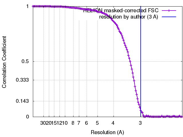
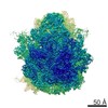
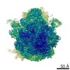
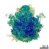
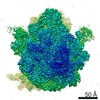
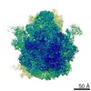
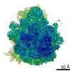
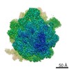
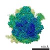
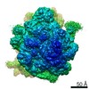
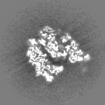
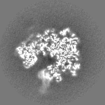
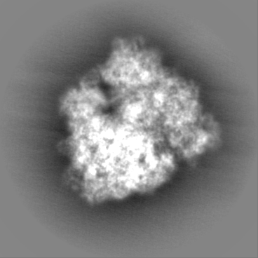
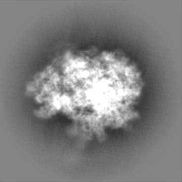
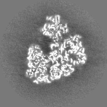
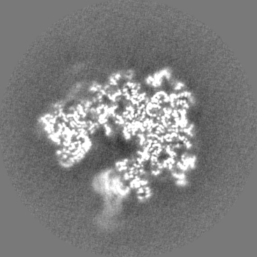
ジャーナル: Nat Microbiol / 年: 2018 タイトル: Structure of a hibernating 100S ribosome reveals an inactive conformation of the ribosomal protein S1. 著者: Bertrand Beckert / Martin Turk / Andreas Czech / Otto Berninghausen / Roland Beckmann / Zoya Ignatova / Jürgen M Plitzko / Daniel N Wilson / 要旨: To survive under conditions of stress, such as nutrient deprivation, bacterial 70S ribosomes dimerize to form hibernating 100S particles. In γ-proteobacteria, such as Escherichia coli, 100S ...To survive under conditions of stress, such as nutrient deprivation, bacterial 70S ribosomes dimerize to form hibernating 100S particles. In γ-proteobacteria, such as Escherichia coli, 100S formation requires the ribosome modulation factor (RMF) and the hibernation promoting factor (HPF). Here we present single-particle cryo-electron microscopy structures of hibernating 70S and 100S particles isolated from stationary-phase E. coli cells at 3.0 Å and 7.9 Å resolution, respectively. The structures reveal the binding sites for HPF and RMF as well as the unexpected presence of deacylated E-site transfer RNA and ribosomal protein bS1. HPF interacts with the anticodon-stem-loop of the E-tRNA and occludes the binding site for the messenger RNA as well as A- and P-site tRNAs. RMF facilitates stabilization of a compact conformation of bS1, which together sequester the anti-Shine-Dalgarno sequence of the 16S ribosomal RNA (rRNA), thereby inhibiting translation initiation. At the dimerization interface, the C-terminus of uS2 probes the mRNA entrance channel of the symmetry-related particle, thus suggesting that dimerization inactivates ribosomes by blocking the binding of mRNA within the channel. The back-to-back E. coli 100S arrangement is distinct from 100S particles observed previously in Gram-positive bacteria, and reveals a unique role for bS1 in translation regulation.
全体 : Structure of a hibernating 100S ribosome reveals an inactive conf...
全体
名称: Structure of a hibernating 100S ribosome reveals an inactive conformation of the ribosomal protein S1
要素
複合体: Structure of a hibernating 100S ribosome reveals an inactive conformation of the ribosomal protein S1
RNA: 16S ribosomal RNA
タンパク質・ペプチド: 30S ribosomal protein S2
タンパク質・ペプチド: 30S ribosomal protein S3
タンパク質・ペプチド: 30S ribosomal protein S4
タンパク質・ペプチド: 30S ribosomal protein S5
タンパク質・ペプチド: 30S ribosomal protein S6
タンパク質・ペプチド: 30S ribosomal protein S7
タンパク質・ペプチド: 30S ribosomal protein S8
タンパク質・ペプチド: 30S ribosomal protein S9
タンパク質・ペプチド: 30S ribosomal protein S10
タンパク質・ペプチド: 30S ribosomal protein S11
タンパク質・ペプチド: 30S ribosomal protein S12
タンパク質・ペプチド: 30S ribosomal protein S13
タンパク質・ペプチド: 30S ribosomal protein S14
タンパク質・ペプチド: 30S ribosomal protein S15
タンパク質・ペプチド: 30S ribosomal protein S16
タンパク質・ペプチド: 30S ribosomal protein S17
タンパク質・ペプチド: 30S ribosomal protein S18
タンパク質・ペプチド: 30S ribosomal protein S19
タンパク質・ペプチド: 30S ribosomal protein S20
タンパク質・ペプチド: 30S ribosomal protein S21
RNA: 23S ribosomal RNA
RNA: 5S ribosomal RNA
タンパク質・ペプチド: 50S ribosomal protein L2
タンパク質・ペプチド: 50S ribosomal protein L3
タンパク質・ペプチド: 50S ribosomal protein L4
タンパク質・ペプチド: 50S ribosomal protein L5
タンパク質・ペプチド: 50S ribosomal protein L6
タンパク質・ペプチド: 50S ribosomal protein L9
タンパク質・ペプチド: 50S ribosomal protein L13
タンパク質・ペプチド: 50S ribosomal protein L14
タンパク質・ペプチド: 50S ribosomal protein L15
タンパク質・ペプチド: 50S ribosomal protein L16
タンパク質・ペプチド: 50S ribosomal protein L17
タンパク質・ペプチド: 50S ribosomal protein L18
タンパク質・ペプチド: 50S ribosomal protein L19
タンパク質・ペプチド: 50S ribosomal protein L20
タンパク質・ペプチド: 50S ribosomal protein L21
タンパク質・ペプチド: 50S ribosomal protein L22
タンパク質・ペプチド: 50S ribosomal protein L23
タンパク質・ペプチド: 50S ribosomal protein L24
タンパク質・ペプチド: 50S ribosomal protein L25
タンパク質・ペプチド: 50S ribosomal protein L27
タンパク質・ペプチド: 50S ribosomal protein L28
タンパク質・ペプチド: 50S ribosomal protein L29
タンパク質・ペプチド: 50S ribosomal protein L30
タンパク質・ペプチド: 50S ribosomal protein L32
タンパク質・ペプチド: 50S ribosomal protein L33
タンパク質・ペプチド: 50S ribosomal protein L34
タンパク質・ペプチド: 50S ribosomal protein L35
タンパク質・ペプチド: 50S ribosomal protein L36
タンパク質・ペプチド: 50S ribosomal protein L31
RNA: tRNA
タンパク質・ペプチド: Ribosome modulation factor
タンパク質・ペプチド: 30S ribosomal protein S1
タンパク質・ペプチド: Ribosome hibernation promoting factor
RNA: RNA (5'-R(P*CP*UP*CP*CP*U)-3')
+
超分子 #1: Structure of a hibernating 100S ribosome reveals an inactive conf...
超分子
名称: Structure of a hibernating 100S ribosome reveals an inactive conformation of the ribosomal protein S1 タイプ: complex / ID: 1 / 親要素: 0 / 含まれる分子: #1-#57 詳細: Here we present single particle cryo-electron microscopy structures of hibernating 70S and 100S particles isolated from stationary phase E. coli cells at 3.0 and 7.9 resolution, respectively. ...詳細: Here we present single particle cryo-electron microscopy structures of hibernating 70S and 100S particles isolated from stationary phase E. coli cells at 3.0 and 7.9 resolution, respectively. The structures reveal the binding sites for HPF and RMF as well as the unexpected presence of deacylated E-site tRNA and ribosomal protein bS1. HPF interacts with the anticodon-stem-loop of the E-tRNA and occludes the binding site for the mRNA as well as A- and P-site tRNAs. RMF facilitates stabilization of a compact conformation of bS1, which together sequester the anti-Shine-Dalgarno sequence of the 16S ribosomal RNA (rRNA), thereby inhibiting translation initiation. At the dimerization interface, the C-terminus of uS2 probes the mRNA entrance channel of the symmetry related particle, thus suggesting that dimerization inactivates ribosomes by blocking the binding of mRNA within the channel.
 ムービー
ムービー コントローラー


 データを開く
 基本情報
基本情報 マップデータ
 試料
試料 キーワード
キーワード 機能・相同性情報
 データ登録者
 引用
引用
 構造の表示
構造の表示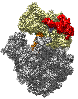
 ダウンロードとリンク
 emd_0137.png
 http://ftp.pdbj.org/pub/emdb/structures/EMD-0137


 Z (Sec.)
 Y (Row.)
 X (Col.)


 試料の構成要素
 解析
解析 電子顕微鏡法
 FIELD EMISSION GUN