 Movie
Movie Controller


+ Open data

- Basic information
| Entry |  | |||||||||
|---|---|---|---|---|---|---|---|---|---|---|
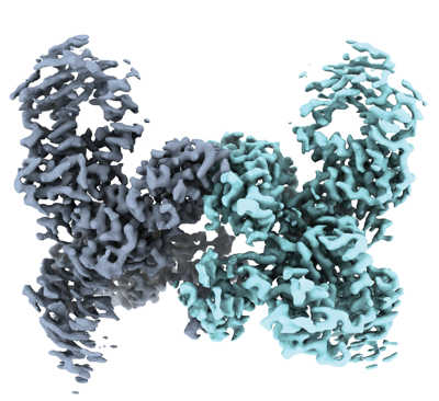
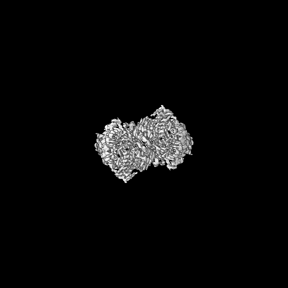
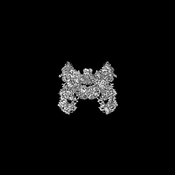
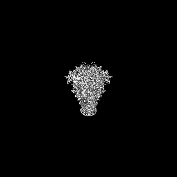
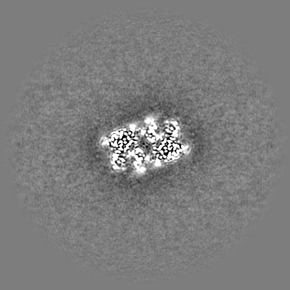
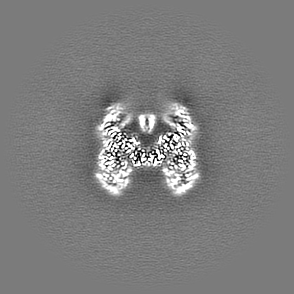
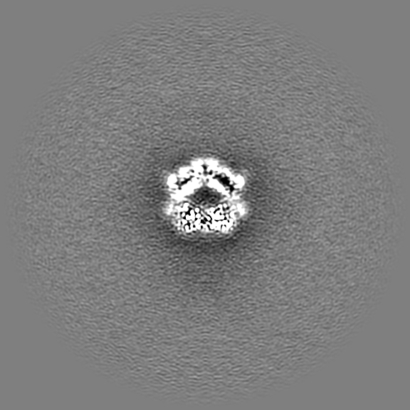
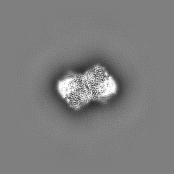
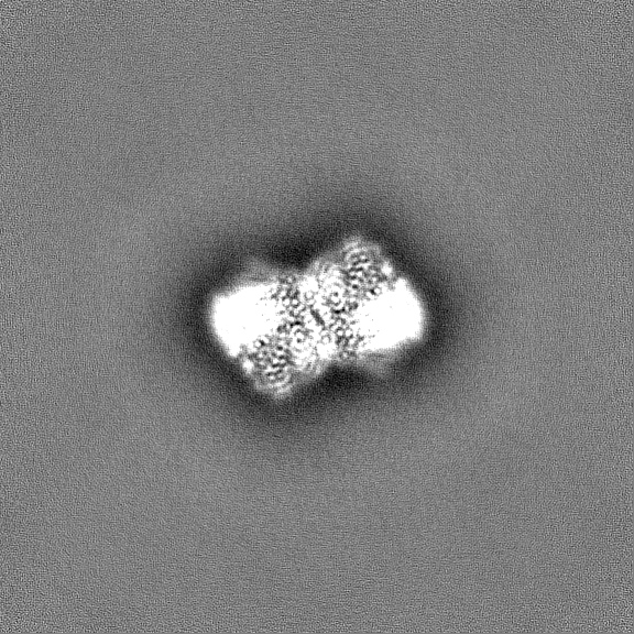
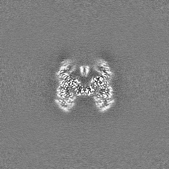
| Title | Vibrio cholerae DdmD apo complex | |||||||||
 Map data Map data | Unsharpened map | |||||||||
 Sample Sample |
| |||||||||
 Keywords Keywords | Helicase / Nuclease / Complex / Effector / IMMUNE SYSTEM | |||||||||
| Function / homology | Helicase/UvrB N-terminal domain-containing protein Function and homology information Function and homology information | |||||||||
| Biological species |   Vibrio cholerae (bacteria) Vibrio cholerae (bacteria) | |||||||||
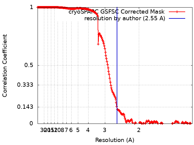
| Method | single particle reconstruction / cryo EM / Resolution: 2.55 Å | |||||||||
 Authors Authors | Loeff L / Jinek M | |||||||||
| Funding support | European Union, 1 items
| |||||||||
 Citation Citation | Journal: Science / Year: 2024 Title: Molecular mechanism of plasmid elimination by the DdmDE defense system. Authors: Luuk Loeff / David W Adams / Christelle Chanez / Sandrine Stutzmann / Laurie Righi / Melanie Blokesch / Martin Jinek /  Abstract: Seventh-pandemic strains contain two pathogenicity islands that encode the DNA defense modules DdmABC and DdmDE. In this study, we used cryogenic electron microscopy to determine the mechanistic ...Seventh-pandemic strains contain two pathogenicity islands that encode the DNA defense modules DdmABC and DdmDE. In this study, we used cryogenic electron microscopy to determine the mechanistic basis for plasmid defense by DdmDE. The helicase-nuclease DdmD adopts an autoinhibited dimeric architecture. The prokaryotic Argonaute protein DdmE uses a DNA guide to target plasmid DNA. The structure of the DdmDE complex, validated by in vivo mutational studies, shows that DNA binding by DdmE triggers disassembly of the DdmD dimer and loading of monomeric DdmD onto the nontarget DNA strand. In vitro studies indicate that DdmD translocates in the 5'-to-3' direction, while partially degrading the plasmid DNA. These findings provide critical insights into the mechanism of DdmDE systems in plasmid elimination. | |||||||||
| History |
|
- Structure visualization
| Supplemental images |
|---|
- Downloads & links
-EMDB archive
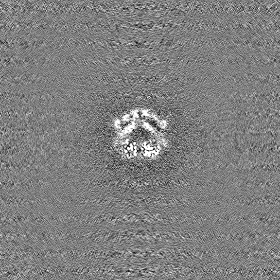
| Map data | emd_50090.map.gz | 361.6 MB | EMDB map data format | |
|---|---|---|---|---|
| Header (meta data) | emd-50090-v30.xmlemd-50090.xml | 18.9 KB 18.9 KB | Display Display | EMDB header |
| FSC (resolution estimation) | emd_50090_fsc.xml | 19 KB | Display | FSC data file |
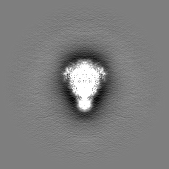
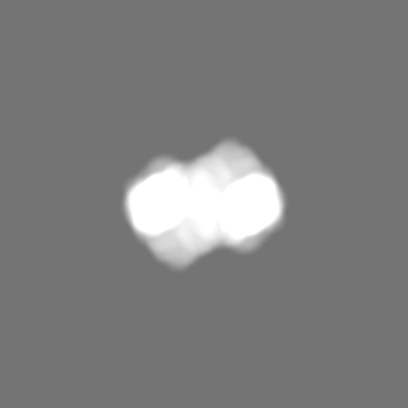
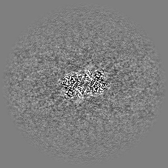
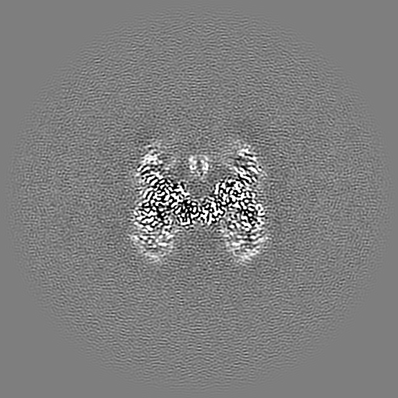
| Images |  emd_50090.png emd_50090.png | 165.5 KB | ||
| Masks | emd_50090_msk_1.map | 729 MB | Mask map | |
| Filedesc metadata | emd-50090.cif.gz | 6.5 KB | ||
| Others | emd_50090_additional_1.map.gzemd_50090_half_map_1.map.gzemd_50090_half_map_2.map.gz | 684.8 MB 675.9 MB 675.9 MB | ||
| Archive directory |  http://ftp.pdbj.org/pub/emdb/structures/EMD-50090ftp://ftp.pdbj.org/pub/emdb/structures/EMD-50090 http://ftp.pdbj.org/pub/emdb/structures/EMD-50090ftp://ftp.pdbj.org/pub/emdb/structures/EMD-50090 | HTTPS FTP |
-Related structure data
| Related structure data |  9ezxMC  9ezyC M: atomic model generated by this map C: citing same article ( |
|---|---|
| Similar structure data |
-Links
| EMDB pages | EMDB (EBI/PDBe) / EMDataResource |
|---|
-Map
| File | Download / File: emd_50090.map.gz / Format: CCP4 / Size: 729 MB / Type: IMAGE STORED AS FLOATING POINT NUMBER (4 BYTES) | ||||||||||||||||||||||||||||||||||||
|---|---|---|---|---|---|---|---|---|---|---|---|---|---|---|---|---|---|---|---|---|---|---|---|---|---|---|---|---|---|---|---|---|---|---|---|---|---|
| Annotation | Unsharpened map | ||||||||||||||||||||||||||||||||||||
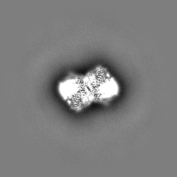
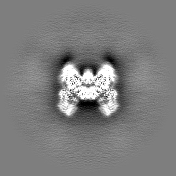
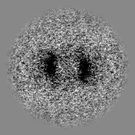
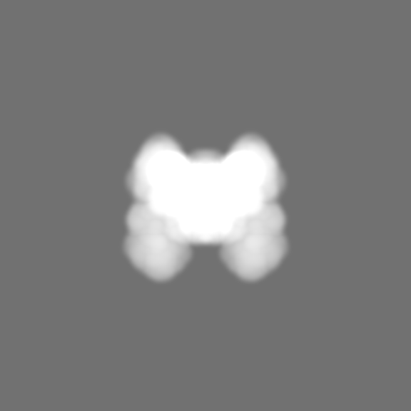
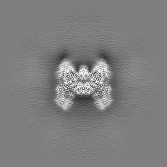
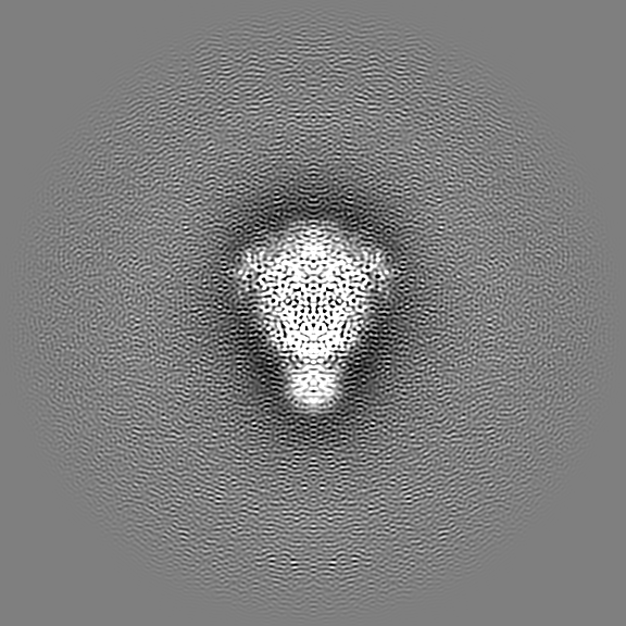
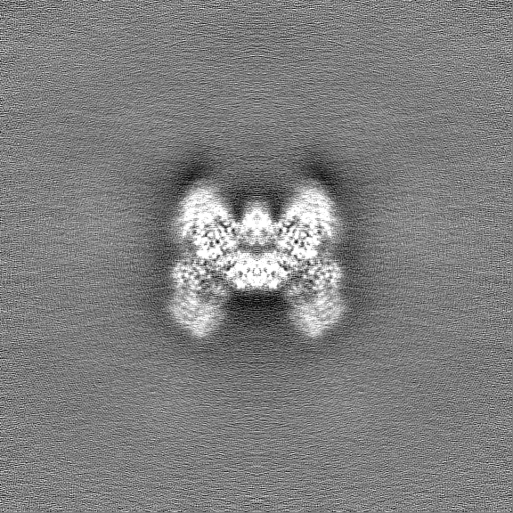
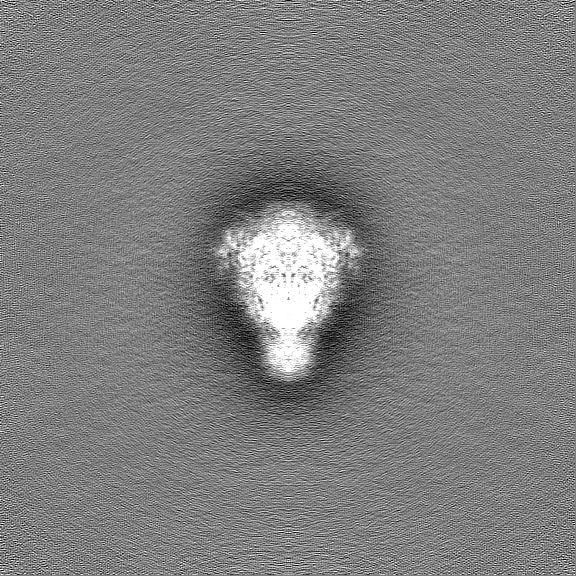
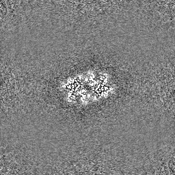
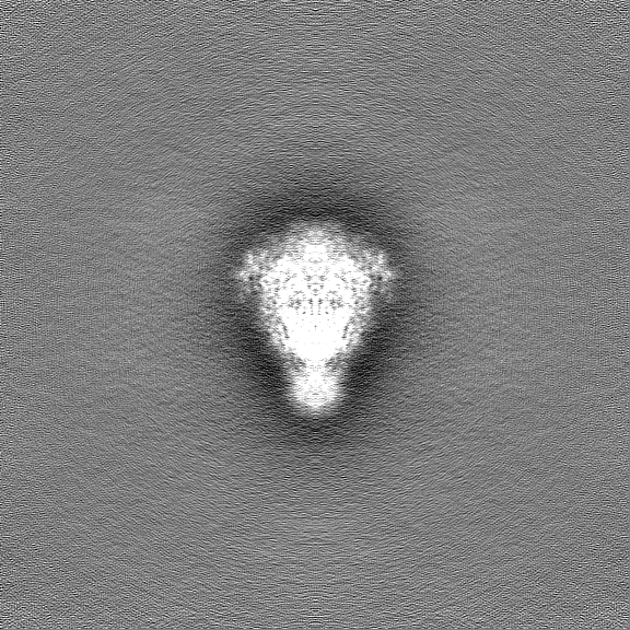
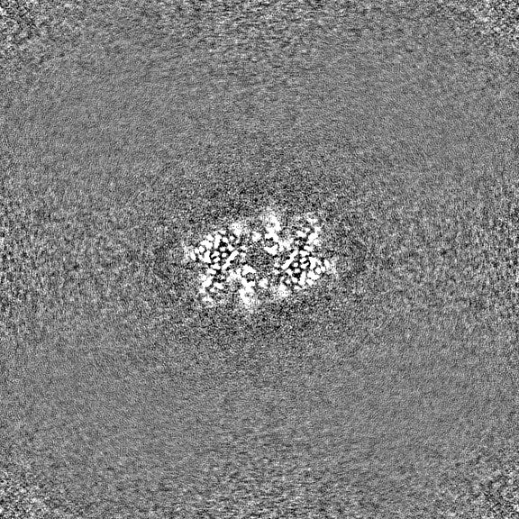
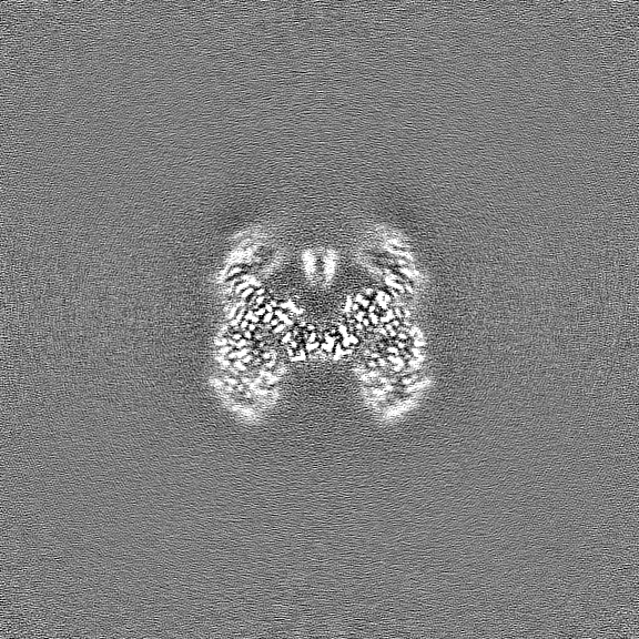
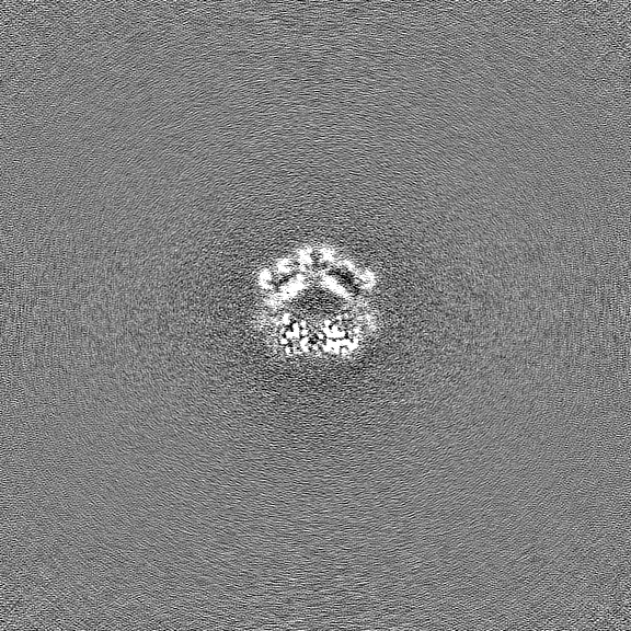
| Projections & slices | Image control
Images are generated by Spider. | ||||||||||||||||||||||||||||||||||||
| Voxel size | X=Y=Z: 0.65 Å | ||||||||||||||||||||||||||||||||||||
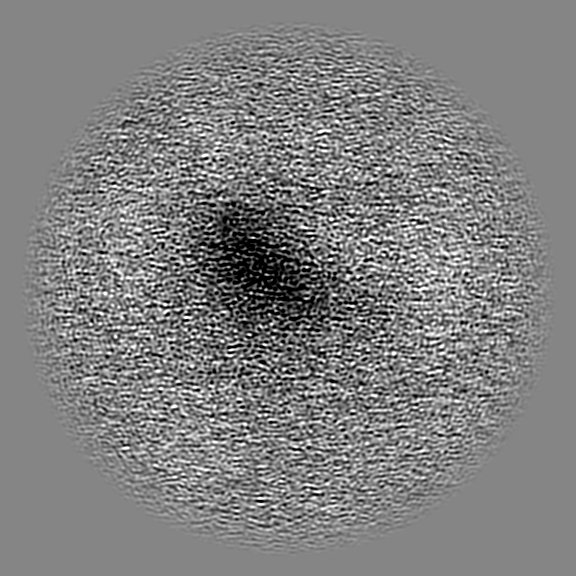
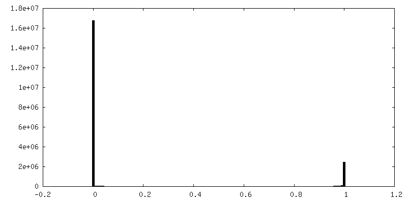
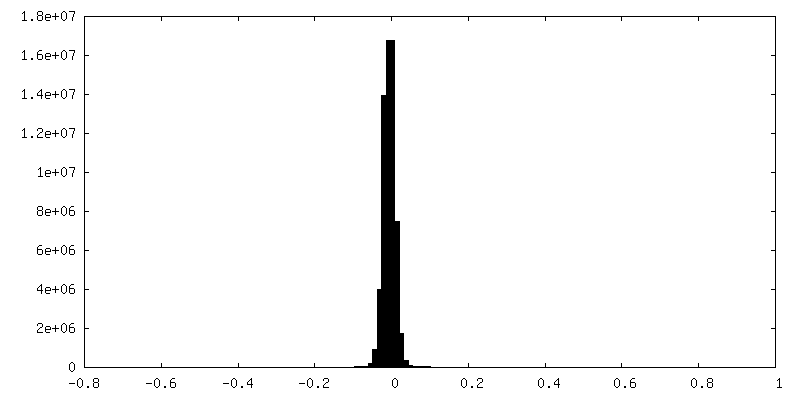
| Density |
| ||||||||||||||||||||||||||||||||||||
| Symmetry | Space group: 1 | ||||||||||||||||||||||||||||||||||||
| Details | EMDB XML:
|
 Z (Sec.)
 Y (Row.)
 X (Col.)


-Supplemental data
-Mask #1
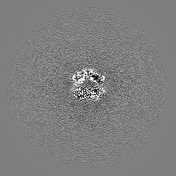
| File | emd_50090_msk_1.map | ||||||||||||
|---|---|---|---|---|---|---|---|---|---|---|---|---|---|
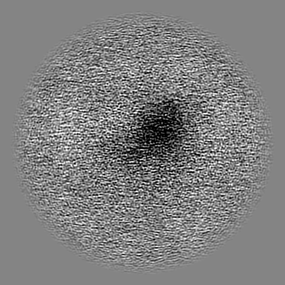
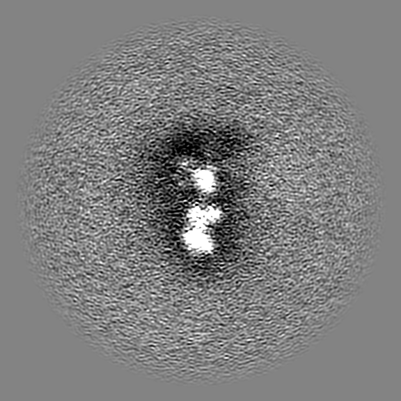
| Projections & Slices |
| ||||||||||||
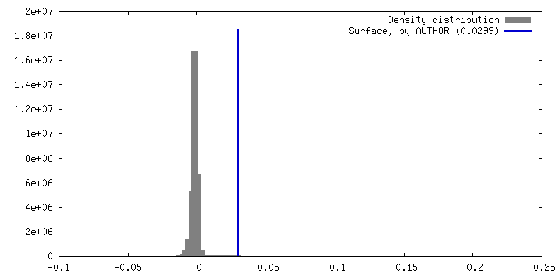
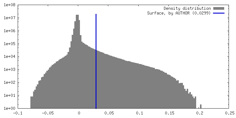
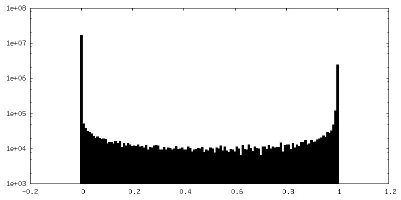
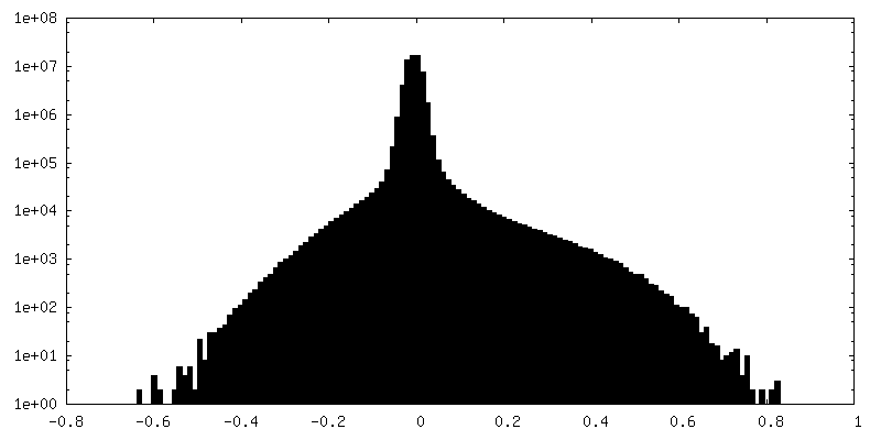
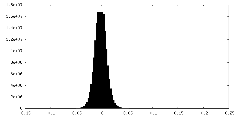
| Density Histograms |


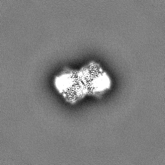
-Additional map: Sharpened EM map
| File | emd_50090_additional_1.map | ||||||||||||
|---|---|---|---|---|---|---|---|---|---|---|---|---|---|
| Annotation | Sharpened EM map | ||||||||||||
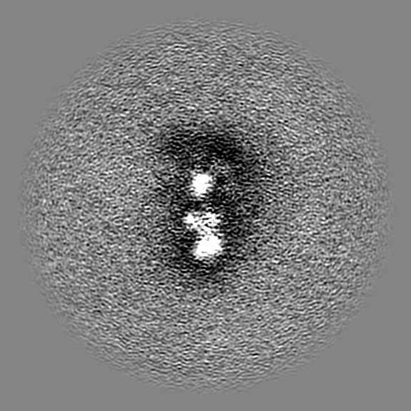
| Projections & Slices |
| ||||||||||||
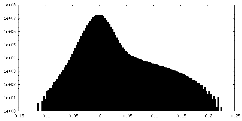
| Density Histograms |
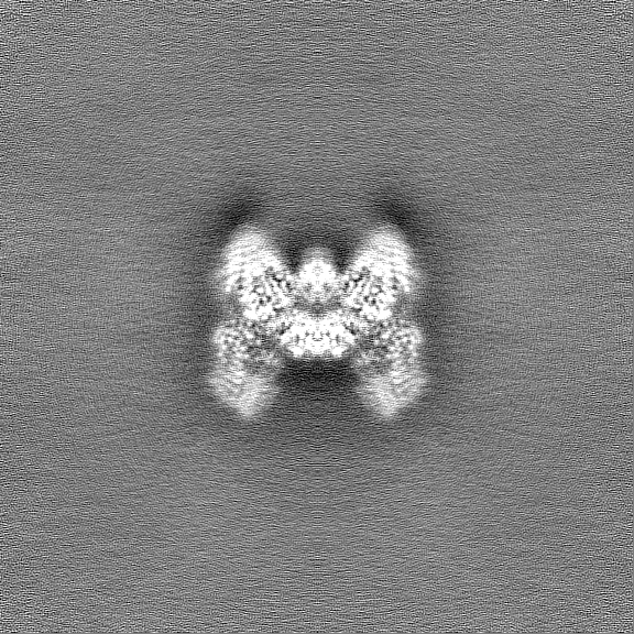
-Half map: Half map A
| File | emd_50090_half_map_1.map | ||||||||||||
|---|---|---|---|---|---|---|---|---|---|---|---|---|---|
| Annotation | Half map A | ||||||||||||
| Projections & Slices |
| ||||||||||||
| Density Histograms |


-Half map: Half map A
| File | emd_50090_half_map_2.map | ||||||||||||
|---|---|---|---|---|---|---|---|---|---|---|---|---|---|
| Annotation | Half map A | ||||||||||||
| Projections & Slices |
| ||||||||||||
| Density Histograms |
- Sample components
-Entire : Dimeric complex of the DdmD protein
| Entire | Name: Dimeric complex of the DdmD protein |
|---|---|
| Components |
|
-Supramolecule #1: Dimeric complex of the DdmD protein
| Supramolecule | Name: Dimeric complex of the DdmD protein / type: complex / ID: 1 / Parent: 0 / Macromolecule list: all |
|---|---|
| Source (natural) | Organism: Vibrio cholerae (bacteria) |
| Molecular weight | Theoretical: 266 KDa |
-Macromolecule #1: Helicase/UvrB N-terminal domain-containing protein
| Macromolecule | Name: Helicase/UvrB N-terminal domain-containing protein / type: protein_or_peptide / ID: 1 / Number of copies: 2 / Enantiomer: LEVO |
|---|---|
| Source (natural) | Organism: Vibrio cholerae (bacteria) |
| Molecular weight | Theoretical: 136.427594 KDa |
| Recombinant expression | Organism: |
| Sequence | String: SNAMNVSIEE FTHFDFQLVP EPSPLDLVIT EPLKNHIEVN GVKSGALLPL PFQTGIGKTY TALNFLLQQM LEQVRSELKE ENTGKKSKR LLYYVTDSVD NVVSAKADLL KLIEKQTVKG EPRFTLEQQE YLKAQIVHLP NQSEQLLQCS DAVLNDVLIG F NLNAERDV ...String: SNAMNVSIEE FTHFDFQLVP EPSPLDLVIT EPLKNHIEVN GVKSGALLPL PFQTGIGKTY TALNFLLQQM LEQVRSELKE ENTGKKSKR LLYYVTDSVD NVVSAKADLL KLIEKQTVKG EPRFTLEQQE YLKAQIVHLP NQSEQLLQCS DAVLNDVLIG F NLNAERDV QAEWSAISGL RRHASNPEVK ISLNRQAGYF YRNLIDRLQK KQKGADRVLL SGSLLASVET LLPGEKIRNG SA HVAFLTT SKFLKGFHNT RSRYSPLRDL SGAVLIIDEI DKQNQVILSE LCKQQAQDLI WAIRTLRANF RDHQLESSPR YDK IEDLFE PLRERLEEFG TNWNLAFAFN TEGANLNERP VRLFSDRSFT HVSSATHKLS LKSDFLRRKN LIFSDEKVEG SLIE KHGLL TRFVNEADVI YQWFLGTMRK AVFQYWENVR GLEIEVRENR SLEGTFQEAV QSLLTHFNLQ EFESAVYESF DTRGL RQSA GGKANKLSSS KSYHHTGLKL VEVAHNQGTR DTVNCKASFL NTSPSGVLAD MVDAGAVILG ISATARADTV IHNFDF KYL NERLGNKLLS LSREQKQRVN NYYHSRRNYK DNGVVLTVKY LNSRDAFLDA LLEEYKPEAR SSHFILNHYL GIAESEQ AF VRSWLSKLLA SIKAFISSPD NRYMLSLLNR TLDTTRQNIN DFIQFCCDKW AKEFNVKTKT FFGVNADWMR LVGYDEIS K HLNTELGKVV VFSTYASMGA GKNPDYAVNL ALEGESLISV ADVTYSTQLR SDIDSIYLEK PTQLLLSDDY SHTANQLCQ FHQILSLQEN GELSPKSAEN WCRQQLMGMS RERSLQQYHQ TSDYQSAVRK YIEQAVGRAG RTSLKRKQIL LFVDSGLKEI LAEESRDPS LFSHEYVALV NKAKSAGKSI VEDRAVRRLF NLAQRNNKDG MLSIKALVHR LHNQPASKSD IQEWQDIRTQ L LRYPTVAF QPERFNRLYL QSMTKGYYRY QGNLDGDPNS FEFFDRVPYG DMVSEEDCSL ATLVQNQYVR PWFERKGFAC SW QKEANVM TPIMFTNIYK GALGEQAVEA VLTAFDFTFE EVPNSIYERF DNRVIFAGIE QPIWLDSKYW KHEGNESSEG YSS KIALVE EEFGPSKFIY VNALGDTSKP IRYLNSCFVE TSPQLAKVIE IPALIDDSNA DTNRTAVQEL IKWLHHS UniProtKB: Helicase/UvrB N-terminal domain-containing protein |
-Experimental details
-Structure determination
| Method | cryo EM |
|---|---|
 Processing Processing | single particle reconstruction |
| Aggregation state | particle |
-Sample preparation
| Concentration | 1.5 mg/mL | |||||||||
|---|---|---|---|---|---|---|---|---|---|---|
| Buffer | pH: 8 Component:
| |||||||||
| Vitrification | Cryogen name: ETHANE / Instrument: FEI VITROBOT MARK IV | |||||||||
| Details | This sample was monodisperse. |
- Electron microscopy
| Microscope | TFS KRIOS |
|---|---|
| Image recording | Film or detector model: GATAN K3 (6k x 4k) / Average electron dose: 59.98 e/Å2 |
| Electron beam | Acceleration voltage: 300 kV / Electron source:  FIELD EMISSION GUN FIELD EMISSION GUN |
| Electron optics | C2 aperture diameter: 50.0 µm / Calibrated magnification: 130000 / Illumination mode: FLOOD BEAM / Imaging mode: BRIGHT FIELD / Cs: 2.7 mm / Nominal defocus max: 2.4 µm / Nominal defocus min: 1.0 µm |
| Sample stage | Specimen holder model: FEI TITAN KRIOS AUTOGRID HOLDER / Cooling holder cryogen: NITROGEN |
| Experimental equipment |  Model: Titan Krios / Image courtesy: FEI Company |
+Image processing
-Atomic model buiding 1
| Initial model | Chain - Source name: AlphaFold / Chain - Initial model type: in silico model / Details: Initial model was generated with alpha fold |
|---|---|
| Output model | PDB-9ezx: |
