 Movie
Movie Controller


+ Open data

- Basic information
| Entry | Database: PDB / ID: 9ezy | ||||||
|---|---|---|---|---|---|---|---|
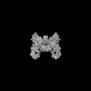
| Title | Vibrio cholerae DdmD-DdmE holo complex | ||||||
 Components Components |
| ||||||
 Keywords Keywords | IMMUNE SYSTEM / Helicase / Nuclease / Complex / Effector / Argonaute | ||||||
| Function / homology | DNA / DNA (> 10) / Helicase/UvrB N-terminal domain-containing protein / Uncharacterized protein Function and homology information Function and homology information | ||||||
| Biological species |   Vibrio cholerae (bacteria) Vibrio cholerae (bacteria)synthetic construct (others) | ||||||
| Method | ELECTRON MICROSCOPY / single particle reconstruction / cryo EM / Resolution: 2.56 Å | ||||||
 Authors Authors | Loeff, L. / Jinek, M. | ||||||
| Funding support | European Union, 1items
| ||||||
 Citation Citation | Journal: Science / Year: 2024 Title: Molecular mechanism of plasmid elimination by the DdmDE defense system. Authors: Luuk Loeff / David W Adams / Christelle Chanez / Sandrine Stutzmann / Laurie Righi / Melanie Blokesch / Martin Jinek /  Abstract: Seventh-pandemic strains contain two pathogenicity islands that encode the DNA defense modules DdmABC and DdmDE. In this study, we used cryogenic electron microscopy to determine the mechanistic ...Seventh-pandemic strains contain two pathogenicity islands that encode the DNA defense modules DdmABC and DdmDE. In this study, we used cryogenic electron microscopy to determine the mechanistic basis for plasmid defense by DdmDE. The helicase-nuclease DdmD adopts an autoinhibited dimeric architecture. The prokaryotic Argonaute protein DdmE uses a DNA guide to target plasmid DNA. The structure of the DdmDE complex, validated by in vivo mutational studies, shows that DNA binding by DdmE triggers disassembly of the DdmD dimer and loading of monomeric DdmD onto the nontarget DNA strand. In vitro studies indicate that DdmD translocates in the 5'-to-3' direction, while partially degrading the plasmid DNA. These findings provide critical insights into the mechanism of DdmDE systems in plasmid elimination. | ||||||
| History |
|
- Structure visualization
| Structure viewer | Molecule: MolmilJmol/JSmol |
|---|
- Downloads & links
-Download
| PDBx/mmCIF format | 9ezy.cif.gz | 405.2 KB | Display | PDBx/mmCIF format |
|---|---|---|---|---|
| PDB format | pdb9ezy.ent.gz | 315.1 KB | Display | PDB format |
| PDBx/mmJSON format | 9ezy.json.gz | Tree view | PDBx/mmJSON format | |
| Others |  Other downloads Other downloads |
-Validation report
| Arichive directory | https://data.pdbj.org/pub/pdb/validation_reports/ez/9ezyftp://data.pdbj.org/pub/pdb/validation_reports/ez/9ezy | HTTPS FTP |
|---|
-Related structure data
| Related structure data | 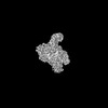 50091MC  9ezxC M: map data used to model this data C: citing same article ( |
|---|---|
| Similar structure data |
-Links
 PDBj
PDBj


- Assembly
| Deposited unit | 
|
|---|---|
| 1 |
|
-Components
-Protein , 2 types, 2 molecules AB
| #1: Protein | Mass: 136427.594 Da / Num. of mol.: 1 Source method: isolated from a genetically manipulated source Source: (gene. exp.) Vibrio cholerae (bacteria) / Gene: VC_1771 / Production host: |
|---|---|
| #2: Protein | Mass: 79468.156 Da / Num. of mol.: 1 Source method: isolated from a genetically manipulated source Source: (gene. exp.) Vibrio cholerae (bacteria) / Gene: VC_1770 / Production host: |
-DNA chain , 3 types, 3 molecules CDE
| #3: DNA chain | Mass: 4567.998 Da / Num. of mol.: 1 / Source method: obtained synthetically Details: The first adenosine nucleotide was added to obtain three oxygen groups on the 5' phosphate of the DNA guide. This nucleotide was not present on the DNA guide used for structure determination. Source: (synth.) synthetic construct (others) |
|---|---|
| #4: DNA chain | Mass: 20341.029 Da / Num. of mol.: 1 / Source method: obtained synthetically / Source: (synth.) synthetic construct (others) |
| #5: DNA chain | Mass: 20305.029 Da / Num. of mol.: 1 / Source method: obtained synthetically / Source: (synth.) synthetic construct (others) |
-Non-polymers , 1 types, 1 molecules 
| #6: Chemical | ChemComp-MG /  Mass: 24.305 Da / Num. of mol.: 1 / Source method: obtained synthetically / Formula: Mg / Feature type: SUBJECT OF INVESTIGATION Mass: 24.305 Da / Num. of mol.: 1 / Source method: obtained synthetically / Formula: Mg / Feature type: SUBJECT OF INVESTIGATION |
|---|
-Details
| Has ligand of interest | Y |
|---|
-Experimental details
-Experiment
| Experiment | Method: ELECTRON MICROSCOPY |
|---|---|
| EM experiment | Aggregation state: PARTICLE / 3D reconstruction method: single particle reconstruction |
- Sample preparation
| Component | Name: DdmD-DdmE Holocomplex / Type: COMPLEX / Entity ID: #1-#5 / Source: RECOMBINANT | ||||||||||||||||||||
|---|---|---|---|---|---|---|---|---|---|---|---|---|---|---|---|---|---|---|---|---|---|
| Molecular weight | Experimental value: NO | ||||||||||||||||||||
| Source (natural) | Organism: Vibrio cholerae (bacteria) | ||||||||||||||||||||
| Source (recombinant) | Organism: | ||||||||||||||||||||
| Buffer solution | pH: 8 | ||||||||||||||||||||
| Buffer component |
| ||||||||||||||||||||
| Specimen | Conc.: 2.5 mg/ml / Embedding applied: NO / Shadowing applied: NO / Staining applied: NO / Vitrification applied: YES | ||||||||||||||||||||
| Vitrification | Instrument: FEI VITROBOT MARK IV / Cryogen name: ETHANE |
- Electron microscopy imaging
| Experimental equipment |  Model: Titan Krios / Image courtesy: FEI Company |
|---|---|
| Microscopy | Model: TFS KRIOS |
| Electron gun | Electron source:  FIELD EMISSION GUN / Accelerating voltage: 300 kV / Illumination mode: FLOOD BEAM FIELD EMISSION GUN / Accelerating voltage: 300 kV / Illumination mode: FLOOD BEAM |
| Electron lens | Mode: BRIGHT FIELD / Calibrated magnification: 130000 X / Nominal defocus max: 2400 nm / Nominal defocus min: 1000 nm / Cs: 2.7 mm / C2 aperture diameter: 50 µm / Alignment procedure: COMA FREE |
| Specimen holder | Cryogen: NITROGEN / Specimen holder model: FEI TITAN KRIOS AUTOGRID HOLDER |
| Image recording | Electron dose: 60.99 e/Å2 / Film or detector model: GATAN K3 (6k x 4k) |
- Processing
| EM software |
| ||||||||||||||||||||||||||||||||||||
|---|---|---|---|---|---|---|---|---|---|---|---|---|---|---|---|---|---|---|---|---|---|---|---|---|---|---|---|---|---|---|---|---|---|---|---|---|---|
| CTF correction | Type: PHASE FLIPPING AND AMPLITUDE CORRECTION | ||||||||||||||||||||||||||||||||||||
| Particle selection | Num. of particles selected: 1283702 | ||||||||||||||||||||||||||||||||||||
| 3D reconstruction | Resolution: 2.56 Å / Resolution method: FSC 0.143 CUT-OFF / Num. of particles: 229173 / Symmetry type: POINT | ||||||||||||||||||||||||||||||||||||
| Atomic model building | Details: Initial model was generated with alpha fold / Source name: AlphaFold / Type: in silico model |
