 Movie
Movie Controller

[English] 日本語
 Yorodumi
- EMDB-34369: Cryo-EM Structure of Membrane-Bound Aldehyde Dehydrogenase from G... -
+ Open data

- Basic information
| Entry |  | |||||||||
|---|---|---|---|---|---|---|---|---|---|---|
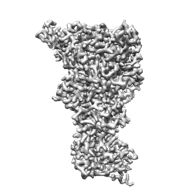
| Title | Cryo-EM Structure of Membrane-Bound Aldehyde Dehydrogenase from Gluconobacter oxydans | |||||||||
 Map data Map data | ||||||||||
 Sample Sample |
| |||||||||
 Keywords Keywords | Complex / Oxidoreductase / Membrane-bound protein | |||||||||
| Function / homology |  Function and homology information Function and homology informationoxidoreductase activity, acting on CH-OH group of donors / 2 iron, 2 sulfur cluster binding / electron transfer activity / oxidoreductase activity / iron ion binding / heme binding / metal ion binding / plasma membrane Similarity search - Function | |||||||||
| Biological species |  Gluconobacter oxydans (bacteria) Gluconobacter oxydans (bacteria) | |||||||||
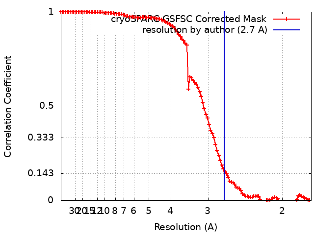
| Method | single particle reconstruction / cryo EM / Resolution: 2.7 Å | |||||||||
 Authors Authors | Adachi T / Miyata T / Makino F / Tanaka H / Namba K / Sowa K / Kitazumi Y / Shirai O | |||||||||
| Funding support |  Japan, 2 items Japan, 2 items
| |||||||||
 Citation Citation | Journal: Acs Catalysis / Year: 2023 Title: Experimental and Theoretical Insights into Bienzymatic Cascade for Mediatorless Bioelectrochemical Ethanol Oxidation with Alcohol and Aldehyde Dehydrogenases Authors: Adachi T / Miyata T / Makino F / Tanaka H / Namba K / Kano K / Sowa K / Kitazumi Y / Shirai O | |||||||||
| History |
|
- Structure visualization
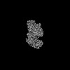
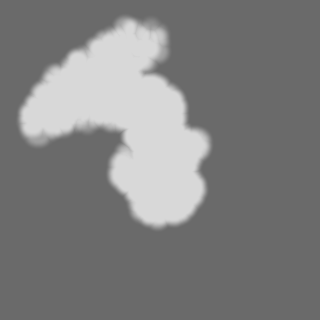
| Supplemental images |
|---|
- Downloads & links
-EMDB archive
| Map data | emd_34369.map.gz | 62.6 MB | EMDB map data format | |
|---|---|---|---|---|
| Header (meta data) | emd-34369-v30.xmlemd-34369.xml | 24.1 KB 24.1 KB | Display Display | EMDB header |
| FSC (resolution estimation) | emd_34369_fsc.xml | 10.5 KB | Display | FSC data file |
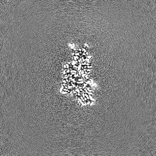
| Images |  emd_34369.png emd_34369.png | 87.4 KB | ||
| Masks | emd_34369_msk_1.map | 125 MB | Mask map | |
| Filedesc metadata | emd-34369.cif.gz | 7.5 KB | ||
| Others | emd_34369_half_map_1.map.gzemd_34369_half_map_2.map.gz | 116.1 MB 116.1 MB | ||
| Archive directory |  http://ftp.pdbj.org/pub/emdb/structures/EMD-34369ftp://ftp.pdbj.org/pub/emdb/structures/EMD-34369 http://ftp.pdbj.org/pub/emdb/structures/EMD-34369ftp://ftp.pdbj.org/pub/emdb/structures/EMD-34369 | HTTPS FTP |
-Related structure data
| Related structure data |  8gy3MC  8gy2C M: atomic model generated by this map C: citing same article ( |
|---|---|
| Similar structure data |
-Links
| EMDB pages | EMDB (EBI/PDBe) / EMDataResource |
|---|---|
| Related items in Molecule of the Month |


-Map
| File | Download / File: emd_34369.map.gz / Format: CCP4 / Size: 125 MB / Type: IMAGE STORED AS FLOATING POINT NUMBER (4 BYTES) | ||||||||||||||||||||||||||||||||||||
|---|---|---|---|---|---|---|---|---|---|---|---|---|---|---|---|---|---|---|---|---|---|---|---|---|---|---|---|---|---|---|---|---|---|---|---|---|---|
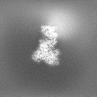
| Projections & slices | Image control
Images are generated by Spider. | ||||||||||||||||||||||||||||||||||||
| Voxel size | X=Y=Z: 0.87 Å | ||||||||||||||||||||||||||||||||||||
| Density |
| ||||||||||||||||||||||||||||||||||||
| Symmetry | Space group: 1 | ||||||||||||||||||||||||||||||||||||
| Details | EMDB XML:
|
 Z (Sec.)
 Y (Row.)
 X (Col.)


-Supplemental data
-Mask #1
| File | emd_34369_msk_1.map | ||||||||||||
|---|---|---|---|---|---|---|---|---|---|---|---|---|---|
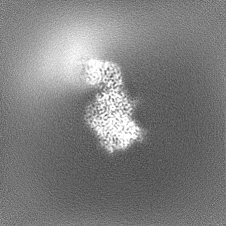
| Projections & Slices |
| ||||||||||||
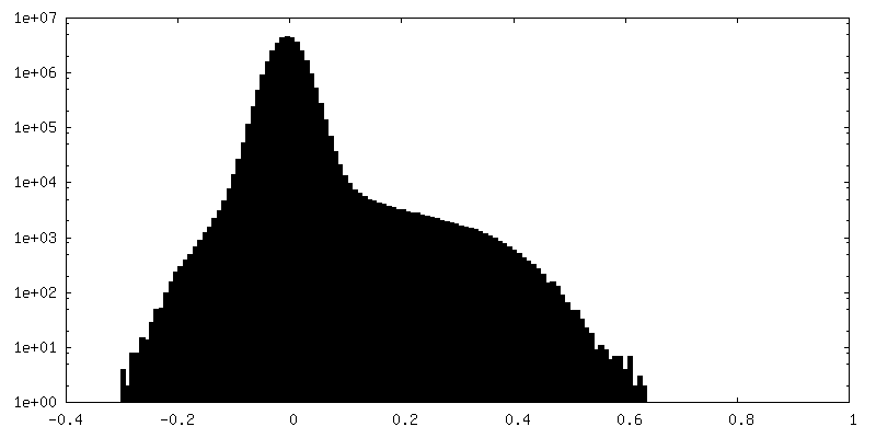
| Density Histograms |


-Half map: #2
| File | emd_34369_half_map_1.map | ||||||||||||
|---|---|---|---|---|---|---|---|---|---|---|---|---|---|
| Projections & Slices |
| ||||||||||||
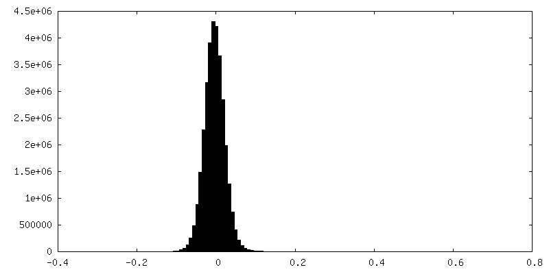
| Density Histograms |


-Half map: #1
| File | emd_34369_half_map_2.map | ||||||||||||
|---|---|---|---|---|---|---|---|---|---|---|---|---|---|
| Projections & Slices |
| ||||||||||||
| Density Histograms |


- Sample components
-Entire : Aldehyde dehydrogenase from Gluconobacter oxydans
| Entire | Name: Aldehyde dehydrogenase from Gluconobacter oxydans |
|---|---|
| Components |
|
-Supramolecule #1: Aldehyde dehydrogenase from Gluconobacter oxydans
| Supramolecule | Name: Aldehyde dehydrogenase from Gluconobacter oxydans / type: complex / ID: 1 / Parent: 0 / Macromolecule list: #1-#3 |
|---|---|
| Source (natural) | Organism: Gluconobacter oxydans (bacteria) |
| Molecular weight | Theoretical: 150 KDa |
-Macromolecule #1: Cytochrome c subunit of aldehyde dehydrogenase
| Macromolecule | Name: Cytochrome c subunit of aldehyde dehydrogenase / type: protein_or_peptide / ID: 1 / Number of copies: 1 / Enantiomer: LEVO / EC number: aldehyde dehydrogenase (FAD-independent) |
|---|---|
| Source (natural) | Organism: Gluconobacter oxydans (bacteria) |
| Molecular weight | Theoretical: 48.5195 KDa |
| Recombinant expression | Organism: Gluconobacter oxydans (bacteria) |
| Sequence | String: MLKRIAAAVI GLGAVGGIGF LAYAWYPAIA PIPRPAASSF SADAISRGEI VANGGYCAEC HTRVDGKPGP ELAGDFKMAT PFGDIFSSN ITPDEEWGIG NWSLAAFKRA MNKGIARDGS QLYPAFPFDH FTKVSDQDVS DLYAYLMTRP AVHLKPRDNT V PFPINIRL ...String: MLKRIAAAVI GLGAVGGIGF LAYAWYPAIA PIPRPAASSF SADAISRGEI VANGGYCAEC HTRVDGKPGP ELAGDFKMAT PFGDIFSSN ITPDEEWGIG NWSLAAFKRA MNKGIARDGS QLYPAFPFDH FTKVSDQDVS DLYAYLMTRP AVHLKPRDNT V PFPINIRL IGQGFWKLLF FTPGRYQNDP KHDAQWNRGA YLAEGNEHCG ACHTPRNLLG AEKMSSVYDG AVIDGWIAPP LN DHNPTPV VWTEDELFQY LRFGVAPLHG SAAGPMSPVP HRFLSKIPEE DVHAIAHYYA DVDKAAQRSS GDQAAITRAM QMS GRDLTG PQPLDEDARL YQGACGACHY NSGPNPVLGR PELALNNALW LDEPNNLYQV MLHGITAEEG QDHISMPSFY SGLS DHDMA RIAAYLRRTR TTLPPWTDLE KKAASARATL EAPPVNASH UniProtKB: Aldehyde dehydrogenase |
-Macromolecule #2: Small subunit of aldehyde dehydrogenase
| Macromolecule | Name: Small subunit of aldehyde dehydrogenase / type: protein_or_peptide / ID: 2 / Number of copies: 1 / Enantiomer: LEVO / EC number: aldehyde dehydrogenase (FAD-independent) |
|---|---|
| Source (natural) | Organism: Gluconobacter oxydans (bacteria) |
| Molecular weight | Theoretical: 16.799998 KDa |
| Recombinant expression | Organism: Gluconobacter oxydans (bacteria) |
| Sequence | String: MTTKFELNGQ PVTVDAPADT PLLWVIRDDL NLTGTKFGCG IGECGACTVH VGGRATRSCI TPLSAVEGAS ITTIEGLDPA GNHVVQVAW RDQQVPQCGY CQSGQIMQAA SLLKDYPNPT DDQIDGVMGG SLCRCMTYIR IRKAIKEAAS RQQEGANNG UniProtKB: Isoquinoline 1-oxidoreductase alpha subunit |
-Macromolecule #3: Large subunit of aldehyde dehydrogenase
| Macromolecule | Name: Large subunit of aldehyde dehydrogenase / type: protein_or_peptide / ID: 3 / Number of copies: 1 / Enantiomer: LEVO / EC number: aldehyde dehydrogenase (FAD-independent) |
|---|---|
| Source (natural) | Organism: Gluconobacter oxydans (bacteria) |
| Molecular weight | Theoretical: 83.302164 KDa |
| Recombinant expression | Organism: Gluconobacter oxydans (bacteria) |
| Sequence | String: MAKIEQIAKK SDATRLSRRN FLMTAAGAGL MFGFARKAGA ATTLPSAMPP EAAFEPNIWC AIAPDGSINV NIVRAEMGQH VGTALARII ADEMDADWDK IKITQVDTAP KWAGKYVTGG SWSVWDTWDT FRQAGAAARS VMIEEGAKLL GTTPDRCTAH E SVVSAGSK ...String: MAKIEQIAKK SDATRLSRRN FLMTAAGAGL MFGFARKAGA ATTLPSAMPP EAAFEPNIWC AIAPDGSINV NIVRAEMGQH VGTALARII ADEMDADWDK IKITQVDTAP KWAGKYVTGG SWSVWDTWDT FRQAGAAARS VMIEEGAKLL GTTPDRCTAH E SVVSAGSK SISFGDIVAR AKPTRTFTPE EMAKLPLKPT GNRRLISKQV PALDIPDKTT GKAIYGIDVK LDGMVYGRPK MP PTRYAAK VISVDDSAAK KIPGYLRYVV LDDPSGIVPG WVVALAKTYP AAIRAADALK VQWNPGPTIN VSEADIIEHG RKL AADPKN GTRVFNDKGV DEALTIHPGQ VFERSYTCAS VAHYQLEPVN AVARHIDGMW EIHTGNQWQS LILPQLAKSL QVPE EQVVM RTYMLGGGFG RRLNGDYCIP AALASKAIGG APVKLILTRS DDMELDSIRS PSIQTIKVAL DNDRKKIVGM DYVAV AGWP TQVMAPAFLA TGEDGKKYDP FAIAGADHWY ETGPTRVRAI SNDLANATFR PGWLRSVSAG WTPWALECFL DELAHS TKQ DPLAFRLSMF TAQGRNAGQA PNSVGGAKRQ AAVLQRLADK IGYANKQLPA DTGIGIATSF GQERGMPTWT AAAAQIH VD RKTGVVTCQK LWLVLDAGTI VDPGGALAQT EGAALWGFSM ALFEGTEIVN GTIKDRNLNT YTPLRIPDVP DIDIEFIQ N TEKPTGLGEP GVTVVAPAIG NAIFNAVGIR LRHMPMRPAD VRRELQQHTS UniProtKB: CO/xanthine dehydrogenase Mo-binding subunit |
-Macromolecule #4: HEME C
| Macromolecule | Name: HEME C / type: ligand / ID: 4 / Number of copies: 3 / Formula: HEC |
|---|---|
| Molecular weight | Theoretical: 618.503 Da |
| Chemical component information |  ChemComp-HEC: |
-Macromolecule #5: UBIQUINONE-10
| Macromolecule | Name: UBIQUINONE-10 / type: ligand / ID: 5 / Number of copies: 1 / Formula: U10 |
|---|---|
| Molecular weight | Theoretical: 863.343 Da |
| Chemical component information |  ChemComp-U10: |
-Macromolecule #6: FE2/S2 (INORGANIC) CLUSTER
| Macromolecule | Name: FE2/S2 (INORGANIC) CLUSTER / type: ligand / ID: 6 / Number of copies: 2 / Formula: FES |
|---|---|
| Molecular weight | Theoretical: 175.82 Da |
| Chemical component information |  ChemComp-FES: |
-Macromolecule #7: (MOLYBDOPTERIN-CYTOSINE DINUCLEOTIDE-S,S)-DIOXO-AQUA-MOLYBDENUM(V)
| Macromolecule | Name: (MOLYBDOPTERIN-CYTOSINE DINUCLEOTIDE-S,S)-DIOXO-AQUA-MOLYBDENUM(V) type: ligand / ID: 7 / Number of copies: 1 / Formula: PCD |
|---|---|
| Molecular weight | Theoretical: 844.471 Da |
| Chemical component information |  ChemComp-PCD: |
-Experimental details
-Structure determination
| Method | cryo EM |
|---|---|
 Processing Processing | single particle reconstruction |
| Aggregation state | particle |
-Sample preparation
| Concentration | 10 mg/mL | |||||||||||||||
|---|---|---|---|---|---|---|---|---|---|---|---|---|---|---|---|---|
| Buffer | pH: 6 Component:
| |||||||||||||||
| Grid | Model: Quantifoil R1.2/1.3 / Material: COPPER / Mesh: 200 / Pretreatment - Type: GLOW DISCHARGE / Pretreatment - Time: 30 sec. | |||||||||||||||
| Vitrification | Cryogen name: ETHANE / Chamber humidity: 100 % / Chamber temperature: 277 K / Instrument: FEI VITROBOT MARK IV |
- Electron microscopy
| Microscope | JEOL CRYO ARM 300 |
|---|---|
| Temperature | Min: 80.0 K / Max: 80.0 K |
| Alignment procedure | Coma free - Residual tilt: 0.01 mrad |
| Specialist optics | Energy filter - Name: In-column Omega Filter / Energy filter - Slit width: 20 eV |
| Image recording | Film or detector model: GATAN K3 (6k x 4k) / Number grids imaged: 1 / Number real images: 12747 / Average exposure time: 3.0 sec. / Average electron dose: 2.0 e/Å2 |
| Electron beam | Acceleration voltage: 300 kV / Electron source:  FIELD EMISSION GUN FIELD EMISSION GUN |
| Electron optics | C2 aperture diameter: 50.0 µm / Calibrated defocus max: 2.5 µm / Calibrated defocus min: 0.5 µm / Calibrated magnification: 56754 / Illumination mode: FLOOD BEAM / Imaging mode: BRIGHT FIELD / Cs: 2.7 mm / Nominal defocus max: 2.5 µm / Nominal defocus min: 0.5 µm / Nominal magnification: 60000 |
| Sample stage | Specimen holder model: JEOL CRYOSPECPORTER / Cooling holder cryogen: NITROGEN |
+Image processing
-Atomic model buiding 1
| Refinement | Space: REAL / Protocol: FLEXIBLE FIT |
|---|---|
| Output model | PDB-8gy3: |
