National Institutes of Health/National Institute of General Medical Sciences (NIH/NIGMS)
GM073791
米国
National Institutes of Health/National Institute of General Medical Sciences (NIH/NIGMS)
GM136511
米国
National Institutes of Health/National Heart, Lung, and Blood Institute (NIH/NHLBI)
HL156431
米国
引用
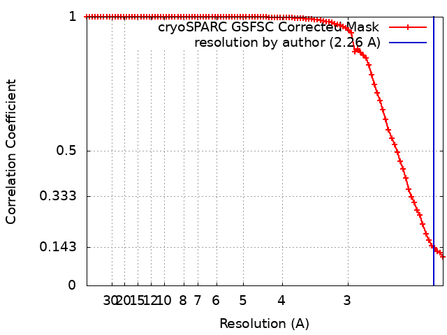
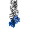
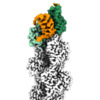
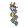
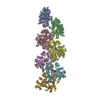
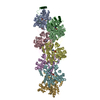
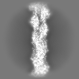
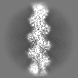
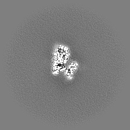
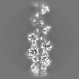
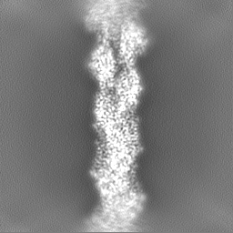
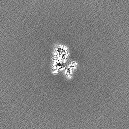
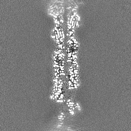
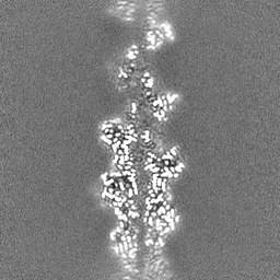
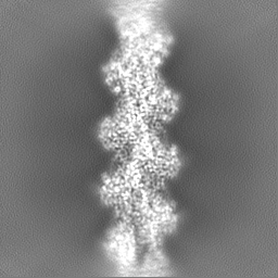
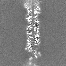
ジャーナル: Science / 年: 2023 タイトル: Structures of the free and capped ends of the actin filament. 著者: Peter J Carman / Kyle R Barrie / Grzegorz Rebowski / Roberto Dominguez / 要旨: The barbed and pointed ends of the actin filament (F-actin) are the sites of growth and shrinkage and the targets of capping proteins that block subunit exchange, including CapZ at the barbed end and ...The barbed and pointed ends of the actin filament (F-actin) are the sites of growth and shrinkage and the targets of capping proteins that block subunit exchange, including CapZ at the barbed end and tropomodulin at the pointed end. We describe cryo-electron microscopy structures of the free and capped ends of F-actin. Terminal subunits at the free barbed end adopt a "flat" F-actin conformation. CapZ binds with minor changes to the barbed end but with major changes to itself. By contrast, subunits at the free pointed end adopt a "twisted" monomeric actin (G-actin) conformation. Tropomodulin binding forces the second subunit into an F-actin conformation. The structures reveal how the ends differ from the middle in F-actin and how these differences control subunit addition, dissociation, capping, and interactions with end-binding proteins.
 ムービー
ムービー コントローラー


 データを開く
 基本情報
基本情報
 マップデータ
 試料
試料 キーワード
キーワード 機能・相同性情報
 データ登録者
 米国, 3件
  引用
引用 構造の表示
構造の表示
 ダウンロードとリンク
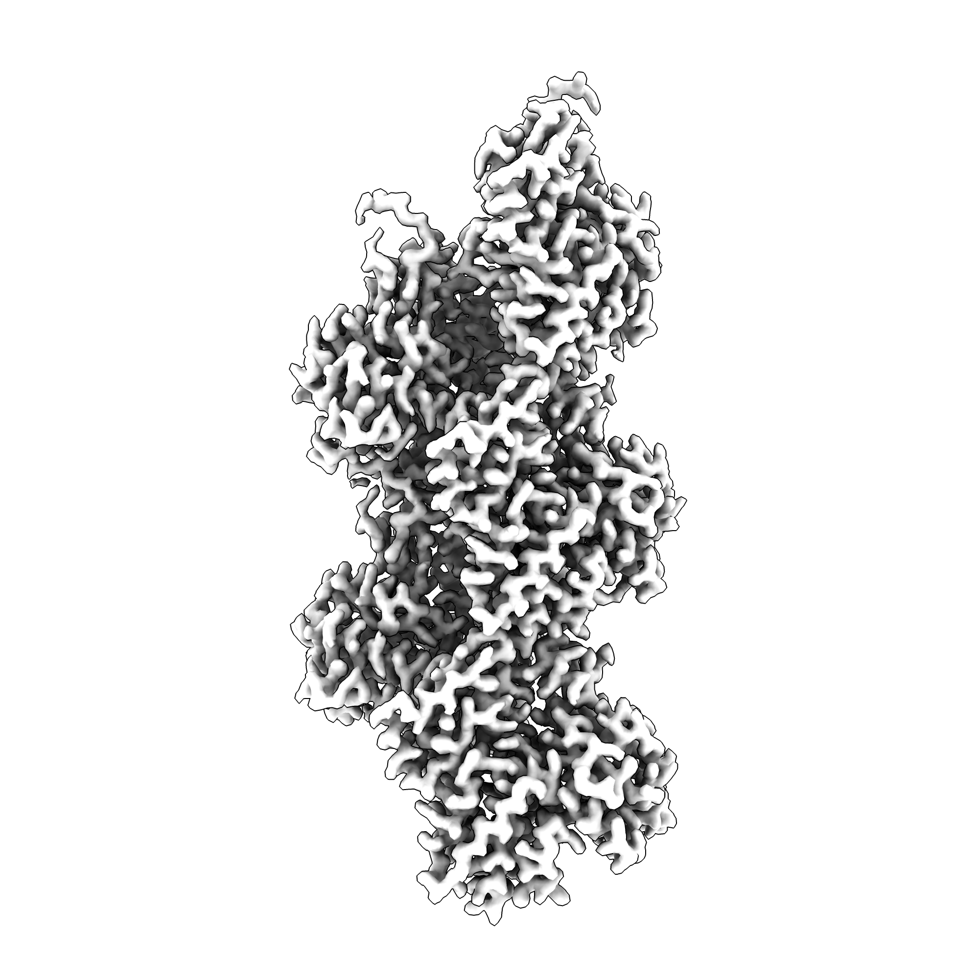 emd_28932.png
 http://ftp.pdbj.org/pub/emdb/structures/EMD-28932
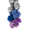


 Z
Z Y
Y X
X


 試料の構成要素
 解析
解析 電子顕微鏡法
 FIELD EMISSION GUN