+データを開く
-基本情報
登録情報 | データベース: EMDB / ID: EMD-22877 | |||||||||
---|---|---|---|---|---|---|---|---|---|---|
タイトル | E. coli ribosome structure | |||||||||
マップデータ | E. coli ribosome structure | |||||||||
試料 |
| |||||||||
生物種 | Escherichia coli (大腸菌) | |||||||||
手法 | サブトモグラム平均法 / クライオ電子顕微鏡法 / 解像度: 20.0 Å | |||||||||
データ登録者 | Chang YJ / Liu J / Xiang YJ / Jacobs-Wagner C | |||||||||
資金援助 | 米国, 2件
| |||||||||
引用 | 
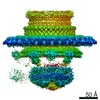
ジャーナル: Cell / 年: 2021 タイトル: Interconnecting solvent quality, transcription, and chromosome folding in Escherichia coli. 著者: Yingjie Xiang / Ivan V Surovtsev / Yunjie Chang / Sander K Govers / Bradley R Parry / Jun Liu / Christine Jacobs-Wagner / 要旨: All cells fold their genomes, including bacterial cells, where the chromosome is compacted into a domain-organized meshwork called the nucleoid. How compaction and domain organization arise is not ...All cells fold their genomes, including bacterial cells, where the chromosome is compacted into a domain-organized meshwork called the nucleoid. How compaction and domain organization arise is not fully understood. Here, we describe a method to estimate the average mesh size of the nucleoid in Escherichia coli. Using nucleoid mesh size and DNA concentration estimates, we find that the cytoplasm behaves as a poor solvent for the chromosome when the cell is considered as a simple semidilute polymer solution. Monte Carlo simulations suggest that a poor solvent leads to chromosome compaction and DNA density heterogeneity (i.e., domain formation) at physiological DNA concentration. Fluorescence microscopy reveals that the heterogeneous DNA density negatively correlates with ribosome density within the nucleoid, consistent with cryoelectron tomography data. Drug experiments, together with past observations, suggest the hypothesis that RNAs contribute to the poor solvent effects, connecting chromosome compaction and domain formation to transcription and intracellular organization. | |||||||||
履歴 |
|
-構造の表示
ムービー |
ムービービューア |
---|---|
構造ビューア | EMマップ: SurfViewMolmilJmol/JSmol |
添付画像 |
-ダウンロードとリンク
-EMDBアーカイブ
マップデータ | emd_22877.map.gz | 1.3 MB | EMDBマップデータ形式 | |
---|---|---|---|---|
ヘッダ (付随情報) | emd-22877-v30.xml emd-22877.xml | 8.7 KB 8.7 KB | 表示 表示 | EMDBヘッダ |
FSC (解像度算出) | emd_22877_fsc.xml | 2.6 KB | 表示 | FSCデータファイル |
画像 | emd_22877.png | 90.2 KB | ||
アーカイブディレクトリ | http://ftp.pdbj.org/pub/emdb/structures/EMD-22877 ftp://ftp.pdbj.org/pub/emdb/structures/EMD-22877 | HTTPS FTP |
-検証レポート
文書・要旨 | emd_22877_validation.pdf.gz | 431.1 KB | 表示 | EMDB検証レポート |
---|---|---|---|---|
文書・詳細版 | emd_22877_full_validation.pdf.gz | 430.7 KB | 表示 | |
XML形式データ | emd_22877_validation.xml.gz | 6.9 KB | 表示 | |
CIF形式データ | emd_22877_validation.cif.gz | 8.2 KB | 表示 | |
アーカイブディレクトリ | https://ftp.pdbj.org/pub/emdb/validation_reports/EMD-22877 ftp://ftp.pdbj.org/pub/emdb/validation_reports/EMD-22877 | HTTPS FTP |
-関連構造データ
関連構造データ | C: 同じ文献を引用 (文献) |
---|---|
類似構造データ | |
電子顕微鏡画像生データ | EMPIAR-10589 (タイトル: cryo-FIB and cryo-ET study of ribosome and polysome structures in E. coli Data size: 9.7 Data #1: Unaligned tilt series of cryo-FIB milling E. coli cells [tilt series]) |
-リンク
EMDBのページ | EMDB (EBI/PDBe) / EMDataResource |
---|---|
「今月の分子」の関連する項目 |
-マップ
ファイル | ダウンロード / ファイル: emd_22877.map.gz / 形式: CCP4 / 大きさ: 1.4 MB / タイプ: IMAGE STORED AS FLOATING POINT NUMBER (4 BYTES) | ||||||||||||||||||||||||||||||||||||||||||||||||||||||||||||||||||||
---|---|---|---|---|---|---|---|---|---|---|---|---|---|---|---|---|---|---|---|---|---|---|---|---|---|---|---|---|---|---|---|---|---|---|---|---|---|---|---|---|---|---|---|---|---|---|---|---|---|---|---|---|---|---|---|---|---|---|---|---|---|---|---|---|---|---|---|---|---|
注釈 | E. coli ribosome structure | ||||||||||||||||||||||||||||||||||||||||||||||||||||||||||||||||||||
ボクセルのサイズ | X=Y=Z: 5.457 Å | ||||||||||||||||||||||||||||||||||||||||||||||||||||||||||||||||||||
密度 |
| ||||||||||||||||||||||||||||||||||||||||||||||||||||||||||||||||||||
対称性 | 空間群: 1 | ||||||||||||||||||||||||||||||||||||||||||||||||||||||||||||||||||||
詳細 | EMDB XML:
CCP4マップ ヘッダ情報:
|
-添付データ
-試料の構成要素
-全体 : subtomogram average structure of E. coli ribosome
全体 | 名称: subtomogram average structure of E. coli ribosome |
---|---|
要素 |
|
-超分子 #1: subtomogram average structure of E. coli ribosome
超分子 | 名称: subtomogram average structure of E. coli ribosome / タイプ: complex / ID: 1 / 親要素: 0 |
---|---|
由来(天然) | 生物種: Escherichia coli (大腸菌) |
-実験情報
-構造解析
手法 | クライオ電子顕微鏡法 |
---|---|
解析 | サブトモグラム平均法 |
試料の集合状態 | cell |
-試料調製
緩衝液 | pH: 7.2 |
---|---|
グリッド | モデル: Quantifoil / 材質: COPPER / メッシュ: 200 / 支持フィルム - 材質: CARBON / 支持フィルム - トポロジー: HOLEY / 前処理 - タイプ: GLOW DISCHARGE / 前処理 - 時間: 30 sec. |
凍結 | 凍結剤: ETHANE |
-電子顕微鏡法
顕微鏡 | FEI TITAN KRIOS |
---|---|
特殊光学系 | エネルギーフィルター - スリット幅: 20 eV |
撮影 | フィルム・検出器のモデル: GATAN K2 SUMMIT (4k x 4k) 検出モード: COUNTING / 平均電子線量: 100.0 e/Å2 |
電子線 | 加速電圧: 300 kV / 電子線源: FIELD EMISSION GUN |
電子光学系 | 照射モード: FLOOD BEAM / 撮影モード: BRIGHT FIELD / Cs: 2.7 mm |
実験機器 | モデル: Titan Krios / 画像提供: FEI Company |