National Institutes of Health/National Institute of General Medical Sciences (NIH/NIGMS)
R01-GM081871
United States
Other private
Simons Foundation/SF349247
United States
National Institutes of Health/National Institute of General Medical Sciences (NIH/NIGMS)
F32GM128303
United States
Other private
Agouron Institute/F00316
United States
National Institutes of Health/National Institute of General Medical Sciences (NIH/NIGMS)
GM103310
United States
National Institutes of Health/Office of the Director
OD019994
United States
Citation
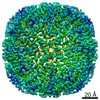
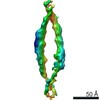
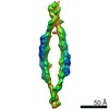
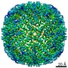
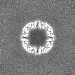
Journal: Nat Commun / Year: 2020 Title: Topaz-Denoise: general deep denoising models for cryoEM and cryoET. Authors: Tristan Bepler / Kotaro Kelley / Alex J Noble / Bonnie Berger / Abstract: Cryo-electron microscopy (cryoEM) is becoming the preferred method for resolving protein structures. Low signal-to-noise ratio (SNR) in cryoEM images reduces the confidence and throughput of ...Cryo-electron microscopy (cryoEM) is becoming the preferred method for resolving protein structures. Low signal-to-noise ratio (SNR) in cryoEM images reduces the confidence and throughput of structure determination during several steps of data processing, resulting in impediments such as missing particle orientations. Denoising cryoEM images can not only improve downstream analysis but also accelerate the time-consuming data collection process by allowing lower electron dose micrographs to be used for analysis. Here, we present Topaz-Denoise, a deep learning method for reliably and rapidly increasing the SNR of cryoEM images and cryoET tomograms. By training on a dataset composed of thousands of micrographs collected across a wide range of imaging conditions, we are able to learn models capturing the complexity of the cryoEM image formation process. The general model we present is able to denoise new datasets without additional training. Denoising with this model improves micrograph interpretability and allows us to solve 3D single particle structures of clustered protocadherin, an elongated particle with previously elusive views. We then show that low dose collection, enabled by Topaz-Denoise, improves downstream analysis in addition to reducing data collection time. We also present a general 3D denoising model for cryoET. Topaz-Denoise and pre-trained general models are now included in Topaz. We expect that Topaz-Denoise will be of broad utility to the cryoEM community for improving micrograph and tomogram interpretability and accelerating analysis.
History
Deposition
May 26, 2020
-
Header (metadata) release
Aug 26, 2020
-
Map release
Aug 26, 2020
-
Update
Dec 2, 2020
-
Current status
Dec 2, 2020
Processing site: RCSB / Status: Released
-
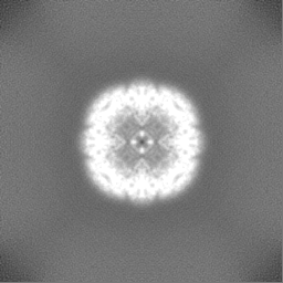
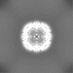
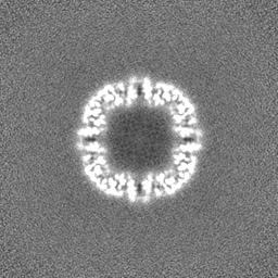
Structure visualization
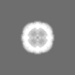
Movie
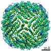
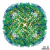
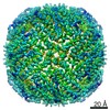
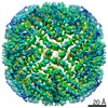
Surface view with section colored by density value
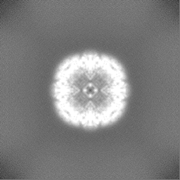
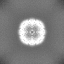
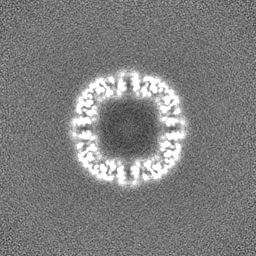
EMPIAR-10473 (Title: Micrograph frames from 110 internal SEMC/NYSBC test datasets used for Topaz-Denoise model generation & analysis Data size: 33.9 TB Data #1: Unaligned micrograph frames from 110 internal SEMC/NYSBC test datasets [micrographs - single frame])
In the structure databanks used in Yorodumi, some data are registered as the other names, "COVID-19 virus" and "2019-nCoV". Here are the details of the virus and the list of structure data.
Jan 31, 2019. EMDB accession codes are about to change! (news from PDBe EMDB page)
EMDB accession codes are about to change! (news from PDBe EMDB page)
The allocation of 4 digits for EMDB accession codes will soon come to an end. Whilst these codes will remain in use, new EMDB accession codes will include an additional digit and will expand incrementally as the available range of codes is exhausted. The current 4-digit format prefixed with “EMD-” (i.e. EMD-XXXX) will advance to a 5-digit format (i.e. EMD-XXXXX), and so on. It is currently estimated that the 4-digit codes will be depleted around Spring 2019, at which point the 5-digit format will come into force.
The EM Navigator/Yorodumi systems omit the EMD- prefix.
Related info.:Q: What is EMD? / ID/Accession-code notation in Yorodumi/EM Navigator
Yorodumi is a browser for structure data from EMDB, PDB, SASBDB, etc.
This page is also the successor to EM Navigator detail page, and also detail information page/front-end page for Omokage search.
The word "yorodu" (or yorozu) is an old Japanese word meaning "ten thousand". "mi" (miru) is to see.
Related info.:EMDB / PDB / SASBDB / Comparison of 3 databanks / Yorodumi Search / Aug 31, 2016. New EM Navigator & Yorodumi / Yorodumi Papers / Jmol/JSmol / Function and homology information / Changes in new EM Navigator and Yorodumi
 Movie
Movie Controller

 Yorodumi
 Open data
 Basic information
 Map data
 Sample
 Authors
 United States, 6 items
  Citation
 Structure visualization
 Movie viewer
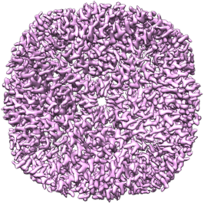
 Downloads & links
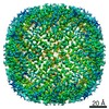
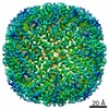
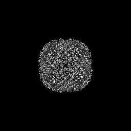
 emd_22058.png
 http://ftp.pdbj.org/pub/emdb/structures/EMD-22058
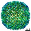

 Z (Sec.)
 Y (Row.)
 X (Col.)


 Sample components
 Processing
 Electron microscopy
 FIELD EMISSION GUN
