Movie
Movie Controller

[English] 日本語
 Yorodumi
- EMDB-21696: SD-like state of human 26S Proteasome with non-cleavable M1-linke... -
+ Open data

- Basic information
| Entry | Database: EMDB / ID: EMD-21696 | ||||||||||||
|---|---|---|---|---|---|---|---|---|---|---|---|---|---|
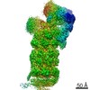
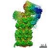
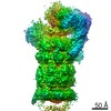
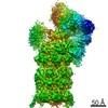
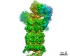
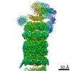
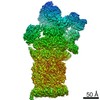
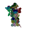
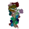
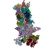
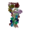
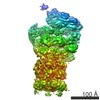
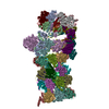
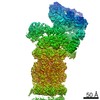
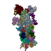
| Title | SD-like state of human 26S Proteasome with non-cleavable M1-linked hexaubiquitin and E3 ubiquitin ligase E6AP/UBE3A | ||||||||||||
 Map data Map data | SD-like state of human 26S Proteasome with non-cleavable M1-linked hexaubiquitin and E3 ubiquitin ligase E6AP/UBE3A | ||||||||||||
 Sample Sample |
| ||||||||||||
 Keywords Keywords | 26S protease / ubiquitin / complex / subunit / HYDROLASE-PROTEIN BINDING complex | ||||||||||||
| Function / homology |  Function and homology information Function and homology informationpositive regulation of inclusion body assembly / thyrotropin-releasing hormone receptor binding / nuclear proteasome complex / host-mediated perturbation of viral transcription / Impaired BRCA2 translocation to the nucleus / Impaired BRCA2 binding to SEM1 (DSS1) / proteasome accessory complex / purine ribonucleoside triphosphate binding / integrator complex / proteasome regulatory particle ...positive regulation of inclusion body assembly / thyrotropin-releasing hormone receptor binding / nuclear proteasome complex / host-mediated perturbation of viral transcription / Impaired BRCA2 translocation to the nucleus / Impaired BRCA2 binding to SEM1 (DSS1) / proteasome accessory complex / purine ribonucleoside triphosphate binding / integrator complex / proteasome regulatory particle / CD8-positive, alpha-beta T cell differentiation / thymic T cell selection / cytosolic proteasome complex / CD8-positive, alpha-beta T cell homeostasis / positive regulation of proteasomal protein catabolic process / proteasome-activating activity / Antigen processing: Ub, ATP-independent proteasomal degradation / proteasome regulatory particle, lid subcomplex / proteasome regulatory particle, base subcomplex / negative regulation of programmed cell death / T-helper 1 cell differentiation / negative regulation of regulatory T cell differentiation / protein K63-linked deubiquitination / metal-dependent deubiquitinase activity / cellular response to type I interferon / Regulation of ornithine decarboxylase (ODC) / proteasome core complex / Proteasome assembly / T-helper 17 cell differentiation / retrograde vesicle-mediated transport, Golgi to endoplasmic reticulum / Cross-presentation of soluble exogenous antigens (endosomes) / transcription factor binding / Somitogenesis / flagellated sperm motility / K63-linked deubiquitinase activity / Homologous DNA Pairing and Strand Exchange / Defective homologous recombination repair (HRR) due to BRCA1 loss of function / Defective HDR through Homologous Recombination Repair (HRR) due to PALB2 loss of BRCA1 binding function / Defective HDR through Homologous Recombination Repair (HRR) due to PALB2 loss of BRCA2/RAD51/RAD51C binding function / Resolution of D-loop Structures through Synthesis-Dependent Strand Annealing (SDSA) / Resolution of D-loop Structures through Holliday Junction Intermediates / proteasome binding / Impaired BRCA2 binding to RAD51 / myofibril / positive regulation of RNA polymerase II transcription preinitiation complex assembly / proteasomal ubiquitin-independent protein catabolic process / general transcription initiation factor binding / Presynaptic phase of homologous DNA pairing and strand exchange / proteasome storage granule / protein deubiquitination / AMPK-induced ERAD and lysosome mediated degradation of PD-L1(CD274) / polyubiquitin modification-dependent protein binding / proteasome endopeptidase complex / NF-kappaB binding / proteasome core complex, beta-subunit complex / endopeptidase activator activity / threonine-type endopeptidase activity / proteasome core complex, alpha-subunit complex / proteasome assembly / mRNA export from nucleus / GSK3B-mediated proteasomal degradation of PD-L1(CD274) / SARS-CoV-1 targets host intracellular signalling and regulatory pathways / SPOP-mediated proteasomal degradation of PD-L1(CD274) / regulation of G1/S transition of mitotic cell cycle / immune system process / enzyme regulator activity / regulation of macroautophagy / positive regulation of interleukin-2 production / ERAD pathway / ciliary tip / Ribosome Quality Control (RQC) complex extracts and degrades nascent peptide / response to type II interferon / inclusion body / TBP-class protein binding / stem cell differentiation / proteasome complex / : / regulation of proteasomal protein catabolic process / sarcomere / sperm end piece / Regulation of activated PAK-2p34 by proteasome mediated degradation / ubiquitin binding / proteasomal protein catabolic process / Autodegradation of Cdh1 by Cdh1:APC/C / negative regulation of inflammatory response to antigenic stimulus / APC/C:Cdc20 mediated degradation of Securin / N-glycan trimming in the ER and Calnexin/Calreticulin cycle / Asymmetric localization of PCP proteins / Ubiquitin-dependent degradation of Cyclin D / lipopolysaccharide binding / SCF-beta-TrCP mediated degradation of Emi1 / NIK-->noncanonical NF-kB signaling / AUF1 (hnRNP D0) binds and destabilizes mRNA / TNFR2 non-canonical NF-kB pathway / Assembly of the pre-replicative complex / Vpu mediated degradation of CD4 / P-body / Cdc20:Phospho-APC/C mediated degradation of Cyclin A / Dectin-1 mediated noncanonical NF-kB signaling / Degradation of DVL Similarity search - Function | ||||||||||||
| Biological species |  Homo sapiens (human) Homo sapiens (human) | ||||||||||||
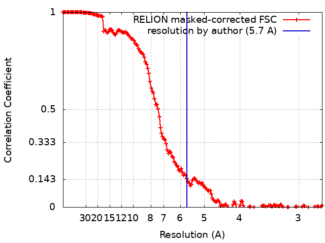
| Method | single particle reconstruction / cryo EM / Resolution: 5.7 Å | ||||||||||||
 Authors Authors | Chen X / Walters KJ | ||||||||||||
| Funding support |  United States, 3 items United States, 3 items
| ||||||||||||
 Citation Citation | Journal: Structure / Year: 2020 Title: Cryo-EM Reveals Unanchored M1-Ubiquitin Chain Binding at hRpn11 of the 26S Proteasome. Authors: Xiang Chen / Zachary Dorris / Dan Shi / Rick K Huang / Htet Khant / Tara Fox / Natalia de Val / Dewight Williams / Ping Zhang / Kylie J Walters / Abstract: The 26S proteasome is specialized for regulated protein degradation and formed by a dynamic regulatory particle (RP) that caps a hollow cylindrical core particle (CP) where substrates are proteolyzed. ...The 26S proteasome is specialized for regulated protein degradation and formed by a dynamic regulatory particle (RP) that caps a hollow cylindrical core particle (CP) where substrates are proteolyzed. Its diverse substrates unify as proteasome targets by ubiquitination. We used cryogenic electron microscopy (cryo-EM) to study how human 26S proteasome interacts with M1-linked hexaubiquitin (M1-Ub) unanchored to a substrate and E3 ubiquitin ligase E6AP/UBE3A. Proteasome structures are available with model substrates extending through the RP ATPase ring and substrate-conjugated K63-linked ubiquitin chains present at inhibited deubiquitinating enzyme hRpn11 and the nearby ATPase hRpt4/hRpt5 coiled coil. In this study, we find M1-Ub at the hRpn11 site despite the absence of conjugated substrate, indicating that ubiquitin binding at this location does not require substrate interaction with the RP. Moreover, unanchored M1-Ub binds to this hRpn11 site of the proteasome with the CP gating residues in both the closed and opened conformational states. | ||||||||||||
| History |
|
- Structure visualization
| Movie |
Movie viewer |
|---|---|
| Structure viewer | EM map: SurfViewMolmilJmol/JSmol |
| Supplemental images |

- Downloads & links
-EMDB archive
| Map data | emd_21696.map.gz | 24 MB | EMDB map data format | |
|---|---|---|---|---|
| Header (meta data) | emd-21696-v30.xmlemd-21696.xml | 52.8 KB 52.8 KB | Display Display | EMDB header |
| FSC (resolution estimation) | emd_21696_fsc.xml | 16 KB | Display | FSC data file |
| Images | 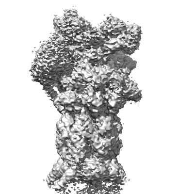 emd_21696.png emd_21696.png | 117.5 KB | ||
| Filedesc metadata | emd-21696.cif.gz | 13.5 KB | ||
| Archive directory |  http://ftp.pdbj.org/pub/emdb/structures/EMD-21696ftp://ftp.pdbj.org/pub/emdb/structures/EMD-21696 http://ftp.pdbj.org/pub/emdb/structures/EMD-21696ftp://ftp.pdbj.org/pub/emdb/structures/EMD-21696 | HTTPS FTP |
-Related structure data
| Related structure data |  6wjnMC 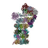 6wjdC C: citing same article ( M: atomic model generated by this map |
|---|---|
| Similar structure data | |
| EM raw data | EMPIAR-10401 (Title: SA-like and SD-like states of human 26S Proteasome with non-cleavable M1-linked hexaubiquitin and E3 ubiquitin ligase E6AP/UBE3A Data size: 330.3 Data #1: Motion-corrected single frame micrographs of human 26S Proteasome with non-cleavable M1-linked hexaubiquitin and E3 ubiquitin ligase E6AP/UBE3A [micrographs - single frame]) |
-Links
| EMDB pages | EMDB (EBI/PDBe) / EMDataResource |
|---|---|
| Related items in Molecule of the Month |


-Map
| File | Download / File: emd_21696.map.gz / Format: CCP4 / Size: 347.6 MB / Type: IMAGE STORED AS FLOATING POINT NUMBER (4 BYTES) | ||||||||||||||||||||||||||||||||||||||||||||||||||||||||||||
|---|---|---|---|---|---|---|---|---|---|---|---|---|---|---|---|---|---|---|---|---|---|---|---|---|---|---|---|---|---|---|---|---|---|---|---|---|---|---|---|---|---|---|---|---|---|---|---|---|---|---|---|---|---|---|---|---|---|---|---|---|---|
| Annotation | SD-like state of human 26S Proteasome with non-cleavable M1-linked hexaubiquitin and E3 ubiquitin ligase E6AP/UBE3A | ||||||||||||||||||||||||||||||||||||||||||||||||||||||||||||
| Projections & slices | Image control
Images are generated by Spider. | ||||||||||||||||||||||||||||||||||||||||||||||||||||||||||||
| Voxel size | X=Y=Z: 1.365 Å | ||||||||||||||||||||||||||||||||||||||||||||||||||||||||||||
| Density |
| ||||||||||||||||||||||||||||||||||||||||||||||||||||||||||||
| Symmetry | Space group: 1 | ||||||||||||||||||||||||||||||||||||||||||||||||||||||||||||
| Details | EMDB XML:
CCP4 map header:
| ||||||||||||||||||||||||||||||||||||||||||||||||||||||||||||
 Z (Sec.)
 Y (Row.)
 X (Col.)


-Supplemental data
- Sample components
+Entire : SD-like state of human 26S Proteasome with non-cleavable M1-linke...
+Supramolecule #1: SD-like state of human 26S Proteasome with non-cleavable M1-linke...
+Macromolecule #1: 26S proteasome non-ATPase regulatory subunit 1
+Macromolecule #2: 26S proteasome non-ATPase regulatory subunit 3
+Macromolecule #3: 26S proteasome non-ATPase regulatory subunit 12
+Macromolecule #4: 26S proteasome non-ATPase regulatory subunit 11
+Macromolecule #5: 26S proteasome non-ATPase regulatory subunit 6
+Macromolecule #6: 26S proteasome non-ATPase regulatory subunit 7
+Macromolecule #7: 26S proteasome non-ATPase regulatory subunit 13
+Macromolecule #8: 26S proteasome non-ATPase regulatory subunit 4
+Macromolecule #9: 26S proteasome non-ATPase regulatory subunit 14
+Macromolecule #10: 26S proteasome non-ATPase regulatory subunit 8
+Macromolecule #11: 26S proteasome complex subunit SEM1
+Macromolecule #12: Proteasome subunit alpha type-6
+Macromolecule #13: Proteasome subunit alpha type-2
+Macromolecule #14: Proteasome subunit alpha type-4
+Macromolecule #15: Proteasome subunit alpha type-7
+Macromolecule #16: Proteasome subunit alpha type-5
+Macromolecule #17: Proteasome subunit alpha type-1
+Macromolecule #18: Proteasome subunit alpha type-3
+Macromolecule #19: Proteasome subunit beta type-6
+Macromolecule #20: Proteasome subunit beta type-7
+Macromolecule #21: Proteasome subunit beta type-3
+Macromolecule #22: Proteasome subunit beta type-2
+Macromolecule #23: Proteasome subunit beta type-5
+Macromolecule #24: Proteasome subunit beta type-1
+Macromolecule #25: Proteasome subunit beta type-4
+Macromolecule #26: 26S proteasome regulatory subunit 7
+Macromolecule #27: 26S proteasome regulatory subunit 4
+Macromolecule #28: 26S proteasome regulatory subunit 8
+Macromolecule #29: 26S proteasome regulatory subunit 6B
+Macromolecule #30: 26S proteasome regulatory subunit 10B
+Macromolecule #31: 26S proteasome regulatory subunit 6A
+Macromolecule #32: 26S proteasome non-ATPase regulatory subunit 2
+Macromolecule #33: ZINC ION
+Macromolecule #34: PHOSPHOTHIOPHOSPHORIC ACID-ADENYLATE ESTER

-Experimental details
-Structure determination
| Method | cryo EM |
|---|---|
 Processing Processing | single particle reconstruction |
| Aggregation state | particle |
-Sample preparation
| Concentration | 1 mg/mL | |||||||||||||||||||||
|---|---|---|---|---|---|---|---|---|---|---|---|---|---|---|---|---|---|---|---|---|---|---|
| Buffer | pH: 7.5 Component:
Details: 50 mM Tris, pH 7.5, 50 mM NaCl, 1.5 mM ATP-gamma-S, 5 mM MgCl2, 2 mM DTT, 10 uM zinc sulfate | |||||||||||||||||||||
| Grid | Model: Quantifoil R1.2/1.3 / Material: COPPER / Mesh: 200 / Pretreatment - Type: GLOW DISCHARGE / Pretreatment - Time: 30 sec. | |||||||||||||||||||||
| Vitrification | Cryogen name: ETHANE / Chamber humidity: 100 % / Chamber temperature: 291.15 K / Instrument: FEI VITROBOT MARK IV |
- Electron microscopy
| Microscope | FEI TITAN KRIOS |
|---|---|
| Image recording | Film or detector model: GATAN K2 SUMMIT (4k x 4k) / Average electron dose: 36.64 e/Å2 |
| Electron beam | Acceleration voltage: 300 kV / Electron source:  FIELD EMISSION GUN FIELD EMISSION GUN |
| Electron optics | Illumination mode: FLOOD BEAM / Imaging mode: BRIGHT FIELD |
| Sample stage | Cooling holder cryogen: NITROGEN |
| Experimental equipment |  Model: Titan Krios / Image courtesy: FEI Company |