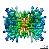
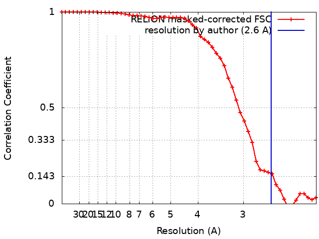
ジャーナル: Proc Natl Acad Sci U S A / 年: 2020 タイトル: High-yield monolayer graphene grids for near-atomic resolution cryoelectron microscopy. 著者: Yimo Han / Xiao Fan / Haozhe Wang / Fang Zhao / Christopher G Tully / Jing Kong / Nan Yao / Nieng Yan / 要旨: Cryogenic electron microscopy (cryo-EM) has become one of the most powerful techniques to reveal the atomic structures and working mechanisms of biological macromolecules. New designs of the cryo-EM ...Cryogenic electron microscopy (cryo-EM) has become one of the most powerful techniques to reveal the atomic structures and working mechanisms of biological macromolecules. New designs of the cryo-EM grids-aimed at preserving thin, uniform vitrified ice and improving protein adsorption-have been considered a promising approach to achieving higher resolution with the minimal amount of materials and data. Here, we describe a method for preparing graphene cryo-EM grids with up to 99% monolayer graphene coverage that allows for more than 70% grid squares for effective data acquisition with improved image quality and protein density. Using our graphene grids, we have achieved 2.6-Å resolution for streptavidin, with a molecular weight of 52 kDa, from 11,000 particles. Our graphene grids increase the density of examined soluble, membrane, and lipoproteins by at least 5-fold, affording the opportunity for structural investigation of challenging proteins which cannot be produced in large quantity. In addition, our method employs only simple tools that most structural biology laboratories can access. Moreover, this approach supports customized grid designs targeting specific proteins, owing to its broad compatibility with a variety of nanomaterials.
 ムービー
ムービー コントローラー


 データを開く
 基本情報
基本情報 マップデータ
 試料
試料 機能・相同性情報
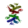
 Streptomyces avidinii (バクテリア)
 データ登録者
 米国, 1件
  引用
引用 構造の表示
構造の表示
 ダウンロードとリンク
 emd_20907.png
 http://ftp.pdbj.org/pub/emdb/structures/EMD-20907


 試料の構成要素
 解析
解析 電子顕微鏡法
 FIELD EMISSION GUN