 Movie
Movie Controller

[English] 日本語
 Yorodumi
- EMDB-16039: Tau Paired Helical Filament from Cellular Fraction of Alzheimer's... -
+ Open data

- Basic information
| Entry |  | |||||||||
|---|---|---|---|---|---|---|---|---|---|---|
| Title | Tau Paired Helical Filament from Cellular Fraction of Alzheimer's disease brain | |||||||||
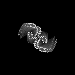
 Map data Map data | ||||||||||
 Sample Sample |
| |||||||||
 Keywords Keywords | PROTEIN FIBRIL / AMYLOID | |||||||||
| Function / homology |  Function and homology information Function and homology informationplus-end-directed organelle transport along microtubule / histone-dependent DNA binding / negative regulation of protein localization to mitochondrion / neurofibrillary tangle / microtubule lateral binding / axonal transport / tubulin complex / positive regulation of protein localization to synapse / phosphatidylinositol bisphosphate binding / generation of neurons ...plus-end-directed organelle transport along microtubule / histone-dependent DNA binding / negative regulation of protein localization to mitochondrion / neurofibrillary tangle / microtubule lateral binding / axonal transport / tubulin complex / positive regulation of protein localization to synapse / phosphatidylinositol bisphosphate binding / generation of neurons / rRNA metabolic process / axonal transport of mitochondrion / regulation of mitochondrial fission / axon development / regulation of microtubule-based movement / intracellular distribution of mitochondria / regulation of chromosome organization / central nervous system neuron development / minor groove of adenine-thymine-rich DNA binding / lipoprotein particle binding / microtubule polymerization / negative regulation of mitochondrial membrane potential / regulation of microtubule polymerization / dynactin binding / apolipoprotein binding / protein polymerization / main axon / Caspase-mediated cleavage of cytoskeletal proteins / regulation of microtubule polymerization or depolymerization / negative regulation of mitochondrial fission / axolemma / glial cell projection / neurofibrillary tangle assembly / positive regulation of axon extension / regulation of cellular response to heat / positive regulation of microtubule polymerization / positive regulation of protein localization / Activation of AMPK downstream of NMDARs / positive regulation of superoxide anion generation / cellular response to brain-derived neurotrophic factor stimulus / regulation of long-term synaptic depression / supramolecular fiber organization / cytoplasmic microtubule organization / regulation of calcium-mediated signaling / axon cytoplasm / somatodendritic compartment / synapse assembly / phosphatidylinositol binding / nuclear periphery / astrocyte activation / enzyme inhibitor activity / protein phosphatase 2A binding / stress granule assembly / regulation of microtubule cytoskeleton organization / regulation of autophagy / cellular response to reactive oxygen species / microglial cell activation / cellular response to nerve growth factor stimulus / Hsp90 protein binding / protein homooligomerization / SH3 domain binding / PKR-mediated signaling / synapse organization / regulation of synaptic plasticity / response to lead ion / microtubule cytoskeleton organization / memory / neuron projection development / cytoplasmic ribonucleoprotein granule / cell-cell signaling / single-stranded DNA binding / cellular response to heat / protein-folding chaperone binding / growth cone / microtubule cytoskeleton / actin binding / cell body / double-stranded DNA binding / microtubule binding / sequence-specific DNA binding / amyloid fibril formation / dendritic spine / microtubule / protein-macromolecule adaptor activity / learning or memory / neuron projection / membrane raft / negative regulation of gene expression / axon / neuronal cell body / DNA damage response / dendrite / protein kinase binding / enzyme binding / mitochondrion / DNA binding / RNA binding / extracellular region / identical protein binding / nucleus Similarity search - Function | |||||||||
| Biological species |  Homo sapiens (human) Homo sapiens (human) | |||||||||
| Method | helical reconstruction / cryo EM / Resolution: 3.27 Å | |||||||||
 Authors Authors | Behr TS / Ryskeldi-Falcon B | |||||||||
| Funding support |  United Kingdom, 1 items United Kingdom, 1 items
| |||||||||
 Citation Citation | Journal: bioRxiv / Year: 2023 Title: Tau filaments are tethered within brain extracellular vesicles in Alzheimer's disease. Authors: S L Fowler / T S Behr / E Turkes / P Maglio Cauhy / M S Foiani / A Schaler / G Crowley / S Bez / E Ficulle / E Tsefou / D P O'Brien / R Fischer / B Geary / P Gaur / C Miller / P D'Acunzo / E ...Authors: S L Fowler / T S Behr / E Turkes / P Maglio Cauhy / M S Foiani / A Schaler / G Crowley / S Bez / E Ficulle / E Tsefou / D P O'Brien / R Fischer / B Geary / P Gaur / C Miller / P D'Acunzo / E Levy / K E Duff / B Ryskeldi-Falcon Abstract: The abnormal assembly of tau protein in neurons is the pathological hallmark of multiple neurodegenerative diseases, including Alzheimer's disease (AD). In addition, assembled tau associates with ...The abnormal assembly of tau protein in neurons is the pathological hallmark of multiple neurodegenerative diseases, including Alzheimer's disease (AD). In addition, assembled tau associates with extracellular vesicles (EVs) in the central nervous system of patients with AD, which is linked to its clearance and prion-like propagation between neurons. However, the identities of the assembled tau species and the EVs, as well as how they associate, are not known. Here, we combined quantitative mass spectrometry, cryo-electron tomography and single-particle cryo-electron microscopy to study brain EVs from AD patients. We found filaments of truncated tau enclosed within EVs enriched in endo-lysosomal proteins. We observed multiple filament interactions, including with molecules that tethered filaments to the EV limiting membrane, suggesting selective packaging. Our findings will guide studies into the molecular mechanisms of EV-mediated secretion of assembled tau and inform the targeting of EV-associated tau as potential therapeutic and biomarker strategies for AD. | |||||||||
| History |
|
- Structure visualization
| Supplemental images |
|---|

- Downloads & links
-EMDB archive
| Map data | emd_16039.map.gz | 34.6 MB | EMDB map data format | |
|---|---|---|---|---|
| Header (meta data) | emd-16039-v30.xmlemd-16039.xml | 14.8 KB 14.8 KB | Display Display | EMDB header |
| FSC (resolution estimation) | emd_16039_fsc.xml | 11 KB | Display | FSC data file |
| Images | 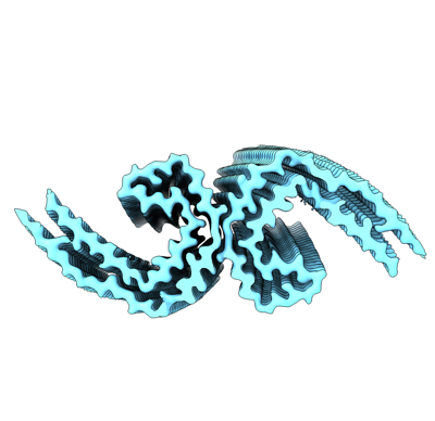 emd_16039.png emd_16039.png | 73.1 KB | ||
| Masks | emd_16039_msk_1.map | 113.6 MB | Mask map | |
| Filedesc metadata | emd-16039.cif.gz | 5.4 KB | ||
| Others | emd_16039_half_map_1.map.gzemd_16039_half_map_2.map.gz | 89.2 MB 89.2 MB | ||
| Archive directory |  http://ftp.pdbj.org/pub/emdb/structures/EMD-16039ftp://ftp.pdbj.org/pub/emdb/structures/EMD-16039 http://ftp.pdbj.org/pub/emdb/structures/EMD-16039ftp://ftp.pdbj.org/pub/emdb/structures/EMD-16039 | HTTPS FTP |
-Related structure data
| Related structure data |  8bgvMC  8bgsC M: atomic model generated by this map C: citing same article ( |
|---|---|
| Similar structure data |

-Links
| EMDB pages | EMDB (EBI/PDBe) / EMDataResource |
|---|---|
| Related items in Molecule of the Month |


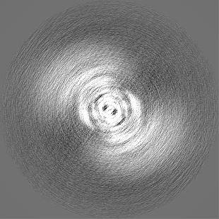
-Map
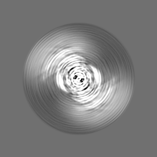
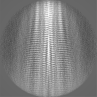
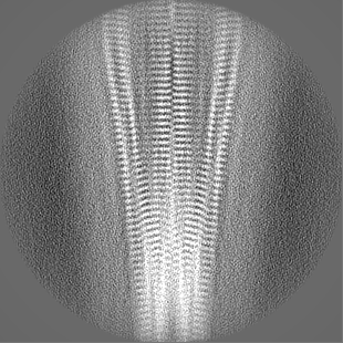
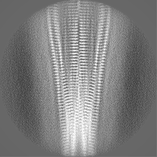
| File | Download / File: emd_16039.map.gz / Format: CCP4 / Size: 113.6 MB / Type: IMAGE STORED AS FLOATING POINT NUMBER (4 BYTES) | ||||||||||||||||||||||||||||||||||||
|---|---|---|---|---|---|---|---|---|---|---|---|---|---|---|---|---|---|---|---|---|---|---|---|---|---|---|---|---|---|---|---|---|---|---|---|---|---|
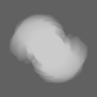
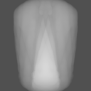
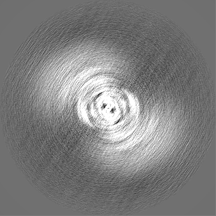
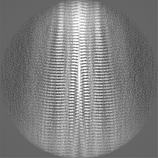
| Projections & slices | Image control
Images are generated by Spider. | ||||||||||||||||||||||||||||||||||||
| Voxel size | X=Y=Z: 0.834 Å | ||||||||||||||||||||||||||||||||||||
| Density |
| ||||||||||||||||||||||||||||||||||||
| Symmetry | Space group: 1 | ||||||||||||||||||||||||||||||||||||
| Details | EMDB XML:
|
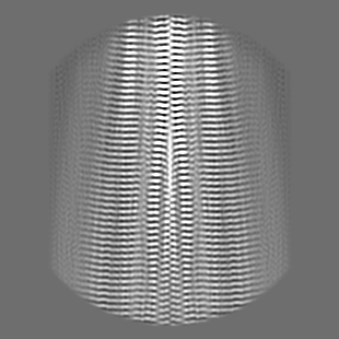
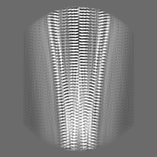
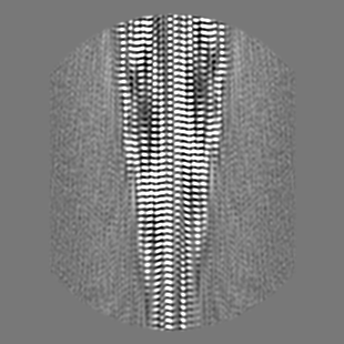
 Z (Sec.)
 Y (Row.)
 X (Col.)


-Supplemental data
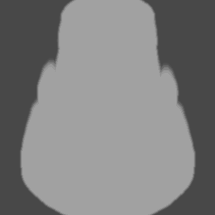
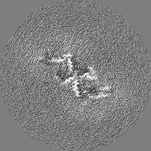
-Mask #1
| File | emd_16039_msk_1.map | ||||||||||||
|---|---|---|---|---|---|---|---|---|---|---|---|---|---|
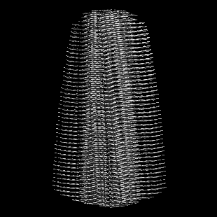
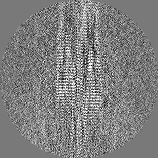
| Projections & Slices |
| ||||||||||||
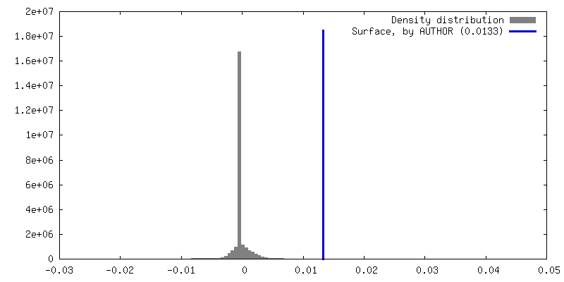
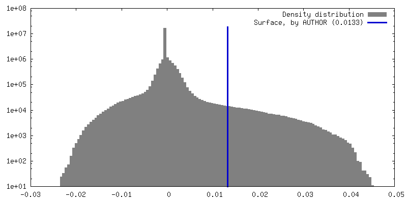
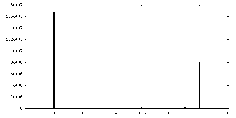
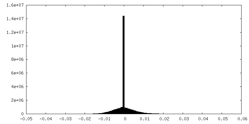
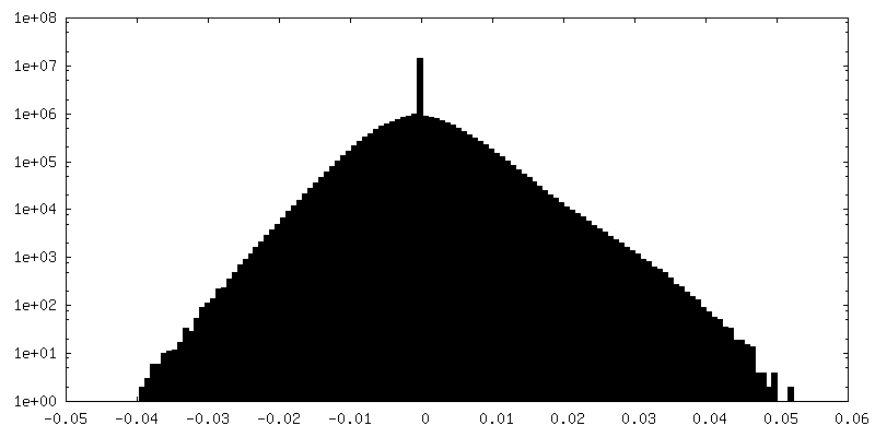
| Density Histograms |


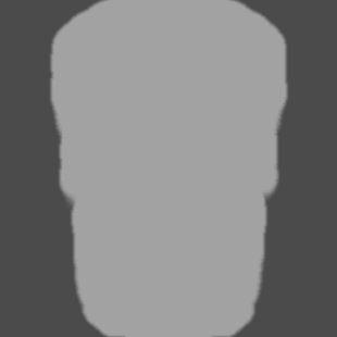

-Half map: #1
| File | emd_16039_half_map_1.map | ||||||||||||
|---|---|---|---|---|---|---|---|---|---|---|---|---|---|
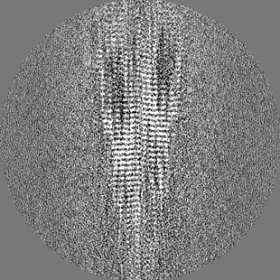
| Projections & Slices |
| ||||||||||||
| Density Histograms |

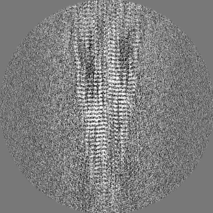
-Half map: #2
| File | emd_16039_half_map_2.map | ||||||||||||
|---|---|---|---|---|---|---|---|---|---|---|---|---|---|
| Projections & Slices |
| ||||||||||||
| Density Histograms |


- Sample components
-Entire : Tau Paired Helical Filament
| Entire | Name: Tau Paired Helical Filament |
|---|---|
| Components |
|
-Supramolecule #1: Tau Paired Helical Filament
| Supramolecule | Name: Tau Paired Helical Filament / type: tissue / ID: 1 / Parent: 0 / Macromolecule list: all Details: Tau Paired Helical Filament extracted from the cellular fraction of the brain of an individual who had Alzheimer's disease. |
|---|---|
| Source (natural) | Organism: Homo sapiens (human) / Organ: Brain / Tissue: Brain |
-Macromolecule #1: Microtubule-associated protein tau
| Macromolecule | Name: Microtubule-associated protein tau / type: protein_or_peptide / ID: 1 / Number of copies: 7 / Enantiomer: LEVO |
|---|---|
| Source (natural) | Organism: Homo sapiens (human) / Organ: Brain / Tissue: Brain |
| Molecular weight | Theoretical: 45.919871 KDa |
| Sequence | String: MAEPRQEFEV MEDHAGTYGL GDRKDQGGYT MHQDQEGDTD AGLKESPLQT PTEDGSEEPG SETSDAKSTP TAEDVTAPLV DEGAPGKQA AAQPHTEIPE GTTAEEAGIG DTPSLEDEAA GHVTQARMVS KSKDGTGSDD KKAKGADGKT KIATPRGAAP P GQKGQANA ...String: MAEPRQEFEV MEDHAGTYGL GDRKDQGGYT MHQDQEGDTD AGLKESPLQT PTEDGSEEPG SETSDAKSTP TAEDVTAPLV DEGAPGKQA AAQPHTEIPE GTTAEEAGIG DTPSLEDEAA GHVTQARMVS KSKDGTGSDD KKAKGADGKT KIATPRGAAP P GQKGQANA TRIPAKTPPA PKTPPSSGEP PKSGDRSGYS SPGSPGTPGS RSRTPSLPTP PTREPKKVAV VRTPPKSPSS AK SRLQTAP VPMPDLKNVK SKIGSTENLK HQPGGGKVQI INKKLDLSNV QSKCGSKDNI KHVPGGGSVQ IVYKPVDLSK VTS KCGSLG NIHHKPGGGQ VEVKSEKLDF KDRVQSKIGS LDNITHVPGG GNKKIETHKL TFRENAKAKT DHGAEIVYKS PVVS GDTSP RHLSNVSSTG SIDMVDSPQL ATLADEVSAS LAKQGL UniProtKB: Microtubule-associated protein tau |
-Experimental details
-Structure determination
| Method | cryo EM |
|---|---|
 Processing Processing | helical reconstruction |
| Aggregation state | filament |
-Sample preparation
| Buffer | pH: 7.4 |
|---|---|
| Vitrification | Cryogen name: ETHANE |
- Electron microscopy
| Microscope | FEI TITAN KRIOS |
|---|---|
| Image recording | Film or detector model: GATAN K3 (6k x 4k) / Average electron dose: 40.0 e/Å2 |
| Electron beam | Acceleration voltage: 300 kV / Electron source:  FIELD EMISSION GUN FIELD EMISSION GUN |
| Electron optics | Illumination mode: FLOOD BEAM / Imaging mode: BRIGHT FIELD / Nominal defocus max: 2.4 µm / Nominal defocus min: 1.0 µm |
| Experimental equipment |  Model: Titan Krios / Image courtesy: FEI Company |
-Image processing
| Final reconstruction | Applied symmetry - Helical parameters - Δz: 2.413 Å Applied symmetry - Helical parameters - Δ&Phi: 174.454 ° Applied symmetry - Helical parameters - Axial symmetry: C1 (asymmetric) Resolution.type: BY AUTHOR / Resolution: 3.27 Å / Resolution method: FSC 0.143 CUT-OFF / Number images used: 9347 |
|---|---|
| Startup model | Type of model: OTHER Details: Initial 3D reference models were generated de novo by producing sinograms from 2D class averages. |
| Final angle assignment | Type: NOT APPLICABLE |
| FSC plot (resolution estimation) | 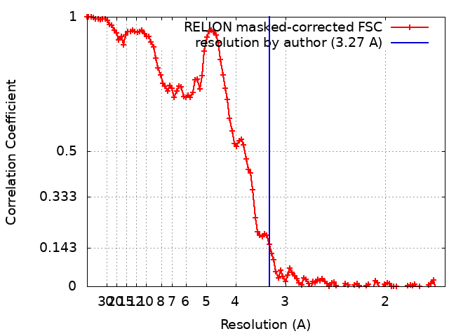 |
