Movie
Movie Controller

[English] 日本語
 Yorodumi
- EMDB-14719: FANCD2 Middle and C-terminal domains with USP1-NTE (focused refin... -
+ Open data

- Basic information
| Entry |  | |||||||||
|---|---|---|---|---|---|---|---|---|---|---|
| Title | FANCD2 Middle and C-terminal domains with USP1-NTE (focused refinement) | |||||||||
 Map data Map data | Locally refined, globally sharpened map | |||||||||
 Sample Sample |
| |||||||||
 Keywords Keywords | Complex / Enzyme-Substrate / HYDROLASE | |||||||||
| Function / homology |  Function and homology information Function and homology informationregulation of CD40 signaling pathway / gamete generation / regulation of regulatory T cell differentiation / homologous chromosome pairing at meiosis / neuronal stem cell population maintenance / double-strand break repair involved in meiotic recombination / brain morphogenesis / DNA repair complex / mitotic intra-S DNA damage checkpoint signaling / interstrand cross-link repair ...regulation of CD40 signaling pathway / gamete generation / regulation of regulatory T cell differentiation / homologous chromosome pairing at meiosis / neuronal stem cell population maintenance / double-strand break repair involved in meiotic recombination / brain morphogenesis / DNA repair complex / mitotic intra-S DNA damage checkpoint signaling / interstrand cross-link repair / condensed chromosome / DNA polymerase binding / response to gamma radiation / TP53 Regulates Transcription of DNA Repair Genes / Fanconi Anemia Pathway / cellular response to oxidative stress / regulation of inflammatory response / nuclear body / chromatin / nucleolus / nucleoplasm / nucleus / cytosol Similarity search - Function | |||||||||
| Biological species |  Homo sapiens (human) Homo sapiens (human) | |||||||||
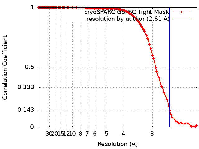
| Method | single particle reconstruction / cryo EM / Resolution: 2.61 Å | |||||||||
 Authors Authors | Rennie ML / Walden H | |||||||||
| Funding support | European Union,  United Kingdom, 2 items United Kingdom, 2 items
| |||||||||
 Citation Citation | Journal: Sci Adv / Year: 2022 Title: Cryo-EM reveals a mechanism of USP1 inhibition through a cryptic binding site. Authors: Martin L Rennie / Connor Arkinson / Viduth K Chaugule / Helen Walden / Abstract: Repair of DNA damage is critical to genomic integrity and frequently disrupted in cancers. Ubiquitin-specific protease 1 (USP1), a nucleus-localized deubiquitinase, lies at the interface of multiple ...Repair of DNA damage is critical to genomic integrity and frequently disrupted in cancers. Ubiquitin-specific protease 1 (USP1), a nucleus-localized deubiquitinase, lies at the interface of multiple DNA repair pathways and is a promising drug target for certain cancers. Although multiple inhibitors of this enzyme, including one in phase 1 clinical trials, have been established, their binding mode is unknown. Here, we use cryo-electron microscopy to study an assembled enzyme-substrate-inhibitor complex of USP1 and the well-established inhibitor, ML323. Achieving 2.5-Å resolution, with and without ML323, we find an unusual binding mode in which the inhibitor disrupts part of the hydrophobic core of USP1. The consequent conformational changes in the secondary structure lead to subtle rearrangements in the active site that underlie the mechanism of inhibition. These structures provide a platform for structure-based drug design targeting USP1. | |||||||||
| History |
|
- Structure visualization
| Supplemental images |
|---|

- Downloads & links
-EMDB archive
| Map data | emd_14719.map.gz | 118 MB | EMDB map data format | |
|---|---|---|---|---|
| Header (meta data) | emd-14719-v30.xmlemd-14719.xml | 18.3 KB 18.3 KB | Display Display | EMDB header |
| FSC (resolution estimation) | emd_14719_fsc.xml | 10.5 KB | Display | FSC data file |
| Images | 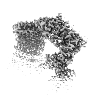 emd_14719.png emd_14719.png | 80.8 KB | ||
| Masks | emd_14719_msk_1.map | 125 MB | Mask map | |
| Filedesc metadata | emd-14719.cif.gz | 5.8 KB | ||
| Others | emd_14719_additional_1.map.gzemd_14719_half_map_1.map.gzemd_14719_half_map_2.map.gz | 35.1 MB 115.9 MB 115.9 MB | ||
| Archive directory |  http://ftp.pdbj.org/pub/emdb/structures/EMD-14719ftp://ftp.pdbj.org/pub/emdb/structures/EMD-14719 http://ftp.pdbj.org/pub/emdb/structures/EMD-14719ftp://ftp.pdbj.org/pub/emdb/structures/EMD-14719 | HTTPS FTP |
-Related structure data


-Links
| EMDB pages | EMDB (EBI/PDBe) / EMDataResource |
|---|---|
| Related items in Molecule of the Month |


-Map
| File | Download / File: emd_14719.map.gz / Format: CCP4 / Size: 125 MB / Type: IMAGE STORED AS FLOATING POINT NUMBER (4 BYTES) | ||||||||||||||||||||||||||||||||||||
|---|---|---|---|---|---|---|---|---|---|---|---|---|---|---|---|---|---|---|---|---|---|---|---|---|---|---|---|---|---|---|---|---|---|---|---|---|---|
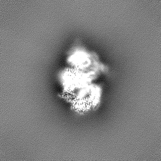
| Annotation | Locally refined, globally sharpened map | ||||||||||||||||||||||||||||||||||||
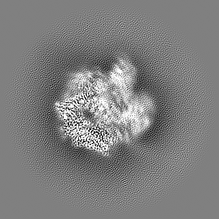
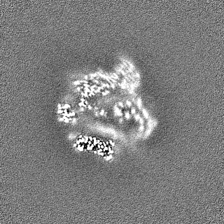
| Projections & slices | Image control
Images are generated by Spider. | ||||||||||||||||||||||||||||||||||||
| Voxel size | X=Y=Z: 1.06 Å | ||||||||||||||||||||||||||||||||||||
| Density |
| ||||||||||||||||||||||||||||||||||||
| Symmetry | Space group: 1 | ||||||||||||||||||||||||||||||||||||
| Details | EMDB XML:
|
 Z (Sec.)
 Y (Row.)
 X (Col.)


-Supplemental data
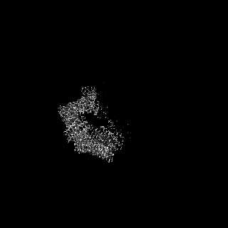
-Mask #1
| File | emd_14719_msk_1.map | ||||||||||||
|---|---|---|---|---|---|---|---|---|---|---|---|---|---|
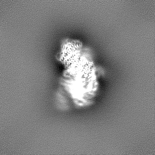
| Projections & Slices |
| ||||||||||||
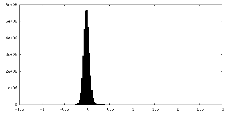
| Density Histograms |


-Additional map: LocSpiral processed map
| File | emd_14719_additional_1.map | ||||||||||||
|---|---|---|---|---|---|---|---|---|---|---|---|---|---|
| Annotation | LocSpiral processed map | ||||||||||||
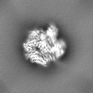
| Projections & Slices |
| ||||||||||||
| Density Histograms |


-Half map: #1
| File | emd_14719_half_map_1.map | ||||||||||||
|---|---|---|---|---|---|---|---|---|---|---|---|---|---|
| Projections & Slices |
| ||||||||||||
| Density Histograms |


-Half map: #2
| File | emd_14719_half_map_2.map | ||||||||||||
|---|---|---|---|---|---|---|---|---|---|---|---|---|---|
| Projections & Slices |
| ||||||||||||
| Density Histograms |


- Sample components
-Entire : FANCD2 mono-ubiquitinated on K561
| Entire | Name: FANCD2 mono-ubiquitinated on K561 |
|---|---|
| Components |
|
-Supramolecule #1: FANCD2 mono-ubiquitinated on K561
| Supramolecule | Name: FANCD2 mono-ubiquitinated on K561 / type: complex / ID: 1 / Parent: 0 / Macromolecule list: all |
|---|---|
| Source (natural) | Organism: Homo sapiens (human) |
-Macromolecule #1: Fanconi anemia group D2 protein
| Macromolecule | Name: Fanconi anemia group D2 protein / type: protein_or_peptide / ID: 1 / Enantiomer: LEVO |
|---|---|
| Source (natural) | Organism: Homo sapiens (human) |
| Sequence | String: GPGSMVSKRR LSKSEDKESL TEDASKTRKQ PLSKKTKKSH IANEVEENDS IFVKLLKISG IILKTGESQN QLAVDQIAFQ KKLFQTLRRH PSYPKIIEEF VSGLESYIED EDSFRNCLLS CERLQDEEAS MGASYSKSLI KLLLGIDILQ PAIIKTLFEK LPEYFFENKN ...String: GPGSMVSKRR LSKSEDKESL TEDASKTRKQ PLSKKTKKSH IANEVEENDS IFVKLLKISG IILKTGESQN QLAVDQIAFQ KKLFQTLRRH PSYPKIIEEF VSGLESYIED EDSFRNCLLS CERLQDEEAS MGASYSKSLI KLLLGIDILQ PAIIKTLFEK LPEYFFENKN SDEINIPRLI VSQLKWLDRV VDGKDLTTKI MQLISIAPEN LQHDIITSLP EILGDSQHAD VGKELSDLLI ENTSLTVPIL DVLSSLRLDP NFLLKVRQLV MDKLSSIRLE DLPVIIKFIL HSVTAMDTLE VISELREKLD LQHCVLPSRL QASQVKLKSK GRASSSGNQE SSGQSCIILL FDVIKSAIRY EKTISEAWIK AIENTASVSE HKVFDLVMLF IIYSTNTQTK KYIDRVLRNK IRSGCIQEQL LQSTFSVHYL VLKDMCSSIL SLAQSLLHSL DQSIISFGSL LYKYAFKFFD TYCQQEVVGA LVTHICSGNE AEVDTALDVL LELVVLNPSA MMMNAVFVKG ILDYLDNISP QQIRKLFYVL STLAFSKQNE ASSHIQDDMH LVIRKQLSST VFKYKLIGII GAVTMAGIMA ADRSESPSLT QERANLSDEQ CTQVTSLLQL VHSCSEQSPQ ASALYYDEFA NLIQHEKLDP KALEWVGHTI CNDFQDAFVV DSCVVPEGDF PFPVKALYGL EEYDTQDGIA INLLPLLFSQ DFAKDGGPVT SQESGQKLVS PLCLAPYFRL LRLCVERQHN GNLEEIDGLL DCPIFLTDLE PGEKLESMSA KERSFMCSLI FLTLNWFREI VNAFCQETSP EMKGKVLTRL KHIVELQIIL EKYLAVTPDY VPPLGNFDVE TLDITPHTVT AISAKIRKKG KIERKQKTDG SKTSSSDTLS EEKNSECDPT PSHRGQLNKE FTGKEEKTSL LLHNSHAFFR ELDIEVFSIL HCGLVTKFIL DTEMHTEATE VVQLGPPELL FLLEDLSQKL ESMLTPPIAR RVPFLKNKGS RNIGFSHLQQ RSAQEIVHCV FQLLTPMCNH LENIHNYFQC LAAENHGVVD GPGVKVQEYH IMSSCYQRLL QIFHGLFAWS GFSQPENQNL LYSALHVLSS RLKQGEHSQP LEELLSQSVH YLQNFHQSIP SFQCALYLIR LLMVILEKST ASAQNKEKIA SLARQFLCRV WPSGDKEKSN ISNDQLHALL CIYLEHTESI LKAIEEIAGV GVPELINSPK DASSSTFPTL TRHTFVVFFR VMMAELEKTV KKIEPGTAAD SQQIHEEKLL YWNMAVRDFS ILINLIKVFD SHPVLHVCLK YGRLFVEAFL KQCMPLLDFS FRKHREDVLS LLETFQLDTR LLHHLCGHSK IHQDTRLTQH VPLLKKTLEL LVCRVKAMLT LNNCREAFWL GNLKNRDLQG EEIKSQNSQE STADESEDDM SSQASKSKAT EDGEEDEVSA GEKEQDSDES YDDSD UniProtKB: Fanconi anemia group D2 protein |
-Experimental details
-Structure determination
| Method | cryo EM |
|---|---|
 Processing Processing | single particle reconstruction |
| Aggregation state | particle |
-Sample preparation
| Buffer | pH: 8 |
|---|---|
| Grid | Model: UltrAuFoil R1.2/1.3 / Mesh: 300 |
| Vitrification | Cryogen name: ETHANE / Chamber humidity: 95 % / Chamber temperature: 288 K / Instrument: FEI VITROBOT MARK IV / Details: blotting for 3.0 seconds. |
| Details | 9.3 uM USP1-UAF1, 1.8 uM FANCI-FANCD2Ub, 2.2 uM dsDNA (61 base-pairs), 18 uM ML323 |
- Electron microscopy
| Microscope | FEI TITAN KRIOS |
|---|---|
| Image recording | Film or detector model: GATAN K3 (6k x 4k) / Average electron dose: 40.0 e/Å2 |
| Electron beam | Acceleration voltage: 300 kV / Electron source:  FIELD EMISSION GUN FIELD EMISSION GUN |
| Electron optics | Illumination mode: FLOOD BEAM / Imaging mode: BRIGHT FIELD / Nominal defocus max: 3.0 µm / Nominal defocus min: 1.0 µm |
| Experimental equipment |  Model: Titan Krios / Image courtesy: FEI Company |