 Movie
Movie Controller


+ Open data

- Basic information
| Entry | Database: EMDB / ID: EMD-13670 | ||||||||||||||||||
|---|---|---|---|---|---|---|---|---|---|---|---|---|---|---|---|---|---|---|---|
| Title | isolated S-layer of Ca.M.Lanthanididphila | ||||||||||||||||||
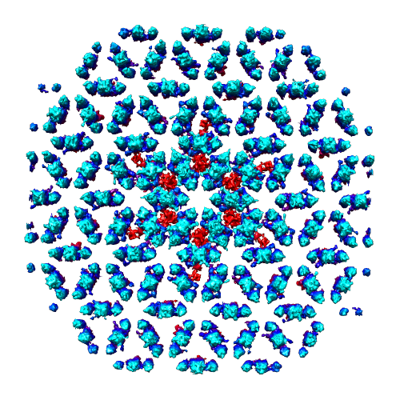
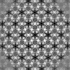
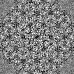
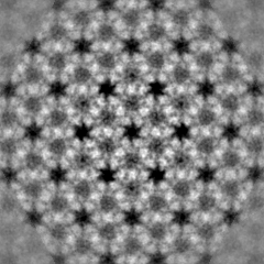
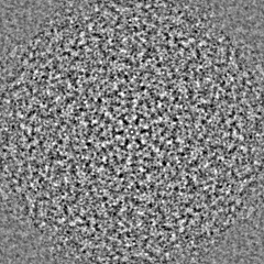
 Map data Map data | Subtomogram average of the Ca.M.lanthanidiphila S-layer obtained from cryo-tomograms of isolated S-layer patches. 6-fold rotational symmetry has been applied. | ||||||||||||||||||
 Sample Sample |
| ||||||||||||||||||
 Keywords Keywords | S-layer / STRUCTURAL PROTEIN | ||||||||||||||||||
| Biological species |  Candidatus Methylomirabilis lanthanidiphila (bacteria) Candidatus Methylomirabilis lanthanidiphila (bacteria) | ||||||||||||||||||
| Method | subtomogram averaging / cryo EM / Resolution: 21.0 Å | ||||||||||||||||||
 Authors Authors | Gambelli L / Mesman R | ||||||||||||||||||
| Funding support | 5 items
| ||||||||||||||||||
 Citation Citation | Journal: Front Microbiol / Year: 2021 Title: The Polygonal Cell Shape and Surface Protein Layer of Anaerobic Methane-Oxidizing Bacteria. Authors: Lavinia Gambelli / Rob Mesman / Wouter Versantvoort / Christoph A Diebolder / Andreas Engel / Wiel Evers / Mike S M Jetten / Martin Pabst / Bertram Daum / Laura van Niftrik /   Abstract: bacteria perform anaerobic methane oxidation coupled to nitrite reduction via an intra-aerobic pathway, producing carbon dioxide and dinitrogen gas. These diderm bacteria possess an unusual ... bacteria perform anaerobic methane oxidation coupled to nitrite reduction via an intra-aerobic pathway, producing carbon dioxide and dinitrogen gas. These diderm bacteria possess an unusual polygonal cell shape with sharp ridges that run along the cell body. Previously, a putative surface protein layer (S-layer) was observed as the outermost cell layer of these bacteria. We hypothesized that this S-layer is the determining factor for their polygonal cell shape. Therefore, we enriched the S-layer from cells and through LC-MS/MS identified a 31 kDa candidate S-layer protein, mela_00855, which had no homology to any other known protein. Antibodies were generated against a synthesized peptide derived from the mela_00855 protein sequence and used in immunogold localization to verify its identity and location. Both on thin sections of cells and in negative-stained enriched S-layer patches, the immunogold localization identified mela_00855 as the S-layer protein. Using electron cryo-tomography and sub-tomogram averaging of S-layer patches, we observed that the S-layer has a hexagonal symmetry. Cryo-tomography of whole cells showed that the S-layer and the outer membrane, but not the peptidoglycan layer and the cytoplasmic membrane, exhibited the polygonal shape. Moreover, the S-layer consisted of multiple rigid sheets that partially overlapped, most likely giving rise to the unique polygonal cell shape. These characteristics make the S-layer of a distinctive and intriguing case to study. | ||||||||||||||||||
| History |
|
- Structure visualization
| Movie |
 Movie viewer Movie viewer |
|---|---|
| Structure viewer | EM map: SurfViewMolmilJmol/JSmol |
| Supplemental images |
- Downloads & links
-EMDB archive
| Map data | emd_13670.map.gz | 46.8 MB | EMDB map data format | |
|---|---|---|---|---|
| Header (meta data) | emd-13670-v30.xmlemd-13670.xml | 11.9 KB 11.9 KB | Display Display | EMDB header |
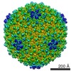
| Images |  emd_13670.png emd_13670.png | 188.6 KB | ||
| Filedesc metadata | emd-13670.cif.gz | 4.4 KB | ||
| Archive directory |  http://ftp.pdbj.org/pub/emdb/structures/EMD-13670ftp://ftp.pdbj.org/pub/emdb/structures/EMD-13670 http://ftp.pdbj.org/pub/emdb/structures/EMD-13670ftp://ftp.pdbj.org/pub/emdb/structures/EMD-13670 | HTTPS FTP |
-Related structure data
| Related structure data | C: citing same article ( |
|---|---|
| Similar structure data | |
| EM raw data | EMPIAR-10822 (Title: Cryo Electron Tomography of isolated S-layer patches from Ca.M.lanthanidiphila Data size: 46.3 Data #1: Reconstructed tomogram of isolated Ca. M.lanthanidiphila S-layer patches [reconstructed volumes] Data #2: Reconstructed tomogram of isolated Ca. M.lanthanidiphila S-layer patches [reconstructed volumes]) |


-Links
| EMDB pages | EMDB (EBI/PDBe) / EMDataResource |
|---|
-Map
| File | Download / File: emd_13670.map.gz / Format: CCP4 / Size: 52.7 MB / Type: IMAGE STORED AS FLOATING POINT NUMBER (4 BYTES) | ||||||||||||||||||||||||||||||||||||||||||||||||||||||||||||||||||||
|---|---|---|---|---|---|---|---|---|---|---|---|---|---|---|---|---|---|---|---|---|---|---|---|---|---|---|---|---|---|---|---|---|---|---|---|---|---|---|---|---|---|---|---|---|---|---|---|---|---|---|---|---|---|---|---|---|---|---|---|---|---|---|---|---|---|---|---|---|---|
| Annotation | Subtomogram average of the Ca.M.lanthanidiphila S-layer obtained from cryo-tomograms of isolated S-layer patches. 6-fold rotational symmetry has been applied. | ||||||||||||||||||||||||||||||||||||||||||||||||||||||||||||||||||||
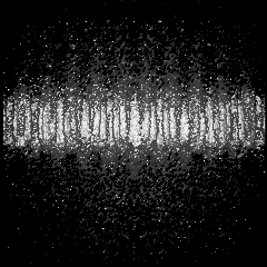
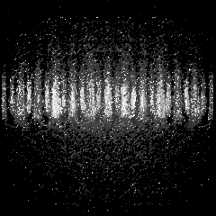
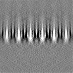
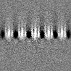
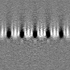
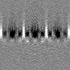
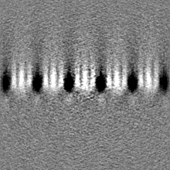
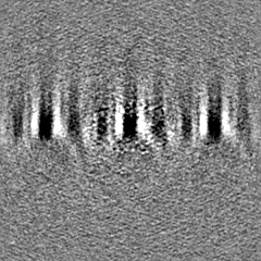
| Projections & slices | Image control
Images are generated by Spider. | ||||||||||||||||||||||||||||||||||||||||||||||||||||||||||||||||||||
| Voxel size | X=Y=Z: 2.62631 Å | ||||||||||||||||||||||||||||||||||||||||||||||||||||||||||||||||||||
| Density |
| ||||||||||||||||||||||||||||||||||||||||||||||||||||||||||||||||||||
| Symmetry | Space group: 1 | ||||||||||||||||||||||||||||||||||||||||||||||||||||||||||||||||||||
| Details | EMDB XML:
CCP4 map header:
| ||||||||||||||||||||||||||||||||||||||||||||||||||||||||||||||||||||
 Z (Sec.)
 Y (Row.)
 X (Col.)


-Supplemental data
- Sample components
-Entire : isolated S-layer of Ca.M.lanthanidiphila
| Entire | Name: isolated S-layer of Ca.M.lanthanidiphila |
|---|---|
| Components |
|
-Supramolecule #1: isolated S-layer of Ca.M.lanthanidiphila
| Supramolecule | Name: isolated S-layer of Ca.M.lanthanidiphila / type: organelle_or_cellular_component / ID: 1 / Parent: 0 Details: S-layer was isolated by boiling disrupted flocks of Ca.M.Lanthanidiphila enrichment culture in 4% SDS. |
|---|---|
| Source (natural) | Organism: Candidatus Methylomirabilis lanthanidiphila (bacteria) Location in cell: on top of outer membrane |
-Experimental details
-Structure determination
| Method | cryo EM |
|---|---|
 Processing Processing | subtomogram averaging |
| Aggregation state | 2D array |
-Sample preparation
| Buffer | pH: 7 |
|---|---|
| Grid | Model: Quantifoil R2/2 / Material: COPPER / Mesh: 200 / Pretreatment - Type: GLOW DISCHARGE / Pretreatment - Time: 40 sec. / Pretreatment - Atmosphere: AIR / Pretreatment - Pressure: 20.0 kPa / Details: glow-discharged at 15 mA current |
| Vitrification | Cryogen name: ETHANE / Chamber humidity: 100 % / Chamber temperature: 293 K / Instrument: FEI VITROBOT MARK IV Details: 2 microlitre sample was mixed with 0.5 microlitre 10 nm ProteinA gold solution Grids were plunge frozen in liquid ethane, blot force 2, blot time 2.5-3 sec.. |
| Details | concentrated isolated S-layer in ultrapure water |
- Electron microscopy
| Microscope | FEI TITAN KRIOS |
|---|---|
| Temperature | Max: 93.0 K |
| Specialist optics | Energy filter - Name: GIF Bioquantum / Energy filter - Slit width: 20 eV |
| Image recording | Film or detector model: GATAN K3 BIOQUANTUM (6k x 4k) / Number grids imaged: 1 / Average exposure time: 0.3 sec. / Average electron dose: 1.71538 e/Å2 |
| Electron beam | Acceleration voltage: 300 kV / Electron source:  FIELD EMISSION GUN FIELD EMISSION GUN |
| Electron optics | C2 aperture diameter: 50.0 µm / Calibrated defocus max: 4.7 µm / Calibrated defocus min: 4.43 µm / Illumination mode: FLOOD BEAM / Imaging mode: BRIGHT FIELD / Cs: 2.7 mm / Nominal defocus max: 5.0 µm / Nominal defocus min: 4.0 µm / Nominal magnification: 33000 |
| Sample stage | Specimen holder model: FEI TITAN KRIOS AUTOGRID HOLDER / Cooling holder cryogen: NITROGEN |
| Experimental equipment |  Model: Titan Krios / Image courtesy: FEI Company |
-Image processing
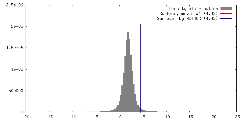
| Final reconstruction | Number classes used: 1 / Applied symmetry - Point group: C6 (6 fold cyclic) / Algorithm: BACK PROJECTION / Resolution.type: BY AUTHOR / Resolution: 21.0 Å / Resolution method: OTHER / Software - Name: PEET (ver. 1.14.0) Details: Resolution of the Ca. M. lanthanidiphila S-layer sub-tomogram average was determined by FFT Number subtomograms used: 8938 |
|---|---|
| Extraction | Number tomograms: 2 / Number images used: 11274 / Method: random grid / Software - Name: PEET (ver. 1.14.0) |
| Final angle assignment | Type: NOT APPLICABLE |
