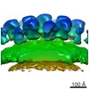
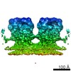
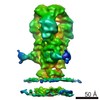
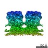
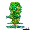
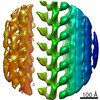
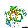
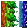
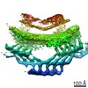
Journal: PLoS Pathog / Year: 2016 Title: Cryo-electron Microscopy Structure of the Native Prototype Foamy Virus Glycoprotein and Virus Architecture. Authors: Grégory Effantin / Leandro F Estrozi / Nick Aschman / Patricia Renesto / Nicole Stanke / Dirk Lindemann / Guy Schoehn / Winfried Weissenhorn / Abstract: Foamy viruses (FV) belong to the genus Spumavirus, which forms a distinct lineage in the Retroviridae family. Although the infection in natural hosts and zoonotic transmission to humans is ...Foamy viruses (FV) belong to the genus Spumavirus, which forms a distinct lineage in the Retroviridae family. Although the infection in natural hosts and zoonotic transmission to humans is asymptomatic, FVs can replicate well in human cells making it an attractive gene therapy vector candidate. Here we present cryo-electron microscopy and (cryo-)electron tomography ultrastructural data on purified prototype FV (PFV) and PFV infected cells. Mature PFV particles have a distinct morphology with a capsid of constant dimension as well as a less ordered shell of density between the capsid and the membrane likely formed by the Gag N-terminal domain and the cytoplasmic part of the Env leader peptide gp18LP. The viral membrane contains trimeric Env glycoproteins partly arranged in interlocked hexagonal assemblies. In situ 3D reconstruction by subtomogram averaging of wild type Env and of a Env gp48TM- gp80SU cleavage site mutant showed a similar spike architecture as well as stabilization of the hexagonal lattice by clear connections between lower densities of neighboring trimers. Cryo-EM was employed to obtain a 9 Å resolution map of the glycoprotein in its pre-fusion state, which revealed extensive trimer interactions by the receptor binding subunit gp80SU at the top of the spike and three central helices derived from the fusion protein subunit gp48TM. The lower part of Env, presumably composed of interlaced parts of gp48TM, gp80SU and gp18LP anchors the spike at the membrane. We propose that the gp48TM density continues into three central transmembrane helices, which interact with three outer transmembrane helices derived from gp18LP. Our ultrastructural data and 9 Å resolution glycoprotein structure provide important new insights into the molecular architecture of PFV and its distinct evolutionary relationship with other members of the Retroviridae.
History
Deposition
Jun 3, 2016
-
Header (metadata) release
Jun 22, 2016
-
Map release
Jun 22, 2016
-
Update
Jul 20, 2016
-
Current status
Jul 20, 2016
Processing site: PDBe / Status: Released
-
Structure visualization
Movie
Surface view with section colored by density value
Name: Human spumaretrovirus / type: virus / ID: 1 / Parent: 0 / Details: Mutant in the Env glycoprotein / NCBI-ID: 11963 / Sci species name: Human spumaretrovirus / Virus type: VIRION / Virus isolate: SPECIES / Virus enveloped: Yes / Virus empty: No
Host system
Organism: Homo sapiens (human) / Recombinant plasmid: pcoPG4, pcoPE32, pcoPP
Molecular weight
Theoretical: 330 KDa
-
Experimental details
-
Structure determination
Method
cryo EM
Processing
subtomogram averaging
Aggregation state
particle
-
Sample preparation
Buffer
pH: 7.5
Vitrification
Cryogen name: ETHANE
Details
The iFuse mutant is a variant of Env where the furine cleavage site between the SUrface (gp80 SU ) and TransMembrane (gp48 TM ) domains of Env has been mutated. This results in a partially processed glycoprotein as the cleavage between the LP and SU domains is preserved. Particles are released at nearly wild type level from cells but are non-infectious
-
Electron microscopy
Microscope
FEI TECNAI F20
Image recording
Film or detector model: FEI EAGLE (4k x 4k) / Average electron dose: 1.0 e/Å2
Electron beam
Acceleration voltage: 200 kV / Electron source: FIELD EMISSION GUN
Electron optics
Illumination mode: FLOOD BEAM / Imaging mode: BRIGHT FIELD
Experimental equipment
Model: Tecnai F20 / Image courtesy: FEI Company
-
Image processing
Final reconstruction
Applied symmetry - Point group: C3 (3 fold cyclic) / Resolution.type: BY AUTHOR / Resolution: 28.0 Å / Resolution method: FSC 0.5 CUT-OFF / Software - Name: PEET / Number subtomograms used: 3000
Extraction
Number tomograms: 7 / Number images used: 3930
Final angle assignment
Type: PROJECTION MATCHING
+
About Yorodumi
-
News
-
Feb 9, 2022. New format data for meta-information of EMDB entries
New format data for meta-information of EMDB entries
Version 3 of the EMDB header file is now the official format.
The previous official version 1.9 will be removed from the archive.
In the structure databanks used in Yorodumi, some data are registered as the other names, "COVID-19 virus" and "2019-nCoV". Here are the details of the virus and the list of structure data.
Jan 31, 2019. EMDB accession codes are about to change! (news from PDBe EMDB page)
EMDB accession codes are about to change! (news from PDBe EMDB page)
The allocation of 4 digits for EMDB accession codes will soon come to an end. Whilst these codes will remain in use, new EMDB accession codes will include an additional digit and will expand incrementally as the available range of codes is exhausted. The current 4-digit format prefixed with “EMD-” (i.e. EMD-XXXX) will advance to a 5-digit format (i.e. EMD-XXXXX), and so on. It is currently estimated that the 4-digit codes will be depleted around Spring 2019, at which point the 5-digit format will come into force.
The EM Navigator/Yorodumi systems omit the EMD- prefix.
Related info.:Q: What is EMD? / ID/Accession-code notation in Yorodumi/EM Navigator
Yorodumi is a browser for structure data from EMDB, PDB, SASBDB, etc.
This page is also the successor to EM Navigator detail page, and also detail information page/front-end page for Omokage search.
The word "yorodu" (or yorozu) is an old Japanese word meaning "ten thousand". "mi" (miru) is to see.
Related info.:EMDB / PDB / SASBDB / Comparison of 3 databanks / Yorodumi Search / Aug 31, 2016. New EM Navigator & Yorodumi / Yorodumi Papers / Jmol/JSmol / Function and homology information / Changes in new EM Navigator and Yorodumi
 Movie
Movie Controller


 Open data
 Basic information
 Map data
 Sample
 Human spumaretrovirus
 Authors
 Citation

 Structure visualization
 Movie viewer
 Downloads & links
 emd_4007.png
 http://ftp.pdbj.org/pub/emdb/structures/EMD-4007
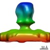
 Z (Sec.)
 Y (Row.)
 X (Col.)


 Sample components
 Homo sapiens (human) / Recombinant plasmid: pcoPG4, pcoPE32, pcoPP
 Processing
 Electron microscopy
 FIELD EMISSION GUN
