 Movie
Movie Controller

[English] 日本語
 Yorodumi
- EMDB-1101: Coat protein fold and maturation transition of bacteriophage P22 ... -
+ Open data

- Basic information
| Entry | Database: EMDB / ID: EMD-1101 | |||||||||
|---|---|---|---|---|---|---|---|---|---|---|
| Title | Coat protein fold and maturation transition of bacteriophage P22 seen at subnanometer resolutions. | |||||||||
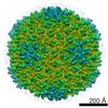
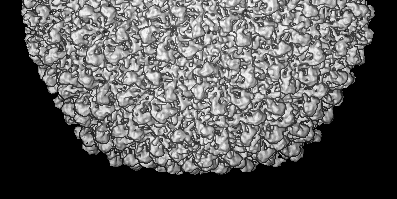
 Map data Map data | Z-axis along 3fold symmetry axis, Y-axis along 2fold symmetry axis | |||||||||
 Sample Sample |
| |||||||||
| Biological species |  Enterobacteria phage P22 (virus) Enterobacteria phage P22 (virus) | |||||||||
| Method | single particle reconstruction / cryo EM / Resolution: 9.5 Å | |||||||||
 Authors Authors | Jiang W / Li Z / Zhang Z / Baker ML / Prevelige PE / Chiu W | |||||||||
 Citation Citation | Journal: Nat Struct Biol / Year: 2003 Title: Coat protein fold and maturation transition of bacteriophage P22 seen at subnanometer resolutions. Authors: Wen Jiang / Zongli Li / Zhixian Zhang / Matthew L Baker / Peter E Prevelige / Wah Chiu /  Abstract: Bacteriophage P22 is a prototypical biological machine used for studying protein complex assembly and capsid maturation. Using cryo-EM, we solved the structures of P22 before and after the capsid ...Bacteriophage P22 is a prototypical biological machine used for studying protein complex assembly and capsid maturation. Using cryo-EM, we solved the structures of P22 before and after the capsid maturation at 8.5 A and 9.5 A resolutions, respectively. These structures allowed visualization of alpha-helices and beta-sheets from which the capsid protein fold is derived. The capsid fold is similar to that of the coat protein of HK97 bacteriophage. The cryo-EM shows that a large conformational change of the P22 capsid during maturation transition involves not only the domain movement of individual subunits, but also refolding of the capsid protein. | |||||||||
| History |
|
- Structure visualization
| Movie |
 Movie viewer Movie viewer |
|---|---|
| Structure viewer | EM map: SurfViewMolmilJmol/JSmol |
| Supplemental images |
 UCSF Chimera
- Downloads & links
-EMDB archive
| Map data | emd_1101.map.gz | 61.3 MB | EMDB map data format | |
|---|---|---|---|---|
| Header (meta data) | emd-1101-v30.xmlemd-1101.xml | 9.9 KB 9.9 KB | Display Display | EMDB header |
| Images | 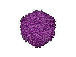 1101.gif 1101.gif | 17.6 KB | ||
| Archive directory |  http://ftp.pdbj.org/pub/emdb/structures/EMD-1101ftp://ftp.pdbj.org/pub/emdb/structures/EMD-1101 http://ftp.pdbj.org/pub/emdb/structures/EMD-1101ftp://ftp.pdbj.org/pub/emdb/structures/EMD-1101 | HTTPS FTP |
-Related structure data
| Similar structure data |
|---|


-Links
| EMDB pages | EMDB (EBI/PDBe) / EMDataResource |
|---|
-Map
| File | Download / File: emd_1101.map.gz / Format: CCP4 / Size: 116.8 MB / Type: IMAGE STORED AS FLOATING POINT NUMBER (4 BYTES) | ||||||||||||||||||||||||||||||||||||||||||||||||||||||||||||||||||||
|---|---|---|---|---|---|---|---|---|---|---|---|---|---|---|---|---|---|---|---|---|---|---|---|---|---|---|---|---|---|---|---|---|---|---|---|---|---|---|---|---|---|---|---|---|---|---|---|---|---|---|---|---|---|---|---|---|---|---|---|---|---|---|---|---|---|---|---|---|---|
| Annotation | Z-axis along 3fold symmetry axis, Y-axis along 2fold symmetry axis | ||||||||||||||||||||||||||||||||||||||||||||||||||||||||||||||||||||
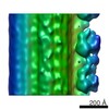
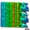
| Projections & slices | Image control
Images are generated by Spider. generated in cubic-lattice coordinate | ||||||||||||||||||||||||||||||||||||||||||||||||||||||||||||||||||||
| Voxel size | X=Y=Z: 1.815 Å | ||||||||||||||||||||||||||||||||||||||||||||||||||||||||||||||||||||
| Density |
| ||||||||||||||||||||||||||||||||||||||||||||||||||||||||||||||||||||
| Symmetry | Space group: 1 | ||||||||||||||||||||||||||||||||||||||||||||||||||||||||||||||||||||
| Details | EMDB XML:
CCP4 map header:
| ||||||||||||||||||||||||||||||||||||||||||||||||||||||||||||||||||||
 Z (Sec.)
 Y (Row.)
 X (Col.)


-Supplemental data
- Sample components
-Entire : P22 Mature Phage
| Entire | Name: P22 Mature Phage |
|---|---|
| Components |
|
-Supramolecule #1000: P22 Mature Phage
| Supramolecule | Name: P22 Mature Phage / type: sample / ID: 1000 / Number unique components: 1 |
|---|---|
| Molecular weight | Experimental: 18 MDa / Theoretical: 18 MDa |
-Supramolecule #1: Enterobacteria phage P22
| Supramolecule | Name: Enterobacteria phage P22 / type: virus / ID: 1 / Name.synonym: P22 mature phage / Details: the icosahedral protein shell / NCBI-ID: 10754 / Sci species name: Enterobacteria phage P22 / Virus type: VIRION / Virus isolate: STRAIN / Virus enveloped: No / Virus empty: No / Syn species name: P22 mature phage |
|---|---|
| Host (natural) | Organism:  Salmonella (bacteria) / synonym: BACTERIA(EUBACTERIA) Salmonella (bacteria) / synonym: BACTERIA(EUBACTERIA) |
| Molecular weight | Experimental: 18 MDa / Theoretical: 18 MDa |
| Virus shell | Shell ID: 1 / Name: capsid shell / Diameter: 700 Å / T number (triangulation number): 7 |
-Experimental details
-Structure determination
| Method | cryo EM |
|---|---|
 Processing Processing | single particle reconstruction |
| Aggregation state | particle |
-Sample preparation
| Grid | Details: 400 mesh copper grid |
|---|---|
| Vitrification | Cryogen name: ETHANE / Chamber temperature: 90 K / Instrument: HOMEMADE PLUNGER / Details: Vitrification instrument: home-made plunger |
- Electron microscopy
| Microscope | JEOL 4000EX |
|---|---|
| Temperature | Min: 95 K / Max: 95 K / Average: 95 K |
| Alignment procedure | Legacy - Astigmatism: by monitoring Thon rings at very close to focus |
| Image recording | Category: FILM / Film or detector model: KODAK SO-163 FILM / Digitization - Scanner: ZEISS SCAI / Digitization - Sampling interval: 7 µm / Number real images: 100 / Average electron dose: 30 e/Å2 Details: scanned at 7micrometer then averaged to 14micrometer step size Bits/pixel: 8 |
| Tilt angle min | 0 |
| Tilt angle max | 0 |
| Electron beam | Acceleration voltage: 400 kV / Electron source: LAB6 |
| Electron optics | Illumination mode: FLOOD BEAM / Imaging mode: BRIGHT FIELD / Cs: 5.6 mm / Nominal defocus max: 3.0 µm / Nominal defocus min: 1.0 µm / Nominal magnification: 40000 |
| Sample stage | Specimen holder: side entry / Specimen holder model: GATAN LIQUID NITROGEN |
-Image processing
| CTF correction | Details: per micrograph |
|---|---|
| Final reconstruction | Applied symmetry - Point group: I (icosahedral) / Algorithm: OTHER / Resolution.type: BY AUTHOR / Resolution: 9.5 Å / Resolution method: FSC 0.5 CUT-OFF / Software - Name: SAVR / Number images used: 5600 |
-Atomic model buiding 1
| Initial model | PDB ID:  1fh6 Chain - Chain ID: A |
|---|---|
| Software | Name: foldhunter |
| Details | PDBEntryID_givenInChain. Protocol: rigid body |
| Refinement | Protocol: RIGID BODY FIT / Target criteria: correlation coefficient |
