 Movie
Movie Controller

[English] 日本語
 Yorodumi
- EMDB-5200: 5.4-Angstrom cryoEM structure of the Bordetella Bacteriophage capsid -
+ Open data

- Basic information
| Entry | Database: EMDB / ID: EMD-5200 | |||||||||
|---|---|---|---|---|---|---|---|---|---|---|
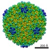
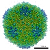
| Title | 5.4-Angstrom cryoEM structure of the Bordetella Bacteriophage capsid | |||||||||
 Map data Map data | This is the cryoEM density map for half of the icosahedral capsid of a bacterial phage, BPP. | |||||||||
 Sample Sample |
| |||||||||
 Keywords Keywords | Bordetella Bacteriophage / capsid / cryoEM / structure / major capsid protein / cementing protein | |||||||||
| Biological species | Bordetella Bacteriophage | |||||||||
| Method | single particle reconstruction / cryo EM / Resolution: 5.4 Å | |||||||||
 Authors Authors | Jin L / Hodes A / Hui WH / Zhang X / Yu X / Miller JF / Zhou ZH | |||||||||
 Citation Citation | Journal: To Be Published Title: 5.4-Angstrom cryoEM structure of the Bordetella Bacteriophage capsid Authors: Jin L / Hodes A / Hui WH / Zhang X / Zhang X / Yu X / Miller JF / Zhou ZH | |||||||||
| History |
|
- Structure visualization
| Movie |
 Movie viewer Movie viewer |
|---|---|
| Structure viewer | EM map: SurfViewMolmilJmol/JSmol |
| Supplemental images |

- Downloads & links
-EMDB archive
| Map data | emd_5200.map.gz | 380.3 MB | EMDB map data format | |
|---|---|---|---|---|
| Header (meta data) | emd-5200-v30.xmlemd-5200.xml | 9.4 KB 9.4 KB | Display Display | EMDB header |
| Images |  emd_5200_1.jpg emd_5200_1.jpg | 478.3 KB | ||
| Archive directory |  http://ftp.pdbj.org/pub/emdb/structures/EMD-5200ftp://ftp.pdbj.org/pub/emdb/structures/EMD-5200 http://ftp.pdbj.org/pub/emdb/structures/EMD-5200ftp://ftp.pdbj.org/pub/emdb/structures/EMD-5200 | HTTPS FTP |
-Related structure data
| Similar structure data |
|---|


-Links
| EMDB pages | EMDB (EBI/PDBe) / EMDataResource |
|---|
-Map
| File | Download / File: emd_5200.map.gz / Format: CCP4 / Size: 763 MB / Type: IMAGE STORED AS FLOATING POINT NUMBER (4 BYTES) | ||||||||||||||||||||||||||||||||||||||||||||||||||||||||||||||||||||
|---|---|---|---|---|---|---|---|---|---|---|---|---|---|---|---|---|---|---|---|---|---|---|---|---|---|---|---|---|---|---|---|---|---|---|---|---|---|---|---|---|---|---|---|---|---|---|---|---|---|---|---|---|---|---|---|---|---|---|---|---|---|---|---|---|---|---|---|---|---|
| Annotation | This is the cryoEM density map for half of the icosahedral capsid of a bacterial phage, BPP. | ||||||||||||||||||||||||||||||||||||||||||||||||||||||||||||||||||||
| Projections & slices | Image control
Images are generated by Spider. generated in cubic-lattice coordinate | ||||||||||||||||||||||||||||||||||||||||||||||||||||||||||||||||||||
| Voxel size | X=Y=Z: 0.9716 Å | ||||||||||||||||||||||||||||||||||||||||||||||||||||||||||||||||||||
| Density |
| ||||||||||||||||||||||||||||||||||||||||||||||||||||||||||||||||||||
| Symmetry | Space group: 1 | ||||||||||||||||||||||||||||||||||||||||||||||||||||||||||||||||||||
| Details | EMDB XML:
CCP4 map header:
| ||||||||||||||||||||||||||||||||||||||||||||||||||||||||||||||||||||
 Z (Sec.)
 Y (Row.)
 X (Col.)


-Supplemental data
- Sample components
-Entire : Bordetella Bacteriophage
| Entire | Name: Bordetella Bacteriophage |
|---|---|
| Components |
|
-Supramolecule #1000: Bordetella Bacteriophage
| Supramolecule | Name: Bordetella Bacteriophage / type: sample / ID: 1000 Details: The whole phage (with portal and tail structures) were imaged initially. After icosahedral averaging in 3D reconstruction, the resulting density map covers only the capsid. Oligomeric state: Icosahedral particle made of the major capsid protein and the cementing protein Number unique components: 2 |
|---|
-Supramolecule #1: Bordetella Bacteriophage
| Supramolecule | Name: Bordetella Bacteriophage / type: virus / ID: 1 / Name.synonym: Bordetella phage / Sci species name: Bordetella Bacteriophage / Database: NCBI / Virus type: VIRION / Virus isolate: STRAIN / Virus enveloped: No / Virus empty: No / Syn species name: Bordetella phage |
|---|---|
| Host (natural) | Organism:  Bordetella (bacteria) / synonym: BACTERIA(EUBACTERIA) Bordetella (bacteria) / synonym: BACTERIA(EUBACTERIA) |
| Virus shell | Shell ID: 1 / Diameter: 685 Å / T number (triangulation number): 7 |
-Experimental details
-Structure determination
| Method | cryo EM |
|---|---|
 Processing Processing | single particle reconstruction |
| Aggregation state | particle |
-Sample preparation
| Buffer | pH: 7.5 / Details: 50mM Tris-HCl, 250mM NaCl |
|---|---|
| Grid | Details: holey carbon films (Quantifoil, Germany) supported on 400-mesh copper grids |
| Vitrification | Cryogen name: NITROGEN / Chamber temperature: 100 K / Instrument: HOMEMADE PLUNGER Details: Vitrification instrument: manual plunger. Vitrification was carried out using a house-made manual plunger. Method: Blot for 2-3 seconds before plunging into liquid ethane |
- Electron microscopy
| Microscope | FEI POLARA 300 |
|---|---|
| Temperature | Min: 90 K / Max: 105 K / Average: 100 K |
| Date | Oct 20, 2006 |
| Image recording | Category: CCD / Film or detector model: GENERIC TVIPS / Average electron dose: 20 e/Å2 |
| Electron beam | Acceleration voltage: 300 kV / Electron source:  FIELD EMISSION GUN FIELD EMISSION GUN |
| Electron optics | Calibrated magnification: 97940 / Illumination mode: FLOOD BEAM / Imaging mode: BRIGHT FIELD / Cs: 2.0 mm / Nominal defocus max: 1.72 µm / Nominal defocus min: 0.5 µm / Nominal magnification: 97940 |
| Sample stage | Specimen holder: Eucentric / Specimen holder model: GATAN LIQUID NITROGEN |
| Experimental equipment |  Model: Tecnai Polara / Image courtesy: FEI Company |
-Image processing
| Details | The partilces were imaged with a 4kx4k TVIPS CCD camera. |
|---|---|
| CTF correction | Details: Each particle |
| Final reconstruction | Algorithm: OTHER / Resolution.type: BY AUTHOR / Resolution: 5.4 Å / Resolution method: FSC 0.5 CUT-OFF / Software - Name: IMIRS / Details: Focal pair method / Number images used: 18524 |
