 Movie
Movie Controller


+ Open data

- Basic information
| Entry |  | ||||||||||||||||||
|---|---|---|---|---|---|---|---|---|---|---|---|---|---|---|---|---|---|---|---|
| Title | Cryo-EM structure of the Xenopus egg 80S ribosome | ||||||||||||||||||
 Map data Map data | |||||||||||||||||||
 Sample Sample |
| ||||||||||||||||||
 Keywords Keywords | Eif5a / Eef2 / Habp4 / DapL1 / Dap1 / egg / Xenopus / ribosome | ||||||||||||||||||
| Function / homology |  Function and homology information Function and homology informationdeath domain binding / positive regulation of translational termination / ribosome hibernation / positive regulation of translational elongation / laminin receptor activity / negative regulation of translational frameshifting / translational elongation / 90S preribosome / endonucleolytic cleavage to generate mature 3'-end of SSU-rRNA from (SSU-rRNA, 5.8S rRNA, LSU-rRNA) / translation elongation factor activity ...death domain binding / positive regulation of translational termination / ribosome hibernation / positive regulation of translational elongation / laminin receptor activity / negative regulation of translational frameshifting / translational elongation / 90S preribosome / endonucleolytic cleavage to generate mature 3'-end of SSU-rRNA from (SSU-rRNA, 5.8S rRNA, LSU-rRNA) / translation elongation factor activity / protein-RNA complex assembly / maturation of LSU-rRNA / translation regulator activity / endonucleolytic cleavage in ITS1 to separate SSU-rRNA from 5.8S rRNA and LSU-rRNA from tricistronic rRNA transcript (SSU-rRNA, 5.8S rRNA, LSU-rRNA) / rough endoplasmic reticulum / laminin binding / translation initiation factor binding / translation initiation factor activity / translation repressor activity / rescue of stalled cytosolic ribosome / class I DNA-(apurinic or apyrimidinic site) endonuclease activity / cytosolic ribosome / cellular response to amino acid starvation / DNA-(apurinic or apyrimidinic site) lyase / protein kinase C binding / negative regulation of autophagy / ribosomal large subunit biogenesis / maturation of LSU-rRNA from tricistronic rRNA transcript (SSU-rRNA, 5.8S rRNA, LSU-rRNA) / maturation of SSU-rRNA from tricistronic rRNA transcript (SSU-rRNA, 5.8S rRNA, LSU-rRNA) / positive regulation of apoptotic signaling pathway / maturation of SSU-rRNA / apoptotic signaling pathway / small-subunit processome / modification-dependent protein catabolic process / spindle / protein tag activity / kinase activity / rRNA processing / regulation of translation / large ribosomal subunit / ribosomal small subunit assembly / ribosome binding / ribosomal small subunit biogenesis / 5S rRNA binding / ribosomal large subunit assembly / small ribosomal subunit / small ribosomal subunit rRNA binding / cytosolic small ribosomal subunit / large ribosomal subunit rRNA binding / cytosolic large ribosomal subunit / Hydrolases; Acting on acid anhydrides; Acting on GTP to facilitate cellular and subcellular movement / cytoplasmic translation / tRNA binding / mitochondrial inner membrane / negative regulation of translation / rRNA binding / structural constituent of ribosome / protein ubiquitination / ribosome / translation / ribonucleoprotein complex / cell division / DNA repair / mRNA binding / GTPase activity / apoptotic process / ubiquitin protein ligase binding / centrosome / endoplasmic reticulum membrane / GTP binding / nucleolus / endoplasmic reticulum / RNA binding / zinc ion binding / nucleus / plasma membrane / cytoplasm / cytosol Similarity search - Function | ||||||||||||||||||
| Biological species | |||||||||||||||||||
| Method | single particle reconstruction / cryo EM / Resolution: 2.4 Å | ||||||||||||||||||
 Authors Authors | Leesch F / Lorenzo-Orts L | ||||||||||||||||||
| Funding support |  Austria, Austria,  Switzerland, 5 items Switzerland, 5 items
| ||||||||||||||||||
 Citation Citation | Journal: Nature / Year: 2023 Title: A molecular network of conserved factors keeps ribosomes dormant in the egg. Authors: Friederike Leesch / Laura Lorenzo-Orts / Carina Pribitzer / Irina Grishkovskaya / Josef Roehsner / Anastasia Chugunova / Manuel Matzinger / Elisabeth Roitinger / Katarina Belačić / Susanne ...Authors: Friederike Leesch / Laura Lorenzo-Orts / Carina Pribitzer / Irina Grishkovskaya / Josef Roehsner / Anastasia Chugunova / Manuel Matzinger / Elisabeth Roitinger / Katarina Belačić / Susanne Kandolf / Tzi-Yang Lin / Karl Mechtler / Anton Meinhart / David Haselbach / Andrea Pauli / Abstract: Ribosomes are produced in large quantities during oogenesis and are stored in the egg. However, the egg and early embryo are translationally repressed. Here, using mass spectrometry and cryo-electron ...Ribosomes are produced in large quantities during oogenesis and are stored in the egg. However, the egg and early embryo are translationally repressed. Here, using mass spectrometry and cryo-electron microscopy analyses of ribosomes isolated from zebrafish (Danio rerio) and Xenopus laevis eggs and embryos, we provide molecular evidence that ribosomes transition from a dormant state to an active state during the first hours of embryogenesis. Dormant ribosomes are associated with four conserved factors that form two modules, consisting of Habp4-eEF2 and death associated protein 1b (Dap1b) or Dap in complex with eIF5a. Both modules occupy functionally important sites and act together to stabilize ribosomes and repress translation. Dap1b (also known as Dapl1 in mammals) is a newly discovered translational inhibitor that stably inserts into the polypeptide exit tunnel. Addition of recombinant zebrafish Dap1b protein is sufficient to block translation and reconstitute the dormant egg ribosome state in a mammalian translation extract in vitro. Thus, a developmentally programmed, conserved ribosome state has a key role in ribosome storage and translational repression in the egg. | ||||||||||||||||||
| History |
|
- Structure visualization
| Supplemental images |
|---|

- Downloads & links
-EMDB archive
| Map data | emd_13113.map.gz | 340.9 MB | EMDB map data format | |
|---|---|---|---|---|
| Header (meta data) | emd-13113-v30.xmlemd-13113.xml | 103.7 KB 103.7 KB | Display Display | EMDB header |
| Images | 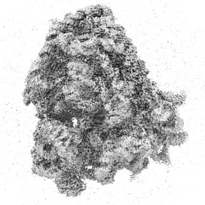 emd_13113.png emd_13113.png | 92 KB | ||
| Filedesc metadata | emd-13113.cif.gz | 20.5 KB | ||
| Archive directory |  http://ftp.pdbj.org/pub/emdb/structures/EMD-13113ftp://ftp.pdbj.org/pub/emdb/structures/EMD-13113 http://ftp.pdbj.org/pub/emdb/structures/EMD-13113ftp://ftp.pdbj.org/pub/emdb/structures/EMD-13113 | HTTPS FTP |
-Related structure data
| Related structure data |  7oycMC  7oyaC  7oybC  7oydC M: atomic model generated by this map C: citing same article ( |
|---|---|
| Similar structure data |


-Links
| EMDB pages | EMDB (EBI/PDBe) / EMDataResource |
|---|---|
| Related items in Molecule of the Month |


-Map
| File | Download / File: emd_13113.map.gz / Format: CCP4 / Size: 421.9 MB / Type: IMAGE STORED AS FLOATING POINT NUMBER (4 BYTES) | ||||||||||||||||||||||||||||||||||||
|---|---|---|---|---|---|---|---|---|---|---|---|---|---|---|---|---|---|---|---|---|---|---|---|---|---|---|---|---|---|---|---|---|---|---|---|---|---|
| Projections & slices | Image control
Images are generated by Spider. | ||||||||||||||||||||||||||||||||||||
| Voxel size | X=Y=Z: 1.06 Å | ||||||||||||||||||||||||||||||||||||
| Density |
| ||||||||||||||||||||||||||||||||||||
| Symmetry | Space group: 1 | ||||||||||||||||||||||||||||||||||||
| Details | EMDB XML:
|
 Z (Sec.)
 Y (Row.)
 X (Col.)


-Supplemental data
- Sample components
+Entire : Xenopus egg 80S ribosome
+Supramolecule #1: Xenopus egg 80S ribosome
+Macromolecule #1: Eukaryotic translation initiation factor 5A
+Macromolecule #6: 60S ribosomal protein L8
+Macromolecule #7: 40S ribosomal protein SA
+Macromolecule #8: Rpl3-prov protein
+Macromolecule #9: 40S ribosomal protein S3a-A
+Macromolecule #10: 60S ribosomal protein L4-B
+Macromolecule #11: 40S ribosomal protein S2
+Macromolecule #12: Rpl5-b protein
+Macromolecule #13: DNA-(apurinic or apyrimidinic site) lyase
+Macromolecule #14: 60S ribosomal protein L6
+Macromolecule #15: 40S ribosomal protein S4
+Macromolecule #16: MGC130910 protein
+Macromolecule #17: Ribosomal_S7 domain-containing protein
+Macromolecule #18: 60S ribosomal protein L7a
+Macromolecule #19: 40S ribosomal protein S6
+Macromolecule #20: 60S ribosomal protein L9
+Macromolecule #21: 40S ribosomal protein S7
+Macromolecule #22: Ribosomal_L16 domain-containing protein
+Macromolecule #23: 40S ribosomal protein S8
+Macromolecule #24: 60S ribosomal protein L11
+Macromolecule #25: 40S ribosomal protein S9
+Macromolecule #26: 40S ribosomal protein S10
+Macromolecule #27: 60S ribosomal protein L13
+Macromolecule #28: 40S ribosomal protein S11
+Macromolecule #29: 60S ribosomal protein L14
+Macromolecule #30: Ribosomal protein L15
+Macromolecule #31: 40S ribosomal protein S13
+Macromolecule #32: 60S ribosomal protein L13a
+Macromolecule #33: Rps14
+Macromolecule #34: 60S ribosomal protein L17
+Macromolecule #35: 40S ribosomal protein S15
+Macromolecule #36: Ribosomal_L18e/L15P domain-containing protein
+Macromolecule #37: Rps16 protein
+Macromolecule #38: 60S ribosomal protein L19
+Macromolecule #39: 40S ribosomal protein S17
+Macromolecule #40: 60S ribosomal protein L18a
+Macromolecule #41: 40S ribosomal protein S18
+Macromolecule #42: 60S ribosomal protein L21
+Macromolecule #43: 40S ribosomal protein S19
+Macromolecule #44: 60S ribosomal protein L22
+Macromolecule #45: 40S ribosomal protein S20
+Macromolecule #46: 60S ribosomal protein L23
+Macromolecule #47: 40S ribosomal protein S21
+Macromolecule #48: TRASH domain-containing protein
+Macromolecule #49: 40S ribosomal protein S15a
+Macromolecule #50: Ribosomal_L23eN domain-containing protein
+Macromolecule #51: 40S ribosomal protein S23
+Macromolecule #52: KOW domain-containing protein
+Macromolecule #53: 40S ribosomal protein S24
+Macromolecule #54: 60S ribosomal protein L27
+Macromolecule #55: 40S ribosomal protein S25
+Macromolecule #56: 60S ribosomal protein L27a
+Macromolecule #57: 40S ribosomal protein S26
+Macromolecule #58: 60S ribosomal protein L29
+Macromolecule #59: 40S ribosomal protein S27
+Macromolecule #60: 60S ribosomal protein L30
+Macromolecule #61: 40S ribosomal protein S28
+Macromolecule #62: 60S ribosomal protein L31
+Macromolecule #63: 40S ribosomal protein S29
+Macromolecule #64: Rpl32
+Macromolecule #65: 40S ribosomal protein S30
+Macromolecule #66: 60S ribosomal protein L35a
+Macromolecule #67: 60S ribosomal protein L34
+Macromolecule #68: Gnb2l1-prov protein
+Macromolecule #69: 60S ribosomal protein L35
+Macromolecule #70: 60S ribosomal protein L36
+Macromolecule #71: HABP4_PAI-RBP1 domain-containing protein
+Macromolecule #72: Ribosomal protein L37
+Macromolecule #73: 60S ribosomal protein L38
+Macromolecule #74: MGC116452 protein
+Macromolecule #75: 60S ribosomal protein L40
+Macromolecule #76: Rpl41
+Macromolecule #77: MGC85428 protein
+Macromolecule #78: Rpl37a
+Macromolecule #79: 60S ribosomal protein L28
+Macromolecule #80: Death-associated protein-like 1-B
+Macromolecule #81: Eef2-prov protein
+Macromolecule #2: 18S rRNA
+Macromolecule #3: 28S rRNA
+Macromolecule #4: 5S rRNA
+Macromolecule #5: 5.8S rRNA
+Macromolecule #82: MAGNESIUM ION
+Macromolecule #83: ZINC ION
-Experimental details
-Structure determination
| Method | cryo EM |
|---|---|
 Processing Processing | single particle reconstruction |
| Aggregation state | particle |
-Sample preparation
| Buffer | pH: 7.6 Component:
| ||||||||||||||||||
|---|---|---|---|---|---|---|---|---|---|---|---|---|---|---|---|---|---|---|---|
| Grid | Model: Quantifoil R3.5/1 / Material: COPPER / Mesh: 200 / Support film - Material: CARBON / Support film - topology: CONTINUOUS / Support film - Film thickness: 2 / Pretreatment - Type: GLOW DISCHARGE / Pretreatment - Time: 60 sec. / Pretreatment - Atmosphere: AIR | ||||||||||||||||||
| Vitrification | Cryogen name: ETHANE |
- Electron microscopy
| Microscope | FEI TITAN KRIOS |
|---|---|
| Image recording | Film or detector model: FEI FALCON III (4k x 4k) / Detector mode: INTEGRATING / Number grids imaged: 1 / Average exposure time: 1.0 sec. / Average electron dose: 40.0 e/Å2 |
| Electron beam | Acceleration voltage: 300 kV / Electron source:  FIELD EMISSION GUN FIELD EMISSION GUN |
| Electron optics | C2 aperture diameter: 70.0 µm / Illumination mode: FLOOD BEAM / Imaging mode: BRIGHT FIELD / Cs: 2.7 mm |
| Sample stage | Specimen holder model: FEI TITAN KRIOS AUTOGRID HOLDER / Cooling holder cryogen: NITROGEN |
| Experimental equipment |  Model: Titan Krios / Image courtesy: FEI Company |
