 Movie
Movie Controller


+ Open data

- Basic information
| Entry | 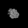 | |||||||||
|---|---|---|---|---|---|---|---|---|---|---|
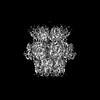
| Title | Structure of Primase-Helicase in SaPI5 | |||||||||
 Map data Map data | ||||||||||
 Sample Sample |
| |||||||||
 Keywords Keywords | Helicase / DNA binding / AMPPNP / REPLICATION | |||||||||
| Function / homology |  Function and homology information Function and homology information | |||||||||
| Biological species |   Staphylococcus aureus (bacteria) Staphylococcus aureus (bacteria) | |||||||||
| Method | single particle reconstruction / cryo EM / Resolution: 3.3 Å | |||||||||
 Authors Authors | Qiao CC / Mir-Sanchis I | |||||||||
| Funding support |  Sweden, 1 items Sweden, 1 items
| |||||||||
 Citation Citation | Journal: Nucleic Acids Res / Year: 2022 Title: Staphylococcal self-loading helicases couple the staircase mechanism with inter domain high flexibility. Authors: Cuncun Qiao / Gianluca Debiasi-Anders / Ignacio Mir-Sanchis / Abstract: Replication is a crucial cellular process. Replicative helicases unwind DNA providing the template strand to the polymerase and promoting replication fork progression. Helicases are multi-domain ...Replication is a crucial cellular process. Replicative helicases unwind DNA providing the template strand to the polymerase and promoting replication fork progression. Helicases are multi-domain proteins which use an ATPase domain to couple ATP hydrolysis with translocation, however the role that the other domains might have during translocation remains elusive. Here, we studied the unexplored self-loading helicases called Reps, present in Staphylococcus aureus pathogenicity islands (SaPIs). Our cryoEM structures of the PriRep5 from SaPI5 (3.3 Å), the Rep1 from SaPI1 (3.9 Å) and Rep1-DNA complex (3.1Å) showed that in both Reps, the C-terminal domain (CTD) undergoes two distinct movements respect the ATPase domain. We experimentally demonstrate both in vitro and in vivo that SaPI-encoded Reps need key amino acids involved in the staircase mechanism of translocation. Additionally, we demonstrate that the CTD's presence is necessary for the maintenance of full ATPase and helicase activities. We speculate that this high interdomain flexibility couples Rep's activities as initiators and as helicases. | |||||||||
| History |
|
- Structure visualization
| Supplemental images |
|---|

- Downloads & links
-EMDB archive
| Map data | emd_12975.map.gz | 165 MB | EMDB map data format | |
|---|---|---|---|---|
| Header (meta data) | emd-12975-v30.xmlemd-12975.xml | 17.3 KB 17.3 KB | Display Display | EMDB header |
| FSC (resolution estimation) | emd_12975_fsc.xml | 16.5 KB | Display | FSC data file |
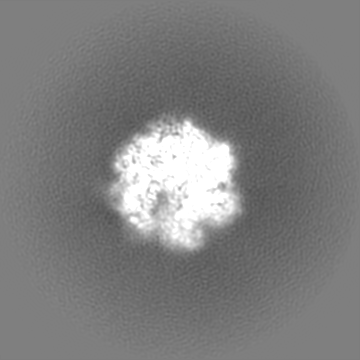
| Images |  emd_12975.png emd_12975.png | 50.1 KB | ||
| Masks | emd_12975_msk_1.map | 178 MB | Mask map | |
| Filedesc metadata | emd-12975.cif.gz | 6.3 KB | ||
| Others | emd_12975_half_map_1.map.gzemd_12975_half_map_2.map.gz | 140.9 MB 140.9 MB | ||
| Archive directory |  http://ftp.pdbj.org/pub/emdb/structures/EMD-12975ftp://ftp.pdbj.org/pub/emdb/structures/EMD-12975 http://ftp.pdbj.org/pub/emdb/structures/EMD-12975ftp://ftp.pdbj.org/pub/emdb/structures/EMD-12975 | HTTPS FTP |
-Related structure data
| Related structure data |  7olaMC  7om0C 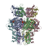 7pdsC M: atomic model generated by this map C: citing same article ( |
|---|---|
| Similar structure data | |
| EM raw data | EMPIAR-10881 (Title: Staphylococcal self-loading helicases couple the staircase mechanism with inter domain high flexibility Data size: 467.6 / Data #1: PriRep5-dsDNA [micrographs - multiframe]) |


-Links
| EMDB pages | EMDB (EBI/PDBe) / EMDataResource |
|---|---|
| Related items in Molecule of the Month |


-Map
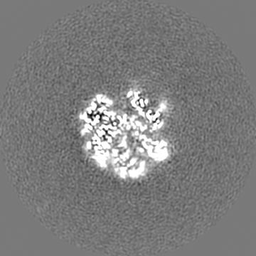
| File | Download / File: emd_12975.map.gz / Format: CCP4 / Size: 178 MB / Type: IMAGE STORED AS FLOATING POINT NUMBER (4 BYTES) | ||||||||||||||||||||||||||||||||||||
|---|---|---|---|---|---|---|---|---|---|---|---|---|---|---|---|---|---|---|---|---|---|---|---|---|---|---|---|---|---|---|---|---|---|---|---|---|---|
| Projections & slices | Image control
Images are generated by Spider. | ||||||||||||||||||||||||||||||||||||
| Voxel size | X=Y=Z: 0.82 Å | ||||||||||||||||||||||||||||||||||||
| Density |
| ||||||||||||||||||||||||||||||||||||
| Symmetry | Space group: 1 | ||||||||||||||||||||||||||||||||||||
| Details | EMDB XML:
|
 Z (Sec.)
 Y (Row.)
 X (Col.)


-Supplemental data
-Mask #1
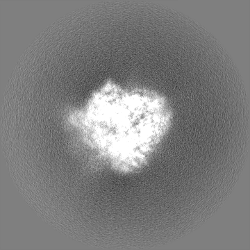
| File | emd_12975_msk_1.map | ||||||||||||
|---|---|---|---|---|---|---|---|---|---|---|---|---|---|
| Projections & Slices |
| ||||||||||||
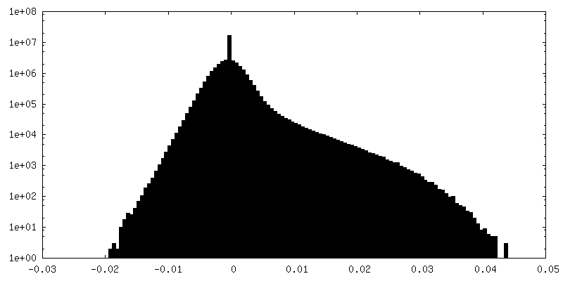
| Density Histograms |


-Half map: #2
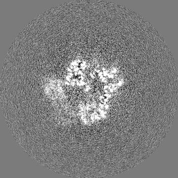
| File | emd_12975_half_map_1.map | ||||||||||||
|---|---|---|---|---|---|---|---|---|---|---|---|---|---|
| Projections & Slices |
| ||||||||||||
| Density Histograms |


-Half map: #1
| File | emd_12975_half_map_2.map | ||||||||||||
|---|---|---|---|---|---|---|---|---|---|---|---|---|---|
| Projections & Slices |
| ||||||||||||
| Density Histograms |


- Sample components
-Entire : Primase-Helicase in SaPI5
| Entire | Name: Primase-Helicase in SaPI5 |
|---|---|
| Components |
|
-Supramolecule #1: Primase-Helicase in SaPI5
| Supramolecule | Name: Primase-Helicase in SaPI5 / type: organelle_or_cellular_component / ID: 1 / Parent: 0 / Macromolecule list: #1 |
|---|---|
| Source (natural) | Organism: Staphylococcus aureus (bacteria) |
| Molecular weight | Theoretical: 669 KDa |
-Macromolecule #1: DNA primase
| Macromolecule | Name: DNA primase / type: protein_or_peptide / ID: 1 / Number of copies: 6 / Enantiomer: LEVO |
|---|---|
| Source (natural) | Organism: Staphylococcus aureus (bacteria) |
| Molecular weight | Theoretical: 93.007031 KDa |
| Recombinant expression | Organism: |
| Sequence | String: MGHHHHHHAI KKKDRIIGVK ELEIPQELKL VPNWVLWRAE WNEKQQNFGK VPYSINGYRA STTNKKTWCD FESVSIEYEV DEQYSGIGF VLSDGNNFVC LDIDNAIDKK GQINSELALK MMQLTYCEKS PSGTGLHCFF KGKLPDNRKK KRTDLDIELY D SARFMTVT ...String: MGHHHHHHAI KKKDRIIGVK ELEIPQELKL VPNWVLWRAE WNEKQQNFGK VPYSINGYRA STTNKKTWCD FESVSIEYEV DEQYSGIGF VLSDGNNFVC LDIDNAIDKK GQINSELALK MMQLTYCEKS PSGTGLHCFF KGKLPDNRKK KRTDLDIELY D SARFMTVT GCTIGQSDIC DNQEVLNTLV DEYFKENLPA NEVVREESNT NIQLSDEDII NIMMKSKQKD KIKDLLQGTY ES YFESSSE AVQSLLHYLA FYTGKNKQQM ERIFLNYNNL TDKWESKRGN TTWGQLELDK AIKNQKTIYT KSIDEFNVIP QGS KDVKQL LNQLGHEERT KMEENWIEEG KRGRKPTTIS PIKCAYILNE HLTFILFDDE ENTKLAMYQF DEGIYTQNTT IIKR VISYL EPKHNSNKAD EVIYHLTNMV DIKEKTNSPY LIPVKNGVFN RKTKQLESFT PDYIFTSKID TSYVRQDIVP EINGW NIDR WIEEIACNDN QVVKLLWQVI NDSMNGNYTR KKAIFFVGDG NNGKGTFQEL LSNVIGYSNI ASLKVNEFDE RFKLSV LEG KTAVIGDDVP VGVYVDDSSN FKSVVTGDPV LVEFKNKPLY RATFKCTVIQ STNGMPKFKD KTGGTLRRLL IVPFNAN FN GIKENFKIKE DYIKNQQVLE YVLYKAINLD FETFDIPDAS KKMLEVFKED NDPVYGFKVN MFDQWTIRKV PKYIVYAF Y KEYCDENGYN ALSSNKFYKQ FEHYLENYWK TDAQRRYDNE ELAKRIYNFN DNRNYIEPIE SGKNYKSYEK VKLKAI UniProtKB: DNA primase |
-Macromolecule #2: PHOSPHOAMINOPHOSPHONIC ACID-ADENYLATE ESTER
| Macromolecule | Name: PHOSPHOAMINOPHOSPHONIC ACID-ADENYLATE ESTER / type: ligand / ID: 2 / Number of copies: 4 / Formula: ANP |
|---|---|
| Molecular weight | Theoretical: 506.196 Da |
| Chemical component information |  ChemComp-ANP: |
-Macromolecule #3: MAGNESIUM ION
| Macromolecule | Name: MAGNESIUM ION / type: ligand / ID: 3 / Number of copies: 3 / Formula: MG |
|---|---|
| Molecular weight | Theoretical: 24.305 Da |
-Experimental details
-Structure determination
| Method | cryo EM |
|---|---|
 Processing Processing | single particle reconstruction |
| Aggregation state | particle |
-Sample preparation
| Concentration | 0.4 mg/mL |
|---|---|
| Buffer | pH: 8 / Component - Concentration: 200.0 mM / Component - Formula: NaCl / Component - Name: Sodium Chloride |
| Grid | Model: Quantifoil R2/2 / Material: COPPER / Mesh: 200 / Support film - Material: CARBON / Support film - topology: CONTINUOUS / Support film - Film thickness: 2 / Pretreatment - Type: GLOW DISCHARGE |
| Vitrification | Cryogen name: ETHANE / Chamber humidity: 100 % / Chamber temperature: 277 K / Instrument: FEI VITROBOT MARK IV |
- Electron microscopy
| Microscope | FEI TITAN KRIOS |
|---|---|
| Image recording | Film or detector model: GATAN K2 QUANTUM (4k x 4k) / Detector mode: COUNTING / Number real images: 4760 / Average exposure time: 4.0 sec. / Average electron dose: 1.16 e/Å2 |
| Electron beam | Acceleration voltage: 300 kV / Electron source:  FIELD EMISSION GUN FIELD EMISSION GUN |
| Electron optics | Illumination mode: SPOT SCAN / Imaging mode: BRIGHT FIELD / Cs: 2.7 mm / Nominal magnification: 165000 |
| Sample stage | Specimen holder model: FEI TITAN KRIOS AUTOGRID HOLDER / Cooling holder cryogen: NITROGEN |
| Experimental equipment |  Model: Titan Krios / Image courtesy: FEI Company |
+Image processing

-Atomic model buiding 1
| Refinement | Space: REAL / Protocol: AB INITIO MODEL / Target criteria: Correlation coefficient |
|---|---|
| Output model | PDB-7ola: |
