 Movie
Movie Controller


+ Open data

- Basic information
| Entry | Database: EMDB / ID: EMD-0721 | |||||||||
|---|---|---|---|---|---|---|---|---|---|---|
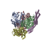
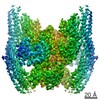
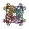
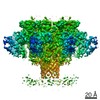
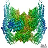
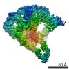
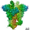
| Title | Pore structure of Iota toxin binding component (Ib) | |||||||||
 Map data Map data | ||||||||||
 Sample Sample |
| |||||||||
 Keywords Keywords | Bacterial binary toxin / Protein translocation channel / ADP-ribosylation / TOXIN | |||||||||
| Function / homology |  Function and homology information Function and homology information | |||||||||
| Biological species |   Clostridium perfringens (bacteria) Clostridium perfringens (bacteria) | |||||||||
| Method | single particle reconstruction / cryo EM / Resolution: 2.9 Å | |||||||||
 Authors Authors | Yoshida T / Yamada T | |||||||||
| Funding support |  Japan, 2 items Japan, 2 items
| |||||||||
 Citation Citation | Journal: Nat Struct Mol Biol / Year: 2020 Title: Cryo-EM structures reveal translocational unfolding in the clostridial binary iota toxin complex. Authors: Tomohito Yamada / Toru Yoshida / Akihiro Kawamoto / Kaoru Mitsuoka / Kenji Iwasaki / Hideaki Tsuge / Abstract: The iota toxin produced by Clostridium perfringens type E is a binary toxin comprising two independent polypeptides: Ia, an ADP-ribosyltransferase, and Ib, which is involved in cell binding and ...The iota toxin produced by Clostridium perfringens type E is a binary toxin comprising two independent polypeptides: Ia, an ADP-ribosyltransferase, and Ib, which is involved in cell binding and translocation of Ia across the cell membrane. Here we report cryo-EM structures of the translocation channel Ib-pore and its complex with Ia. The high-resolution Ib-pore structure demonstrates a similar structural framework to that of the catalytic ϕ-clamp of the anthrax protective antigen pore. However, the Ia-bound Ib-pore structure shows a unique binding mode of Ia: one Ia binds to the Ib-pore, and the Ia amino-terminal domain forms multiple weak interactions with two additional Ib-pore constriction sites. Furthermore, Ib-binding induces tilting and partial unfolding of the Ia N-terminal α-helix, permitting its extension to the ϕ-clamp gate. This new mechanism of N-terminal unfolding is crucial for protein translocation. | |||||||||
| History |
|
- Structure visualization
| Movie |
Movie viewer |
|---|---|
| Structure viewer | EM map: SurfViewMolmilJmol/JSmol |
| Supplemental images |

- Downloads & links
-EMDB archive
| Map data | emd_0721.map.gz | 10 MB | EMDB map data format | |
|---|---|---|---|---|
| Header (meta data) | emd-0721-v30.xmlemd-0721.xml | 14.7 KB 14.7 KB | Display Display | EMDB header |
| FSC (resolution estimation) | emd_0721_fsc.xml | 11.2 KB | Display | FSC data file |
| Images |  emd_0721.png emd_0721.png | 142.5 KB | ||
| Filedesc metadata | emd-0721.cif.gz | 6.2 KB | ||
| Archive directory |  http://ftp.pdbj.org/pub/emdb/structures/EMD-0721ftp://ftp.pdbj.org/pub/emdb/structures/EMD-0721 http://ftp.pdbj.org/pub/emdb/structures/EMD-0721ftp://ftp.pdbj.org/pub/emdb/structures/EMD-0721 | HTTPS FTP |
-Related structure data
| Related structure data |  6klxMC  0713C  0720C  6kloC  6klwC M: atomic model generated by this map C: citing same article ( |
|---|---|
| Similar structure data |

-Links
| EMDB pages | EMDB (EBI/PDBe) / EMDataResource |
|---|---|
| Related items in Molecule of the Month |

-Map
| File | Download / File: emd_0721.map.gz / Format: CCP4 / Size: 125 MB / Type: IMAGE STORED AS FLOATING POINT NUMBER (4 BYTES) | ||||||||||||||||||||||||||||||||||||||||||||||||||||||||||||
|---|---|---|---|---|---|---|---|---|---|---|---|---|---|---|---|---|---|---|---|---|---|---|---|---|---|---|---|---|---|---|---|---|---|---|---|---|---|---|---|---|---|---|---|---|---|---|---|---|---|---|---|---|---|---|---|---|---|---|---|---|---|
| Projections & slices | Image control
Images are generated by Spider. | ||||||||||||||||||||||||||||||||||||||||||||||||||||||||||||
| Voxel size | X=Y=Z: 1.13 Å | ||||||||||||||||||||||||||||||||||||||||||||||||||||||||||||
| Density |
| ||||||||||||||||||||||||||||||||||||||||||||||||||||||||||||
| Symmetry | Space group: 1 | ||||||||||||||||||||||||||||||||||||||||||||||||||||||||||||
| Details | EMDB XML:
CCP4 map header:
| ||||||||||||||||||||||||||||||||||||||||||||||||||||||||||||
 Z (Sec.)
 Y (Row.)
 X (Col.)


-Supplemental data
- Sample components
-Entire : Pore structure of Iota toxin binding component (Ib)
| Entire | Name: Pore structure of Iota toxin binding component (Ib) |
|---|---|
| Components |
|
-Supramolecule #1: Pore structure of Iota toxin binding component (Ib)
| Supramolecule | Name: Pore structure of Iota toxin binding component (Ib) / type: complex / ID: 1 / Parent: 0 / Macromolecule list: #1 |
|---|---|
| Source (natural) | Organism: Clostridium perfringens (bacteria) |
| Molecular weight | Theoretical: 520 KDa |
-Macromolecule #1: Iota toxin component Ib
| Macromolecule | Name: Iota toxin component Ib / type: protein_or_peptide / ID: 1 / Number of copies: 7 / Enantiomer: LEVO |
|---|---|
| Source (natural) | Organism: Clostridium perfringens (bacteria) |
| Molecular weight | Theoretical: 74.37882 KDa |
| Recombinant expression | Organism: |
| Sequence | String: SAAWEDEDLD TDNDNIPDAY EKNGYTIKDS IAVKWNDSFA EQGYKKYVSS YLESNTAGDP YTDYQKASGS IDKAIKLEAR DPLVAAYPV VGVGMENLII STNEHASSDQ GKTVSRATTN SKTDANTVGV SISAGYQNGF TGNITTSYSH TTDNSTAVQD S NGESWNTG ...String: SAAWEDEDLD TDNDNIPDAY EKNGYTIKDS IAVKWNDSFA EQGYKKYVSS YLESNTAGDP YTDYQKASGS IDKAIKLEAR DPLVAAYPV VGVGMENLII STNEHASSDQ GKTVSRATTN SKTDANTVGV SISAGYQNGF TGNITTSYSH TTDNSTAVQD S NGESWNTG LSINKGESAY INANVRYYNT GTAPMYKVTP TTNLVLDGET LATIKAQDNQ IGNNLSPNET YPKKGLSPLA LN TMDQFNA RLIPINYDQL KKLDSGKQIK LETTQVSGNY GTKNSQGQII TEGNSWSNYI SQIDSVSASI ILDTGSQTFE RRV AAKEQG NPEDKTPEIT IGEAIKKAFS ATKNGELLYF NGIPIDESCV ELIFDDNTSE IIKEQLKYLD DKKIYNVKLE RGMN ILIKV PSYFTNFDEY NNFPASWSNI DTKNQDGLQS VANKLSGETK IIIPMSKLKP YKRYVFSGYS KDPSTSNSIT VNIKS KEQK TDYLVPEKDY TKFSYEFETT GKDSSDIEIT LTSSGVIFLD NLSITELNST PEILKEPEIK VPSDQEILDA HNKYYA DIK LDTNTGNTYI DGIYFEPTQT NKEALDYIQK YRVEATLQYS GFKDIGTKDK EIRNYLGDQN QPKTNYINFR SYFTSGE NV MTYKKLRIYA VTPDNRELLV LSVN UniProtKB: Iota toxin component Ib |
-Macromolecule #2: CALCIUM ION
| Macromolecule | Name: CALCIUM ION / type: ligand / ID: 2 / Number of copies: 14 / Formula: CA |
|---|---|
| Molecular weight | Theoretical: 40.078 Da |
-Experimental details
-Structure determination
| Method | cryo EM |
|---|---|
 Processing Processing | single particle reconstruction |
| Aggregation state | particle |
-Sample preparation
| Concentration | 0.38 mg/mL | ||||||||||||
|---|---|---|---|---|---|---|---|---|---|---|---|---|---|
| Buffer | pH: 7.5 Component:
| ||||||||||||
| Grid | Model: Quantifoil R1.2/1.3 / Material: COPPER / Mesh: 200 / Pretreatment - Type: GLOW DISCHARGE / Pretreatment - Time: 30 sec. | ||||||||||||
| Vitrification | Cryogen name: ETHANE / Chamber humidity: 100 % / Chamber temperature: 277 K / Instrument: FEI VITROBOT MARK IV |
- Electron microscopy
| Microscope | FEI TITAN KRIOS |
|---|---|
| Temperature | Min: 78.9 K / Max: 78.9 K |
| Specialist optics | Spherical aberration corrector: Microscope was modified with a Cs corrector |
| Image recording | Film or detector model: FEI FALCON III (4k x 4k) / Detector mode: COUNTING / Digitization - Dimensions - Width: 4096 pixel / Digitization - Dimensions - Height: 4096 pixel / Number grids imaged: 1 / Number real images: 2120 / Average exposure time: 84.09 sec. / Average electron dose: 50.0 e/Å2 |
| Electron beam | Acceleration voltage: 300 kV / Electron source:  FIELD EMISSION GUN FIELD EMISSION GUN |
| Electron optics | C2 aperture diameter: 50.0 µm / Illumination mode: FLOOD BEAM / Imaging mode: BRIGHT FIELD / Nominal defocus max: 2.5 µm / Nominal defocus min: 0.8 µm / Nominal magnification: 59000 |
| Sample stage | Specimen holder model: FEI TITAN KRIOS AUTOGRID HOLDER / Cooling holder cryogen: NITROGEN |
| Experimental equipment |  Model: Titan Krios / Image courtesy: FEI Company |


