 Movie
Movie Controller


+ Open data

- Basic information
| Entry |  Database: PDB chemical components / ID: ADK Database: PDB chemical components / ID: ADK |
|---|---|
| Name | Name: |
-Chemical information
| Composition |  | ||||||||
|---|---|---|---|---|---|---|---|---|---|
| Others | Type: NON-POLYMER / PDB classification: HETAIN / Three letter code: ADK / Model coordinates PDB-ID: 1P7M | ||||||||
| History |
| ||||||||
 External links External links | UniChem / ChemSpider / BindingDB / Brenda / ChEBI / ChEMBL / CompTox / DrugBank / HMDB / SureChEMBL / Wikipedia search / Google search |
- Structure visualization
| Structure viewer | Molecule:  MolmilJmol/JSmol MolmilJmol/JSmol |
|---|
-Details
-SMILES
| ACDLabs 10.04 | | CACTVS 3.341 | OpenEye OEToolkits 1.5.0 | |
|---|
-SMILES CANONICAL
| CACTVS 3.341 | | OpenEye OEToolkits 1.5.0 | |
|---|
-InChI
| InChI 1.03 |
|---|
-InChIKey
| InChI 1.03 |
|---|
-SYSTEMATIC NAME
| ACDLabs 10.04 | | OpenEye OEToolkits 1.5.0 | |
|---|
-PDB entries
Showing all 6 items

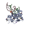
PDB-1p7m: 
SOLUTION STRUCTURE AND BASE PERTURBATION STUDIES REVEAL A NOVEL MODE OF ALKYLATED BASE RECOGNITION BY 3-METHYLADENINE DNA GLYCOSYLASE I
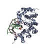
PDB-2ofi:
Crystal Structure of 3-methyladenine DNA Glycosylase I (TAG) bound to DNA/3mA

PDB-4ai5:
Crystal structure of Y16F of 3-methyladenine DNA glycosylase I (TAG) in complex with 3-methyladenine

PDB-4aia:
The structural basis of 3-methyladenine recognition by 3- methyladenine DNA glycosylase I (TAG) from Staphylococcus aureus

PDB-5cld:
Alkylpurine DNA glycosylase AlkD bound to DNA containing an oxocarbenium-intermediate analog and a free 3-methyladenine nucleobase
PDB-5cle:
Alkylpurine DNA glycosylase AlkD bound to DNA containing an abasic-site analog and a free 3-methyladenine nucleobase
