 Movie
Movie Controller


+ Open data

- Basic information
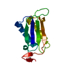
| Entry | Database: PDB / ID: 1bja | ||||||
|---|---|---|---|---|---|---|---|
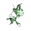
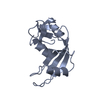
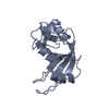
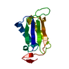
| Title | ACTIVATION DOMAIN OF THE PHAGE T4 TRANSCRIPTION FACTOR MOTA | ||||||
 Components Components | TRANSCRIPTION REGULATORY PROTEIN MOTA | ||||||
 Keywords Keywords | ACTIVATION DOMAIN / PHAGE T4 / MIDDLE MODE TRANSCRIPTION / ALPHA HELICAL STRUCTURE / TRANSCRIPTION REGULATION | ||||||
| Function / homology |  Function and homology information Function and homology information | ||||||
| Biological species |  Enterobacteria phage T4 (virus) Enterobacteria phage T4 (virus) | ||||||
| Method |  X-RAY DIFFRACTION / MIR / Resolution: 2.19 Å X-RAY DIFFRACTION / MIR / Resolution: 2.19 Å | ||||||
 Authors Authors | Finnin, M.S. / Cicero, M.P. / Davies, C. / Porter, S.J. / White, S.W. / Kreuzer, K.N. | ||||||
 Citation Citation | Journal: EMBO J. / Year: 1997 Title: The activation domain of the MotA transcription factor from bacteriophage T4. Authors: Finnin, M.S. / Cicero, M.P. / Davies, C. / Porter, S.J. / White, S.W. / Kreuzer, K.N. | ||||||
| History |
|
- Structure visualization
| Structure viewer | Molecule: MolmilJmol/JSmol |
|---|
- Downloads & links
-Download
| PDBx/mmCIF format | 1bja.cif.gz | 47.2 KB | Display | PDBx/mmCIF format |
|---|---|---|---|---|
| PDB format | pdb1bja.ent.gz | 34.2 KB | Display | PDB format |
| PDBx/mmJSON format | 1bja.json.gz | Tree view | PDBx/mmJSON format | |
| Others |  Other downloads Other downloads |
-Validation report
| Arichive directory | https://data.pdbj.org/pub/pdb/validation_reports/bj/1bjaftp://data.pdbj.org/pub/pdb/validation_reports/bj/1bja | HTTPS FTP |
|---|
-Related structure data
| Similar structure data |
|---|

-Links
 PDBj
PDBj- Assembly
| Deposited unit | 
| |||||||||||||||||
|---|---|---|---|---|---|---|---|---|---|---|---|---|---|---|---|---|---|---|
| 1 | 
| |||||||||||||||||
| 2 | 
| |||||||||||||||||
| Unit cell |
| |||||||||||||||||
| Noncrystallographic symmetry (NCS) | NCS domain:
NCS oper: (Code: given Matrix: (0.9042, -0.381, 0.1928), Vector: Details | IT IS UNCERTAIN WHETHER THE DIMER IN THE CRYSTAL STRUCTURE HAS BIOLOGICAL SIGNIFICANCE. A POSSIBILITY IS THAT THE DIMERIC STRUCTURE IS ALSO FORMED WHEN THE INTACT MOTA PROTEIN BINDS TO ITS TARGET DNA SITE. | |
-Components
| #1: Protein | Mass: 10114.576 Da / Num. of mol.: 2 / Fragment: N-TERMINAL ACTIVATION DOMAIN Source method: isolated from a genetically manipulated source Source: (gene. exp.) Enterobacteria phage T4 (virus) / Genus: T4-like viruses / Species: Enterobacteria phage T4 sensu lato / Gene: MOTA / Plasmid: PET3C / Species (production host): Escherichia coli / Gene (production host): MOTNF / Production host:  #2: Chemical | ChemComp-SO4 / |   Mass: 96.063 Da / Num. of mol.: 1 / Source method: obtained synthetically / Formula: SO4 Mass: 96.063 Da / Num. of mol.: 1 / Source method: obtained synthetically / Formula: SO4#3: Water | ChemComp-HOH / |  Mass: 18.015 Da / Num. of mol.: 60 / Source method: isolated from a natural source / Formula: H2O Mass: 18.015 Da / Num. of mol.: 60 / Source method: isolated from a natural source / Formula: H2O |
|---|
-Experimental details
-Experiment
| Experiment | Method: X-RAY DIFFRACTION / Number of used crystals: 2 |
|---|
- Sample preparation
| Crystal | Density Matthews: 2.12 Å3/Da / Density % sol: 43.37 % |
|---|---|
| Crystal grow | pH: 8.5 Details: PROTEIN WAS CRYSTALLIZED FROM 60% SATURATED AMMONIUM SULPHATE, 100 MM BIS-TRIS PROPANE, PH 8.0 - 9.0, pH 8.5 PH range: 8.0-9.0 |
| Crystal grow | *PLUS Temperature: 22 ℃ / Method: vapor diffusion, hanging drop / Details: Finnin, M.S., (1993) J. Mol. Biol., 232, 301. / PH range low: 9 / PH range high: 8 |
| Components of the solutions | *PLUS Conc.: 60 %sat / Common name: ammonium sulfate |
-Data collection
| Diffraction | Mean temperature: 293 K |
|---|---|
| Diffraction source | Source: ROTATING ANODE / Type: RIGAKU RUH3R / Wavelength: 1.5418 |
| Detector | Type: RIGAKU / Detector: IMAGE PLATE / Date: Jun 1, 1994 / Details: COLLIMATOR |
| Radiation | Monochromator: GRAPHITE(002) / Monochromatic (M) / Laue (L): M / Scattering type: x-ray |
| Radiation wavelength | Wavelength: 1.5418 Å / Relative weight: 1 |
| Reflection | Resolution: 2.19→10 Å / Num. obs: 9394 / % possible obs: 98.5 % / Observed criterion σ(I): 0 / Redundancy: 7 % / Biso Wilson estimate: 31 Å2 / Rmerge(I) obs: 0.048 / Rsym value: 0.053 / Net I/σ(I): 11 |
| Reflection shell | Resolution: 2.19→2.3 Å / Redundancy: 5 % / Rmerge(I) obs: 0.081 / Mean I/σ(I) obs: 2.5 / Rsym value: 0.101 / % possible all: 80 |
| Reflection | *PLUS Num. measured all: 66237 / Rmerge(I) obs: 0.0536 |
- Processing
| Software |
| ||||||||||||||||||||||||||||||||||||||||||||||||||||||||||||
|---|---|---|---|---|---|---|---|---|---|---|---|---|---|---|---|---|---|---|---|---|---|---|---|---|---|---|---|---|---|---|---|---|---|---|---|---|---|---|---|---|---|---|---|---|---|---|---|---|---|---|---|---|---|---|---|---|---|---|---|---|---|
| Refinement | Method to determine structure: MIR / Resolution: 2.19→6 Å / Rfactor Rfree error: 0.017 / Data cutoff high absF: 1000000 / Data cutoff low absF: 0.001 / Isotropic thermal model: 0 / Cross valid method: THROUGHOUT / σ(F): 2
| ||||||||||||||||||||||||||||||||||||||||||||||||||||||||||||
| Displacement parameters | Biso mean: 30 Å2 | ||||||||||||||||||||||||||||||||||||||||||||||||||||||||||||
| Refine analyze |
| ||||||||||||||||||||||||||||||||||||||||||||||||||||||||||||
| Refinement step | Cycle: LAST / Resolution: 2.19→6 Å
| ||||||||||||||||||||||||||||||||||||||||||||||||||||||||||||
| Refine LS restraints |
| ||||||||||||||||||||||||||||||||||||||||||||||||||||||||||||
| Refine LS restraints NCS | NCS model details: 0 | ||||||||||||||||||||||||||||||||||||||||||||||||||||||||||||
| LS refinement shell | Resolution: 2.19→2.29 Å / Rfactor Rfree error: 0.04 / Total num. of bins used: 6
| ||||||||||||||||||||||||||||||||||||||||||||||||||||||||||||
| Xplor file |
| ||||||||||||||||||||||||||||||||||||||||||||||||||||||||||||
| Software | *PLUS Name: X-PLOR / Version: 3.8 / Classification: refinement | ||||||||||||||||||||||||||||||||||||||||||||||||||||||||||||
| Refine LS restraints | *PLUS
|
