 Movie
Movie Controller


+ Open data

- Basic information
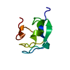
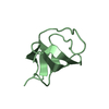
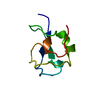
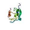
| Entry | Database: PDB / ID: 1ame | ||||||
|---|---|---|---|---|---|---|---|
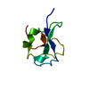
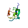
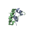
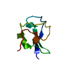
| Title | CRYSTAL STRUCTURE OF TYPE III ANTIFREEZE PROTEIN AT 4 C | ||||||
 Components Components | TYPE III ANTIFREEZE PROTEIN ISOFORM HPLC 12 | ||||||
 Keywords Keywords | ANTIFREEZE PROTEIN / CRYO-CRYSTALLOGRAPHY / COLD-ADAPTATION / CRYSTALLIZATION / FREEZING POINT | ||||||
| Function / homology |  Function and homology information Function and homology information | ||||||
| Biological species |  Macrozoarces americanus (ocean pout) Macrozoarces americanus (ocean pout) | ||||||
| Method |  X-RAY DIFFRACTION / ISOMORPHOUS (ROOM TEMPERATURE STARTING MODEL) / Resolution: 1.65 Å X-RAY DIFFRACTION / ISOMORPHOUS (ROOM TEMPERATURE STARTING MODEL) / Resolution: 1.65 Å | ||||||
 Authors Authors | Jia, Z. / Leinala, E. / Ye, Q. | ||||||
 Citation Citation | Journal: Acta Crystallogr.,Sect.D / Year: 1998 Title: Structure of type III antifreeze protein at 277 K. Authors: Ye, Q. / Leinala, E. / Jia, Z. #1: Journal: Nature / Year: 1996Title: Structural Basis for the Binding of a Globular Antifreeze Protein to Ice Authors: Jia, Z. / Deluca, C.I. / Chao, H. / Davies, P.L. | ||||||
| History |
|
- Structure visualization
| Structure viewer | Molecule: MolmilJmol/JSmol |
|---|
- Downloads & links
-Download
| PDBx/mmCIF format | 1ame.cif.gz | 23.7 KB | Display | PDBx/mmCIF format |
|---|---|---|---|---|
| PDB format | pdb1ame.ent.gz | 15 KB | Display | PDB format |
| PDBx/mmJSON format | 1ame.json.gz | Tree view | PDBx/mmJSON format | |
| Others |  Other downloads Other downloads |
-Validation report
| Arichive directory | https://data.pdbj.org/pub/pdb/validation_reports/am/1ameftp://data.pdbj.org/pub/pdb/validation_reports/am/1ame | HTTPS FTP |
|---|
-Related structure data
| Related structure data | 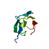 1msiS S: Starting model for refinement |
|---|---|
| Similar structure data |


-Links
 PDBj
PDBj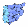
- Assembly
| Deposited unit | 
| ||||||||
|---|---|---|---|---|---|---|---|---|---|
| 1 |
| ||||||||
| Unit cell |
|
-Components
| #1: Protein | Mass: 7050.310 Da / Num. of mol.: 1 / Mutation: INS(0), P64A, P65A Source method: isolated from a genetically manipulated source Source: (gene. exp.) Macrozoarces americanus (ocean pout) / Plasmid: PT7-7F / Production host:  |
|---|---|
| #2: Water | ChemComp-HOH /  Mass: 18.015 Da / Num. of mol.: 45 / Source method: isolated from a natural source / Formula: H2O Mass: 18.015 Da / Num. of mol.: 45 / Source method: isolated from a natural source / Formula: H2O |
-Experimental details
-Experiment
| Experiment | Method: X-RAY DIFFRACTION / Number of used crystals: 1 |
|---|
- Sample preparation
| Crystal | Density Matthews: 2.11 Å3/Da / Density % sol: 31 % / Description: DATA COLLECTED AT 4 DEGREES C | ||||||||||||||||||||
|---|---|---|---|---|---|---|---|---|---|---|---|---|---|---|---|---|---|---|---|---|---|
| Crystal grow | Temperature: 277 K / pH: 4.5 Details: 53% AMMONIUM SULFATE 100 MM SODIUM ACETATE PH 4.5 CRYSTALLIZED AT 4 DEGREES C, temperature 277K | ||||||||||||||||||||
| Crystal grow | *PLUS Temperature: 277 K / Method: vapor diffusion, hanging dropDetails: drop was mixed with an equal volume of reservoir solution | ||||||||||||||||||||
| Components of the solutions | *PLUS
|
-Data collection
| Diffraction | Mean temperature: 277 K |
|---|---|
| Diffraction source | Source: ROTATING ANODE / Type: RIGAKU RUH2R / Wavelength: 1.5418 |
| Detector | Type: MARRESEARCH / Detector: IMAGE PLATE / Date: Feb 1, 1997 |
| Radiation | Monochromator: GRAPHITE(002) / Monochromatic (M) / Laue (L): M / Scattering type: x-ray |
| Radiation wavelength | Wavelength: 1.5418 Å / Relative weight: 1 |
| Reflection | Resolution: 1.65→50 Å / Num. obs: 6600 / % possible obs: 86.6 % / Observed criterion σ(I): 2 / Redundancy: 4.07 % / Biso Wilson estimate: 17.16 Å2 / Rmerge(I) obs: 0.066 / Net I/σ(I): 15.13 |
| Reflection shell | Resolution: 1.65→1.71 Å / Redundancy: 4.03 % / Rmerge(I) obs: 0.397 / Mean I/σ(I) obs: 2.29 / % possible all: 85.8 |
| Reflection | *PLUS Num. measured all: 27105 / Rmerge(I) obs: 0.061 |
| Reflection shell | *PLUS % possible obs: 85.8 % |
- Processing
| Software |
| ||||||||||||||||||||||||||||||||||||||||||||||||||||||||||||
|---|---|---|---|---|---|---|---|---|---|---|---|---|---|---|---|---|---|---|---|---|---|---|---|---|---|---|---|---|---|---|---|---|---|---|---|---|---|---|---|---|---|---|---|---|---|---|---|---|---|---|---|---|---|---|---|---|---|---|---|---|---|
| Refinement | Method to determine structure: ISOMORPHOUS (ROOM TEMPERATURE STARTING MODEL) Starting model: PDB ENTRY 1MSI Resolution: 1.65→8 Å / Rfactor Rfree error: 0.0147 / Data cutoff high absF: 100000 / Data cutoff low absF: 0.1 / Cross valid method: THROUGHOUT / σ(F): 2
| ||||||||||||||||||||||||||||||||||||||||||||||||||||||||||||
| Displacement parameters | Biso mean: 16.7 Å2 | ||||||||||||||||||||||||||||||||||||||||||||||||||||||||||||
| Refinement step | Cycle: LAST / Resolution: 1.65→8 Å
| ||||||||||||||||||||||||||||||||||||||||||||||||||||||||||||
| Refine LS restraints |
| ||||||||||||||||||||||||||||||||||||||||||||||||||||||||||||
| LS refinement shell | Resolution: 1.65→1.72 Å / Rfactor Rfree error: 0.044 / Total num. of bins used: 8
| ||||||||||||||||||||||||||||||||||||||||||||||||||||||||||||
| Xplor file |
| ||||||||||||||||||||||||||||||||||||||||||||||||||||||||||||
| Software | *PLUS Name: X-PLOR / Version: 3.1 / Classification: refinement | ||||||||||||||||||||||||||||||||||||||||||||||||||||||||||||
| Refine LS restraints | *PLUS
|
