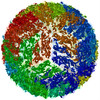
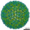
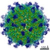
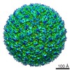
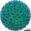
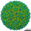
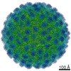
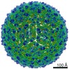
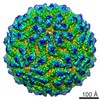
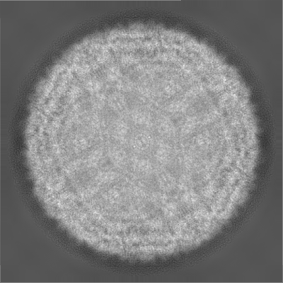
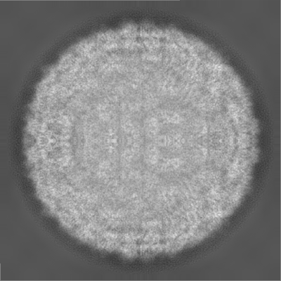
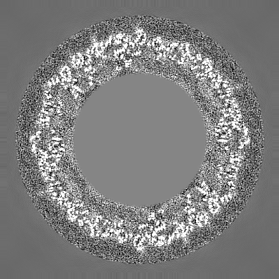
Journal: Nature / Year: 2016 Title: Structure of the thermally stable Zika virus. Authors: Victor A Kostyuchenko / Elisa X Y Lim / Shuijun Zhang / Guntur Fibriansah / Thiam-Seng Ng / Justin S G Ooi / Jian Shi / Shee-Mei Lok / Abstract: Zika virus (ZIKV), formerly a neglected pathogen, has recently been associated with microcephaly in fetuses, and with Guillian-Barré syndrome in adults. Here we present the 3.7 Å resolution cryo- ...Zika virus (ZIKV), formerly a neglected pathogen, has recently been associated with microcephaly in fetuses, and with Guillian-Barré syndrome in adults. Here we present the 3.7 Å resolution cryo-electron microscopy structure of ZIKV, and show that the overall architecture of the virus is similar to that of other flaviviruses. Sequence and structural comparisons of the ZIKV envelope (E) protein with other flaviviruses show that parts of the E protein closely resemble the neurovirulent West Nile and Japanese encephalitis viruses, while others are similar to dengue virus (DENV). However, the contribution of the E protein to flavivirus pathobiology is currently not understood. The virus particle was observed to be structurally stable even when incubated at 40 °C, in sharp contrast to the less thermally stable DENV. This is also reflected in the infectivity of ZIKV compared to DENV serotypes 2 and 4 (DENV2 and DENV4) at different temperatures. The cryo-electron microscopy structure shows a virus with a more compact surface. This structural stability of the virus may help it to survive in the harsh conditions of semen, saliva and urine. Antibodies or drugs that destabilize the structure may help to reduce the disease outcome or limit the spread of the virus.
History
Deposition
May 9, 2016
-
Header (metadata) release
May 25, 2016
-
Map release
May 25, 2016
-
Update
Oct 23, 2019
-
Current status
Oct 23, 2019
Processing site: PDBj / Status: Released
-
Structure visualization
Movie
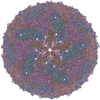
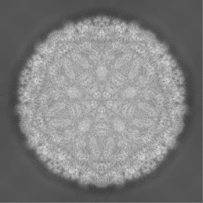
Surface view with section colored by density value
Name: N-ACETYL-D-GLUCOSAMINE / type: ligand / ID: 3 / Number of copies: 3 / Formula: NAG
Molecular weight
Theoretical: 221.208 Da
-
Experimental details
-
Structure determination
Method
cryo EM
Processing
single particle reconstruction
Aggregation state
particle
-
Sample preparation
Buffer
pH: 8 Component:
Concentration
Formula
Name
10.0 mM
C4H12ClNO3
Tris-HCl
120.0 mM
NaCl
Sodium Chloride
1.0 mM
C10H16N2O8
EDTA
Grid
Model: Quantifoil / Material: COPPER / Mesh: 400 / Support film - #0 - Film type ID: 1 / Support film - #0 - Material: CARBON / Support film - #0 - topology: LACEY / Support film - #1 - Film type ID: 2 / Support film - #1 - Material: CARBON / Support film - #1 - topology: CONTINUOUS / Pretreatment - Type: GLOW DISCHARGE
Vitrification
Cryogen name: ETHANE / Chamber humidity: 100 % / Chamber temperature: 298 K / Instrument: FEI VITROBOT MARK IV / Details: blot for 1s.
Details
purified with PEG precipitation, sucrose cushion and potassium tartrate gradient
-
Electron microscopy
Microscope
FEI TITAN KRIOS
Image recording
Film or detector model: FEI FALCON II (4k x 4k) / Detector mode: OTHER / Digitization - Dimensions - Width: 4096 pixel / Digitization - Dimensions - Height: 4096 pixel / Digitization - Sampling interval: 14.0 µm / Digitization - Frames/image: 2-11 / Number grids imaged: 1 / Number real images: 3086 / Average exposure time: 1.8 sec. / Average electron dose: 50.0 e/Å2
Electron beam
Acceleration voltage: 300 kV / Electron source: FIELD EMISSION GUN
Electron optics
Illumination mode: FLOOD BEAM / Imaging mode: BRIGHT FIELD / Cs: 2.7 mm
Experimental equipment
Model: Titan Krios / Image courtesy: FEI Company
+
Image processing
Details
images collected in movie mode, 30 frames per 1.8s exposure; frames aligned with MotionCorr, frames 2-11 averaged to produce images for further processing
Particle selection
Number selected: 11600 / Details: manually picked
CTF correction
Software - Name: ctffind4 (ver. 17) / Details: correction for astigmatism included
In the structure databanks used in Yorodumi, some data are registered as the other names, "COVID-19 virus" and "2019-nCoV". Here are the details of the virus and the list of structure data.
Jan 31, 2019. EMDB accession codes are about to change! (news from PDBe EMDB page)
EMDB accession codes are about to change! (news from PDBe EMDB page)
The allocation of 4 digits for EMDB accession codes will soon come to an end. Whilst these codes will remain in use, new EMDB accession codes will include an additional digit and will expand incrementally as the available range of codes is exhausted. The current 4-digit format prefixed with “EMD-” (i.e. EMD-XXXX) will advance to a 5-digit format (i.e. EMD-XXXXX), and so on. It is currently estimated that the 4-digit codes will be depleted around Spring 2019, at which point the 5-digit format will come into force.
The EM Navigator/Yorodumi systems omit the EMD- prefix.
Related info.:Q: What is EMD? / ID/Accession-code notation in Yorodumi/EM Navigator
Yorodumi is a browser for structure data from EMDB, PDB, SASBDB, etc.
This page is also the successor to EM Navigator detail page, and also detail information page/front-end page for Omokage search.
The word "yorodu" (or yorozu) is an old Japanese word meaning "ten thousand". "mi" (miru) is to see.
Related info.:EMDB / PDB / SASBDB / Comparison of 3 databanks / Yorodumi Search / Aug 31, 2016. New EM Navigator & Yorodumi / Yorodumi Papers / Jmol/JSmol / Function and homology information / Changes in new EM Navigator and Yorodumi
 Movie
Movie Controller

 Yorodumi
 Open data
 Basic information
 Map data
 Sample
 Function and homology information
 ZIKV (virus) /
  Authors
 Singapore, 3 items
  Citation
 Structure visualization
 Downloads & links
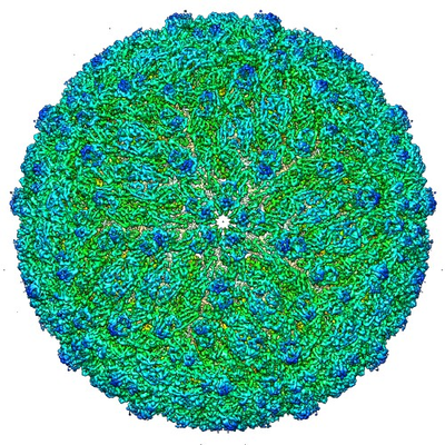 emd_8139.png
 http://ftp.pdbj.org/pub/emdb/structures/EMD-8139


 Z (Sec.)
 Y (Row.)
 X (Col.)


 Sample components
 Homo sapiens (human)
 Processing
 Electron microscopy
 FIELD EMISSION GUN