+ Open data
- Basic information
| Entry | Database: PDB / ID: 7vwx | ||||||
|---|---|---|---|---|---|---|---|
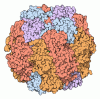
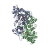
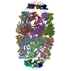
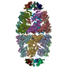
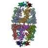
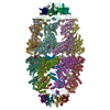
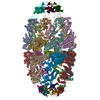
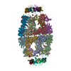
| Title | CryoEM structure of football-shaped GroEL:ES2 with RuBisCO | ||||||
 Components Components |
| ||||||
 Keywords Keywords | CHAPERONE / GroEL/ES system / substrate folding / folding assistance | ||||||
| Function / homology |  Function and homology information Function and homology informationribulose-bisphosphate carboxylase / GroEL-GroES complex / ribulose-bisphosphate carboxylase activity / reductive pentose-phosphate cycle / chaperonin ATPase / virion assembly / : / protein folding chaperone / isomerase activity / ATP-dependent protein folding chaperone ...ribulose-bisphosphate carboxylase / GroEL-GroES complex / ribulose-bisphosphate carboxylase activity / reductive pentose-phosphate cycle / chaperonin ATPase / virion assembly / : / protein folding chaperone / isomerase activity / ATP-dependent protein folding chaperone / monooxygenase activity / response to radiation / unfolded protein binding / protein folding / protein-folding chaperone binding / response to heat / protein refolding / magnesium ion binding / ATP hydrolysis activity / ATP binding / metal ion binding / identical protein binding / membrane / cytosol Similarity search - Function | ||||||
| Biological species |   Rhodospirillum rubrum ATCC 11170 (bacteria) Rhodospirillum rubrum ATCC 11170 (bacteria) | ||||||
| Method | ELECTRON MICROSCOPY / single particle reconstruction / cryo EM / Resolution: 7.6 Å | ||||||
 Authors Authors | Kim, H. / Roh, S.H. | ||||||
| Funding support |  Korea, Republic Of, 1items Korea, Republic Of, 1items
| ||||||
 Citation Citation |  Journal: iScience / Year: 2022 Journal: iScience / Year: 2022Title: Cryo-EM structures of GroEL:ES with RuBisCO visualize molecular contacts of encapsulated substrates in a double-cage chaperonin. Authors: Hyunmin Kim / Junsun Park / Seyeon Lim / Sung-Hoon Jun / Mingyu Jung / Soung-Hun Roh /  Abstract: The GroEL/GroES chaperonin system assists the folding of many proteins, through conformational transitions driven by ATP hydrolysis. Although structural information about bullet-shaped GroEL:ES ...The GroEL/GroES chaperonin system assists the folding of many proteins, through conformational transitions driven by ATP hydrolysis. Although structural information about bullet-shaped GroEL:ES complexes has been extensively reported, the substrate interactions of another functional complex, the football-shaped GroEL:ES, remain elusive. Here, we report single-particle cryo-EM structures of reconstituted wild-type GroEL:ES complexes with a chemically denatured substrate, ribulose-1,5-bisphosphate carboxylase oxygenase (RuBisCO). Our structures demonstrate that native-like folded RuBisCO density is captured at the lower part of the GroEL chamber and that GroEL's bulky hydrophobic residues Phe281, Tyr360, and Phe44 contribute to direct contact with RuBisCO density. In addition, our analysis found that GroEL:ES can be occupied by two substrates simultaneously, one in each chamber. Together, these observations provide insights to the football-shaped GroEL:ES complex as a functional state to assist the substrate folding with visualization. | ||||||
| History |
|
- Structure visualization
| Movie |
 Movie viewer Movie viewer |
|---|---|
| Structure viewer | Molecule:  Molmil Molmil Jmol/JSmol Jmol/JSmol |
- Downloads & links
- Download
| PDBx/mmCIF format |  7vwx.cif.gz 7vwx.cif.gz | 1.9 MB | Display |  PDBx/mmCIF format PDBx/mmCIF format |
|---|---|---|---|---|
| PDB format |  pdb7vwx.ent.gz pdb7vwx.ent.gz | 1.5 MB | Display |  PDB format PDB format |
| PDBx/mmJSON format |  7vwx.json.gz 7vwx.json.gz | Tree view |  PDBx/mmJSON format PDBx/mmJSON format | |
| Others |  Other downloads Other downloads |
-Validation report
| Summary document |  7vwx_validation.pdf.gz 7vwx_validation.pdf.gz | 1.1 MB | Display |  wwPDB validaton report wwPDB validaton report |
|---|---|---|---|---|
| Full document |  7vwx_full_validation.pdf.gz 7vwx_full_validation.pdf.gz | 1.3 MB | Display | |
| Data in XML |  7vwx_validation.xml.gz 7vwx_validation.xml.gz | 241.8 KB | Display | |
| Data in CIF |  7vwx_validation.cif.gz 7vwx_validation.cif.gz | 374 KB | Display | |
| Arichive directory |  https://data.pdbj.org/pub/pdb/validation_reports/vw/7vwx https://data.pdbj.org/pub/pdb/validation_reports/vw/7vwx ftp://data.pdbj.org/pub/pdb/validation_reports/vw/7vwx ftp://data.pdbj.org/pub/pdb/validation_reports/vw/7vwx | HTTPS FTP |
-Related structure data
| Related structure data |  32164MC M: map data used to model this data C: citing same article ( |
|---|---|
| Similar structure data |
- Links
Links
- Assembly
| Deposited unit | 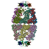
|
|---|---|
| 1 |
|
- Components
| #1: Protein | Mass: 10400.938 Da / Num. of mol.: 14 Source method: isolated from a genetically manipulated source Source: (gene. exp.)   #2: Protein | Mass: 57391.711 Da / Num. of mol.: 14 Source method: isolated from a genetically manipulated source Source: (gene. exp.)   #3: Protein | | Mass: 50538.918 Da / Num. of mol.: 1 Source method: isolated from a genetically manipulated source Source: (gene. exp.)  Rhodospirillum rubrum ATCC 11170 (bacteria) Rhodospirillum rubrum ATCC 11170 (bacteria)Gene: cbbM, Rru_A2400 / Production host:  References: UniProt: Q2RRP5, ribulose-bisphosphate carboxylase Has protein modification | N | |
|---|
-Experimental details
-Experiment
| Experiment | Method: ELECTRON MICROSCOPY |
|---|---|
| EM experiment | Aggregation state: PARTICLE / 3D reconstruction method: single particle reconstruction |
- Sample preparation
| Component |
| ||||||||||||||||||||||||
|---|---|---|---|---|---|---|---|---|---|---|---|---|---|---|---|---|---|---|---|---|---|---|---|---|---|
| Molecular weight | Value: 1 MDa / Experimental value: NO | ||||||||||||||||||||||||
| Source (natural) |
| ||||||||||||||||||||||||
| Source (recombinant) |
| ||||||||||||||||||||||||
| Buffer solution | pH: 7.5 | ||||||||||||||||||||||||
| Specimen | Embedding applied: NO / Shadowing applied: NO / Staining applied: NO / Vitrification applied: YES | ||||||||||||||||||||||||
| Vitrification | Cryogen name: ETHANE |
- Electron microscopy imaging
| Microscopy | Model: JEOL 3200FSC |
|---|---|
| Electron gun | Electron source:  FIELD EMISSION GUN / Accelerating voltage: 300 kV / Illumination mode: FLOOD BEAM FIELD EMISSION GUN / Accelerating voltage: 300 kV / Illumination mode: FLOOD BEAM |
| Electron lens | Mode: BRIGHT FIELD |
| Image recording | Electron dose: 1 e/Å2 / Film or detector model: DIRECT ELECTRON DE-20 (5k x 3k) |
- Processing
| Software | Name: PHENIX / Version: 1.19.1_4122: / Classification: refinement | ||||||||||||||||||||||||
|---|---|---|---|---|---|---|---|---|---|---|---|---|---|---|---|---|---|---|---|---|---|---|---|---|---|
| EM software |
| ||||||||||||||||||||||||
| CTF correction | Type: NONE | ||||||||||||||||||||||||
| Symmetry | Point symmetry: C1 (asymmetric) | ||||||||||||||||||||||||
| 3D reconstruction | Resolution: 7.6 Å / Resolution method: FSC 0.143 CUT-OFF / Num. of particles: 35230 / Symmetry type: POINT | ||||||||||||||||||||||||
| Atomic model building | Protocol: FLEXIBLE FIT / Space: REAL | ||||||||||||||||||||||||
| Atomic model building | 3D fitting-ID: 1 / Source name: PDB / Type: experimental model
| ||||||||||||||||||||||||
| Refine LS restraints |
|
 Movie
Movie Controller


 PDBj
PDBj
