 Movie
Movie Controller

[English] 日本語
 Yorodumi
- PDB-7vh5: Cryo-EM structure of the hexameric plasma membrane H+-ATPase in t... -
+ Open data

- Basic information
| Entry | Database: PDB / ID: 7vh5 | |||||||||||||||||||||||||||
|---|---|---|---|---|---|---|---|---|---|---|---|---|---|---|---|---|---|---|---|---|---|---|---|---|---|---|---|---|
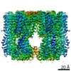
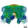
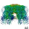
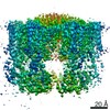
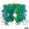
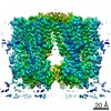
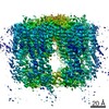
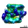
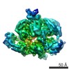
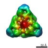
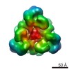
| Title | Cryo-EM structure of the hexameric plasma membrane H+-ATPase in the autoinhibited state (pH 7.4, C1 symmetry) | |||||||||||||||||||||||||||
 Components Components | Plasma membrane ATPase 1 | |||||||||||||||||||||||||||
 Keywords Keywords | TRANSLOCASE / P type ATPase / plasma membrane H+-ATPase / hexamer | |||||||||||||||||||||||||||
| Function / homology |  Function and homology information Function and homology informationP-type H+-exporting transporter / eisosome / proton export across plasma membrane / proteasome storage granule assembly / P-type proton-exporting transporter activity / positive regulation of TORC1 signaling / proton transmembrane transport / regulation of intracellular pH / transmembrane transport / membrane raft ...P-type H+-exporting transporter / eisosome / proton export across plasma membrane / proteasome storage granule assembly / P-type proton-exporting transporter activity / positive regulation of TORC1 signaling / proton transmembrane transport / regulation of intracellular pH / transmembrane transport / membrane raft / ATP hydrolysis activity / mitochondrion / ATP binding / metal ion binding / plasma membrane / cytosol Similarity search - Function | |||||||||||||||||||||||||||
| Biological species |  | |||||||||||||||||||||||||||
| Method | ELECTRON MICROSCOPY / single particle reconstruction / cryo EM / Resolution: 3.2 Å | |||||||||||||||||||||||||||
 Authors Authors | Zhao, P. / Zhao, C. / Chen, D. / Yun, C. / Li, H. / Bai, L. | |||||||||||||||||||||||||||
| Funding support |  China, 1items China, 1items
| |||||||||||||||||||||||||||
 Citation Citation | Journal: Nat Commun / Year: 2021 Title: Structure and activation mechanism of the hexameric plasma membrane H-ATPase. Authors: Peng Zhao / Chaoran Zhao / Dandan Chen / Caihong Yun / Huilin Li / Lin Bai /  Abstract: The S. cerevisiae plasma membrane H-ATPase, Pma1, is a P3A-type ATPase and the primary protein component of the membrane compartment of Pma1 (MCP). Like other plasma membrane H-ATPases, Pma1 ...The S. cerevisiae plasma membrane H-ATPase, Pma1, is a P3A-type ATPase and the primary protein component of the membrane compartment of Pma1 (MCP). Like other plasma membrane H-ATPases, Pma1 assembles and functions as a hexamer, a property unique to this subfamily among the larger family of P-type ATPases. It has been unclear how Pma1 organizes the yeast membrane into MCP microdomains, or why it is that Pma1 needs to assemble into a hexamer to establish the membrane electrochemical proton gradient. Here we report a high-resolution cryo-EM study of native Pma1 hexamers embedded in endogenous lipids. Remarkably, we found that the Pma1 hexamer encircles a liquid-crystalline membrane domain composed of 57 ordered lipid molecules. The Pma1-encircled lipid patch structure likely serves as the building block of the MCP. At pH 7.4, the carboxyl-terminal regulatory α-helix binds to the phosphorylation domains of two neighboring Pma1 subunits, locking the hexamer in the autoinhibited state. The regulatory helix becomes disordered at lower pH, leading to activation of the Pma1 hexamer. The activation process is accompanied by a 6.7 Å downward shift and a 40° rotation of transmembrane helices 1 and 2 that line the proton translocation path. The conformational changes have enabled us to propose a detailed mechanism for ATP-hydrolysis-driven proton pumping across the plasma membrane. Our structures will facilitate the development of antifungal drugs that target this essential protein. | |||||||||||||||||||||||||||
| History |
|
- Structure visualization
| Movie |
Movie viewer |
|---|---|
| Structure viewer | Molecule: MolmilJmol/JSmol |
- Downloads & links
-Download
| PDBx/mmCIF format | 7vh5.cif.gz | 854 KB | Display | PDBx/mmCIF format |
|---|---|---|---|---|
| PDB format | pdb7vh5.ent.gz | 724.3 KB | Display | PDB format |
| PDBx/mmJSON format | 7vh5.json.gz | Tree view | PDBx/mmJSON format | |
| Others |  Other downloads Other downloads |
-Validation report
| Arichive directory | https://data.pdbj.org/pub/pdb/validation_reports/vh/7vh5ftp://data.pdbj.org/pub/pdb/validation_reports/vh/7vh5 | HTTPS FTP |
|---|
-Related structure data
| Related structure data | 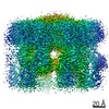 31986MC 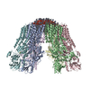 7vh6C M: map data used to model this data C: citing same article ( |
|---|---|
| Similar structure data |

-Links
 PDBj
PDBj


- Assembly
| Deposited unit | 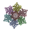
|
|---|---|
| 1 |
|
-Components
| #1: Protein | Mass: 99714.023 Da / Num. of mol.: 6 Source method: isolated from a genetically manipulated source Source: (gene. exp.) Strain: ATCC 204508 / S288c / Gene: PMA1, YGL008C / Production host: References: UniProt: P05030, P-type H+-exporting transporter #2: Chemical | ChemComp-POV / (   Mass: 760.076 Da / Num. of mol.: 51 / Source method: obtained synthetically / Formula: C42H82NO8P / Comment: phospholipid*YM Mass: 760.076 Da / Num. of mol.: 51 / Source method: obtained synthetically / Formula: C42H82NO8P / Comment: phospholipid*YM#3: Chemical | ChemComp-SPH / |   Mass: 299.492 Da / Num. of mol.: 1 / Source method: obtained synthetically / Formula: C18H37NO2 Mass: 299.492 Da / Num. of mol.: 1 / Source method: obtained synthetically / Formula: C18H37NO2Has ligand of interest | N | Has protein modification | N | |
|---|
-Experimental details
-Experiment
| Experiment | Method: ELECTRON MICROSCOPY |
|---|---|
| EM experiment | Aggregation state: PARTICLE / 3D reconstruction method: single particle reconstruction |
- Sample preparation
| Component | Name: Plasma membrane ATPase 1 / Type: COMPLEX / Entity ID: #1 / Source: NATURAL |
|---|---|
| Molecular weight | Value: 99.6 kDa/nm / Experimental value: YES |
| Source (natural) | Organism: |
| Buffer solution | pH: 7.4 |
| Specimen | Embedding applied: NO / Shadowing applied: NO / Staining applied: NO / Vitrification applied: YES |
| Specimen support | Grid material: GOLD / Grid mesh size: 300 divisions/in. / Grid type: Quantifoil R2/1 |
| Vitrification | Cryogen name: ETHANE |
- Electron microscopy imaging
| Experimental equipment |  Model: Titan Krios / Image courtesy: FEI Company |
|---|---|
| Microscopy | Model: FEI TITAN KRIOS |
| Electron gun | Electron source:  FIELD EMISSION GUN / Accelerating voltage: 300 kV / Illumination mode: FLOOD BEAM FIELD EMISSION GUN / Accelerating voltage: 300 kV / Illumination mode: FLOOD BEAM |
| Electron lens | Mode: BRIGHT FIELD |
| Image recording | Electron dose: 1.6 e/Å2 / Film or detector model: GATAN K2 SUMMIT (4k x 4k) |
- Processing
| Software | Name: PHENIX / Version: 1.18.2_3874: / Classification: refinement |
|---|---|
| EM software | Name: PHENIX / Category: model refinement |
| CTF correction | Type: NONE |
| 3D reconstruction | Resolution: 3.2 Å / Resolution method: FSC 0.143 CUT-OFF / Num. of particles: 179392 / Symmetry type: POINT |
