 Movie
Movie Controller


+ Open data

- Basic information
| Entry | Database: PDB / ID: 7sn2 | |||||||||||||||||||||||||||||||||
|---|---|---|---|---|---|---|---|---|---|---|---|---|---|---|---|---|---|---|---|---|---|---|---|---|---|---|---|---|---|---|---|---|---|---|
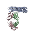
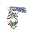
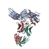
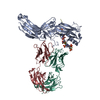
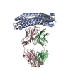
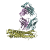
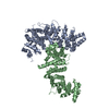
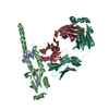
| Title | Structure of human SARS-CoV-2 neutralizing antibody C1C-A3 Fab | |||||||||||||||||||||||||||||||||
 Components Components |
| |||||||||||||||||||||||||||||||||
 Keywords Keywords | IMMUNE SYSTEM / COVID-19 / SARS-CoV-2 / neutralizing antibody / neutralization escape | |||||||||||||||||||||||||||||||||
| Function / homology |  Function and homology information Function and homology informationsymbiont-mediated disruption of host tissue / Maturation of spike protein / Translation of Structural Proteins / Virion Assembly and Release / host cell surface / host extracellular region / symbiont-mediated-mediated suppression of host tetherin activity / Induction of Cell-Cell Fusion / structural constituent of virion / positive regulation of viral entry into host cell ...symbiont-mediated disruption of host tissue / Maturation of spike protein / Translation of Structural Proteins / Virion Assembly and Release / host cell surface / host extracellular region / symbiont-mediated-mediated suppression of host tetherin activity / Induction of Cell-Cell Fusion / structural constituent of virion / positive regulation of viral entry into host cell / membrane fusion / host cell endoplasmic reticulum-Golgi intermediate compartment membrane / Attachment and Entry / entry receptor-mediated virion attachment to host cell / receptor-mediated virion attachment to host cell / host cell surface receptor binding / symbiont-mediated suppression of host innate immune response / endocytosis involved in viral entry into host cell / receptor ligand activity / fusion of virus membrane with host plasma membrane / fusion of virus membrane with host endosome membrane / viral envelope / symbiont entry into host cell / virion attachment to host cell / host cell plasma membrane / SARS-CoV-2 activates/modulates innate and adaptive immune responses / virion membrane / membrane / identical protein binding / plasma membrane Similarity search - Function | |||||||||||||||||||||||||||||||||
| Biological species |   Severe acute respiratory syndrome coronavirus 2 Severe acute respiratory syndrome coronavirus 2 Homo sapiens (human) Homo sapiens (human) | |||||||||||||||||||||||||||||||||
| Method | ELECTRON MICROSCOPY / single particle reconstruction / cryo EM / Resolution: 4.3 Å | |||||||||||||||||||||||||||||||||
 Authors Authors | Pan, J. / Abraham, J. / Yang, P. / Shankar, S. | |||||||||||||||||||||||||||||||||
| Funding support |  United States, 1items United States, 1items
| |||||||||||||||||||||||||||||||||
 Citation Citation | Journal: Science / Year: 2022 Title: Structural basis for continued antibody evasion by the SARS-CoV-2 receptor binding domain. Authors: Katherine G Nabel / Sarah A Clark / Sundaresh Shankar / Junhua Pan / Lars E Clark / Pan Yang / Adrian Coscia / Lindsay G A McKay / Haley H Varnum / Vesna Brusic / Nicole V Tolan / Guohai ...Authors: Katherine G Nabel / Sarah A Clark / Sundaresh Shankar / Junhua Pan / Lars E Clark / Pan Yang / Adrian Coscia / Lindsay G A McKay / Haley H Varnum / Vesna Brusic / Nicole V Tolan / Guohai Zhou / Michaël Desjardins / Sarah E Turbett / Sanjat Kanjilal / Amy C Sherman / Anand Dighe / Regina C LaRocque / Edward T Ryan / Casey Tylek / Joel F Cohen-Solal / Anhdao T Darcy / Davide Tavella / Anca Clabbers / Yao Fan / Anthony Griffiths / Ivan R Correia / Jane Seagal / Lindsey R Baden / Richelle C Charles / Jonathan Abraham /  Abstract: Many studies have examined the impact of severe acute respiratory syndrome coronavirus 2 (SARS-CoV-2) variants on neutralizing antibody activity after they have become dominant strains. Here, we ...Many studies have examined the impact of severe acute respiratory syndrome coronavirus 2 (SARS-CoV-2) variants on neutralizing antibody activity after they have become dominant strains. Here, we evaluate the consequences of further viral evolution. We demonstrate mechanisms through which the SARS-CoV-2 receptor binding domain (RBD) can tolerate large numbers of simultaneous antibody escape mutations and show that pseudotypes containing up to seven mutations, as opposed to the one to three found in previously studied variants of concern, are more resistant to neutralization by therapeutic antibodies and serum from vaccine recipients. We identify an antibody that binds the RBD core to neutralize pseudotypes for all tested variants but show that the RBD can acquire an N-linked glycan to escape neutralization. Our findings portend continued emergence of escape variants as SARS-CoV-2 adapts to humans. | |||||||||||||||||||||||||||||||||
| History |
|
- Structure visualization
| Movie |
Movie viewer |
|---|---|
| Structure viewer | Molecule: MolmilJmol/JSmol |
- Downloads & links
-Download
| PDBx/mmCIF format | 7sn2.cif.gz | 254.8 KB | Display | PDBx/mmCIF format |
|---|---|---|---|---|
| PDB format | pdb7sn2.ent.gz | 189.5 KB | Display | PDB format |
| PDBx/mmJSON format | 7sn2.json.gz | Tree view | PDBx/mmJSON format | |
| Others |  Other downloads Other downloads |
-Validation report
| Arichive directory | https://data.pdbj.org/pub/pdb/validation_reports/sn/7sn2ftp://data.pdbj.org/pub/pdb/validation_reports/sn/7sn2 | HTTPS FTP |
|---|
-Related structure data
| Related structure data |  25209MC  7sn0C  7sn1C  7sn3C M: map data used to model this data C: citing same article ( |
|---|---|
| Similar structure data |

-Links
 PDBj
PDBj


- Assembly
| Deposited unit | 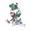
|
|---|---|
| 1 |
|
-Components
| #1: Protein | Mass: 141192.203 Da / Num. of mol.: 1 Mutation: R682G, R683S, R685S, F817P, A892P, A899P, A942P, K986P, V987P Source method: isolated from a genetically manipulated source Details: isolation Wuhan-Hu-1 Source: (gene. exp.) Severe acute respiratory syndrome coronavirus 2Gene: S, 2 / Variant: Wuhan-Hu-1 / Plasmid: pHLSec Cell line (production host): HEK-293 (Thermo Fisher Expi293F) Production host: Homo sapiens (human) / References: UniProt: P0DTC2 |
|---|---|
| #2: Antibody | Mass: 27022.607 Da / Num. of mol.: 1 Source method: isolated from a genetically manipulated source Details: mature antibody (after somatic hypermutation) from germline gene IGHV3-33 Source: (gene. exp.) Homo sapiens (human) / Gene: IGHV3-33 / Plasmid: pVRC8400Cell line (production host): HEK-293 (Thermo Fisher Expi293F) Production host: Homo sapiens (human) |
| #3: Antibody | Mass: 25896.107 Da / Num. of mol.: 1 Source method: isolated from a genetically manipulated source Details: mature antibody (after somatic hypermutation) from germline gene IGKV3-11 Source: (gene. exp.) Homo sapiens (human) / Gene: IGKV3-11 / Plasmid: pVRC8400Cell line (production host): HEK-293T (Thermo Fisher Expi293F) Production host: Homo sapiens (human) |
| #4: Polysaccharide | beta-D-mannopyranose-(1-4)-2-acetamido-2-deoxy-beta-D-glucopyranose-(1-4)-2-acetamido-2-deoxy-beta- ...beta-D-mannopyranose-(1-4)-2-acetamido-2-deoxy-beta-D-glucopyranose-(1-4)-2-acetamido-2-deoxy-beta-D-glucopyranose Source method: isolated from a genetically manipulated source |
| #5: Polysaccharide | 2-acetamido-2-deoxy-beta-D-glucopyranose-(1-2)-alpha-D-mannopyranose-(1-3)-[2-acetamido-2-deoxy- ...2-acetamido-2-deoxy-beta-D-glucopyranose-(1-2)-alpha-D-mannopyranose-(1-3)-[2-acetamido-2-deoxy-beta-D-glucopyranose-(1-2)-alpha-D-mannopyranose-(1-6)]beta-D-mannopyranose-(1-4)-2-acetamido-2-deoxy-beta-D-glucopyranose-(1-4)-2-acetamido-2-deoxy-beta-D-glucopyranose Source method: isolated from a genetically manipulated source |
| Has ligand of interest | N |
| Has protein modification | Y |
-Experimental details
-Experiment
| Experiment | Method: ELECTRON MICROSCOPY |
|---|---|
| EM experiment | Aggregation state: PARTICLE / 3D reconstruction method: single particle reconstruction |
- Sample preparation
| Component |
| ||||||||||||||||||||||||||||
|---|---|---|---|---|---|---|---|---|---|---|---|---|---|---|---|---|---|---|---|---|---|---|---|---|---|---|---|---|---|
| Molecular weight |
| ||||||||||||||||||||||||||||
| Source (natural) |
| ||||||||||||||||||||||||||||
| Source (recombinant) |
| ||||||||||||||||||||||||||||
| Buffer solution | pH: 7.5 | ||||||||||||||||||||||||||||
| Buffer component |
| ||||||||||||||||||||||||||||
| Specimen | Conc.: 0.9 mg/ml / Embedding applied: NO / Shadowing applied: NO / Staining applied: NO / Vitrification applied: YES / Details: this sample was monodisperse. | ||||||||||||||||||||||||||||
| Specimen support | Details: Quantifoil Cu 1.2/1.3 2nm carbon 300 mesh grids glowed discharge at 15 mA in Pelco EasiGlow Grid material: COPPER / Grid mesh size: 300 divisions/in. / Grid type: Quantifoil R1.2/1.3 | ||||||||||||||||||||||||||||
| Vitrification | Instrument: FEI VITROBOT MARK IV / Cryogen name: ETHANE / Humidity: 100 % / Chamber temperature: 293 K / Details: blot for 4-6 seconds |
- Electron microscopy imaging
| Experimental equipment |  Model: Titan Krios / Image courtesy: FEI Company |
|---|---|
| Microscopy | Model: FEI TITAN KRIOS |
| Electron gun | Electron source:  FIELD EMISSION GUN / Accelerating voltage: 300 kV / Illumination mode: FLOOD BEAM FIELD EMISSION GUN / Accelerating voltage: 300 kV / Illumination mode: FLOOD BEAM |
| Electron lens | Mode: BRIGHT FIELD / Nominal magnification: 105000 X / Calibrated magnification: 60606 X / Nominal defocus max: 2500 nm / Nominal defocus min: 1000 nm / Cs: 2.7 mm / C2 aperture diameter: 50 µm / Alignment procedure: COMA FREE |
| Specimen holder | Cryogen: NITROGEN / Specimen holder model: FEI TITAN KRIOS AUTOGRID HOLDER / Temperature (max): 77 K / Temperature (min): 77 K |
| Image recording | Average exposure time: 1.9 sec. / Electron dose: 56.5 e/Å2 / Film or detector model: GATAN K3 BIOQUANTUM (6k x 4k) / Num. of grids imaged: 1 / Num. of real images: 4583 |
| Image scans | Sampling size: 5 µm / Width: 5760 / Height: 4092 |
- Processing
| Software | Name: PHENIX / Version: 1.19_4080: / Classification: refinement | ||||||||||||||||||||||||||||||||
|---|---|---|---|---|---|---|---|---|---|---|---|---|---|---|---|---|---|---|---|---|---|---|---|---|---|---|---|---|---|---|---|---|---|
| EM software |
| ||||||||||||||||||||||||||||||||
| CTF correction | Type: NONE | ||||||||||||||||||||||||||||||||
| Particle selection | Num. of particles selected: 1296929 | ||||||||||||||||||||||||||||||||
| Symmetry | Point symmetry: C1 (asymmetric) | ||||||||||||||||||||||||||||||||
| 3D reconstruction | Resolution: 4.3 Å / Resolution method: FSC 0.143 CUT-OFF / Num. of particles: 344920 / Algorithm: FOURIER SPACE / Num. of class averages: 2 / Symmetry type: POINT | ||||||||||||||||||||||||||||||||
| Refine LS restraints |
|
